Dr Abeera SarfrazDr Muhammad Zubair UTERUS The uterus

Dr Abeera Sarfraz/Dr Muhammad Zubair UTERUS

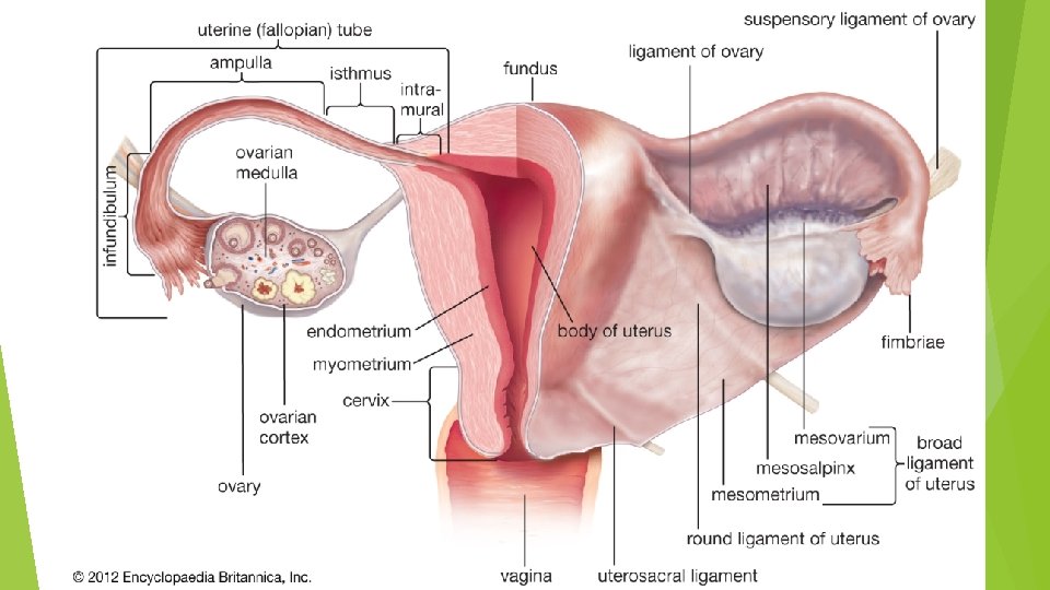
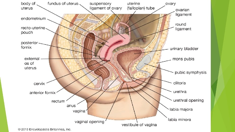
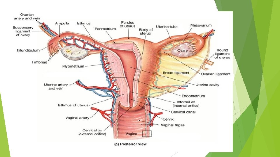
The uterus is a hollow, pear-shaped organ with thick muscular walls. In the young nulliparous adult, it measures about 3 in. (8 cm) long, 2 in. (5 cm) wide, and 1 in. (2. 5 cm) thick. It is divided into the fundus, body, and cervix. The fundus is the part of the uterus that lies above the entrance of the uterine tubes. The body is the part of the uterus that lies below the entrance of the uterine tubes. The cervix is the low, narrow part of the uterus. It pierces the anterior wall of the vagina and is divided into the supravaginal and vaginal parts of the cervix. The cavity of the uterine body is triangular in coronal section, but it is merely a cleft in the sagittal plane. The cavity of the cervix, the cervical canal, communicates with the cavity of the body through the internal os and with that of the vagina through the external os. Before the birth of the first child, the external os is circular. In a parous woman, the vaginal part of the cervix is larger, and the external os becomes a transverse slit so that it possesses an anterior lip and a posterior lip. The uterus is covered with peritoneum except anteriorly, below the level of the internal os, where the peritoneum passes forward onto the bladder. Laterally, a space exists between the attachments of the layers of the broad ligament

The muscular wall, or myometrium, is thick and made up of smooth muscle supported by connective tissue. The endometrium is the mucous membrane lining the body of the uterus. It is continuous above with the mucous membrane lining the uterine tubes and below with the mucous membrane lining the cervix. The endometrium is applied directly to the muscle; there is no submucosa. The endometrium undergoes extensive changes during the menstrual cycle in response to the ovarian hormones. The supravaginal part of the cervix is surrounded by visceral pelvic fascia, which is referred to as the parametrium. The uterine artery crosses the ureter in this fascia on each side of the cervix

RELATIONS Anteriorly: The body of the uterus is related anteriorly to the uterovesical pouch and the superior surface of the bladder. The supravaginal cervix is related to the superior surface of the bladder. The vaginal cervix is related to the anterior fornix of the vagina. Posteriorly: The body of the uterus is related posteriorly to the rectouterine pouch (pouch of Douglas) with coils of ileum or sigmoid colon within it. Laterally: The body of the uterus is related laterally to the broad ligament and the uterine artery and vein. The supravaginal cervix is related to the ureter as it passes forward to enter the bladder. The vaginal cervix is related to the lateral fornix of the vagina. The uterine tubes enter the superolateral angles of the uterus, and the round ligaments of the ovary and of the uterus are attached to the uterine wall just below this level.

UTERINE POSITIONS In most women, the long axis of the uterus is bent forward at about a 90 degree angle to the long axis of the vagina. This position is referred to as anteversion of the uterus. Furthermore, the long axis of the body of the uterus is bent forward at the level of the internal os with the long axis of the cervix. This position is termed anteflexion of the uterus. Thus, in the erect position and with the bladder empty, the uterus lies in an almost horizontal plane. In some women, the fundus and body of the uterus are bent backward on the vagina so that they lie in the rectouterine pouch (pouch of Douglas). In this situation, the uterus is said to be retroverted. If the body of the uterus is, in addition, bent backward on the cervix, it is said to be retroflexed. The uterus serves as a site for the reception, retention, and nutrition of the fertilized ovum.

BLOOD SUPPLY The arterial supply to the uterus is mainly from the uterine artery, a branch of the internal iliac artery. It reaches the uterus by running medially in the base of the broad ligament. It crosses above the ureter at right angles and reaches the cervix at the level of the internal os. The artery then ascends along the lateral margin of the uterus within the broad ligament and ends by anastomosing with the ovarian artery, which also assists in supplying the uterus. The uterine artery gives off a small descending branch that supplies the cervix and the vagina. The uterine vein follows the artery and drains into the internal iliac vein.

LYMPH DRAINAGE The lymph vessels from the fundus of the uterus accompany the ovarian artery and drain into the paraaortic nodes at the level of the first lumbar vertebra. The vessels from the body and cervix drain into the internal and external iliac lymph nodes. A few lymph vessels follow the round ligament of the uterus through the inguinal canal and drain into the superficial inguinal lymph nodes. NERVE SUPPLY Sympathetic and parasympathetic nerves from branches of the inferior hypogastric plexuses innervate the uterus

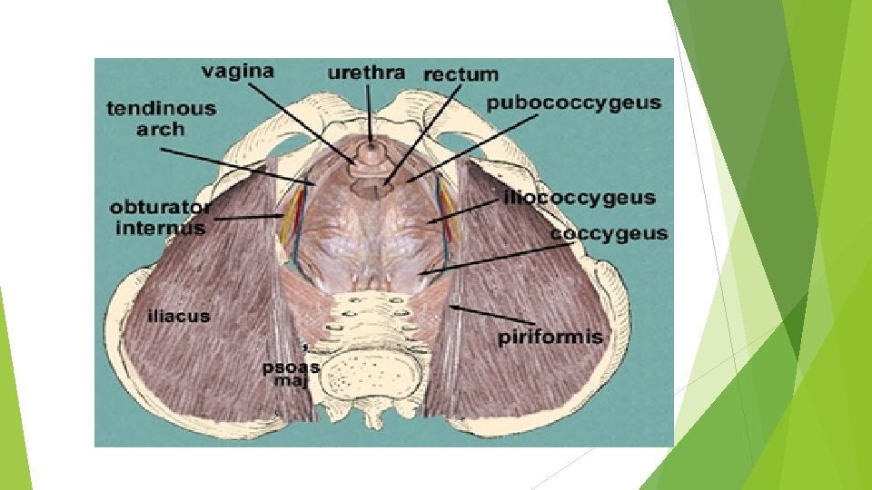
UTERINE SUPPORT The uterus is supported mainly by the tone of the levatores ani muscles and by local condensations of pelvic fascia, which form three important ligaments. Levatores Ani Muscles and Perineal Body Levatores Ani muscles form a broad muscular sheet stretching across the floor of the pelvic cavity, and, together with the pelvic fascia on their upper surface, they effectively support the pelvic viscera and resist the intra-abdominal pressure transmitted downward through the pelvis. The medial edges of the anterior parts of the levatores ani muscles attach to the cervix of the uterus by the pelvic fascia. Some of the fibers of levator ani insert into a fibromuscular structure called the perineal body. This structure is important in maintaining the integrity of the pelvic floor. If the perineal body is damaged during childbirth, prolapse of the pelvic viscera may occur. The perineal body lies in the perineum between the vagina and the anal canal. It is slung up to the pelvic walls by the levatores ani and thus supports the vagina and, indirectly, the uterus.

FASCIAL LIGAMENTS Subperitoneal condensations of pelvic fascia on the upper surface of the levatores ani muscles form three ligaments: transverse cervical, pubocervical, and sacrocervical. These attach to the cervix and the vault of the vagina and play an important role in supporting the uterus and keeping the cervix in its correct position. Transverse Cervical (Cardinal) Ligaments The transverse cervical ligaments are fibromuscular condensations of pelvic fascia that pass to the cervix and the upper end of the vagina from the lateral walls of the pelvis. Pubocervical Ligaments The pubocervical ligaments consist of two firm bands of connective tissue that pass to the cervix from the posterior surface of the pubis. They are positioned on either side of the neck of the bladder, to which they give some support (pubovesical ligaments).

SACROCERVICAL LIGAMENTS The sacrocervical ligaments consist of two firm fibromuscular bands of pelvic fascia that pass to the cervix and the upper end of the vagina from the lower end of the sacrum. They form two ridges, one on either side of the rectouterine pouch. The broad ligaments and the round ligaments of the uterus are lax structures, and the uterus can be pulled up or pushed down for a considerable distance before they become taut. Clinically, they are considered to play a minor role in supporting the uterus. The round ligament of the uterus, which represents the remains of the lower half of the gubernaculum, extends between the superolateral angle of the uterus, through the deep inguinal ring and inguinal canal, to the subcutaneous tissue of the labium majus (see Fig. 9. 11 B). It may help keep the uterus anteverted (tilted forward) and anteflexed (bent forward) but is considerably stretched during pregnancy.

Prepuberty Uterus The fundus and body of the uterus remain small until puberty, when they enlarge greatly in response to the estrogens secreted by the ovaries. Postmenopausal Uterus After menopause, the uterus atrophies and becomes smaller and less vascular. These changes occur because the ovaries no longer produce estrogens and progesterone. Pregnant Uterus During pregnancy, the uterus becomes greatly enlarged as a result of the increasing production of estrogens and progesterone, first by the corpus luteum of the ovary and later by the placenta. At first, it remains as a pelvic organ, but by the 3 rd month, the fundus rises out of the pelvis, and, by the 9 th month, it has reached the xiphoid process (see Fig. 8. 23 B). The increase in size is largely a result of hypertrophy of the smooth muscle fibers of the myometrium, although some hyperplasia takes place.

Role of Uterus in labour Labor, or parturition, is the series of processes by which the baby, the fetal membranes, and the placenta are expelled from the genital tract of the mother. Normally, this process takes place at the end of the 10 th lunar month, at which time, the pregnancy is said to be at term. The cause of the onset of labor is not definitely known. By the end of pregnancy, the contractility of the uterus has been fully developed in response to estrogen, and it is particularly sensitive to the actions of oxytocin at this time. It is possible that the onset of labor is triggered by the sudden withdrawal of progesterone. Once the presenting part (usually the fetal head) starts to stretch the cervix, a nervous reflex mechanism is initiated and increases the force of the contractions of the uterine body. The uterine muscular activity is largely independent of the extrinsic innervation. In women in labor, spinal anesthesia does not interfere with the normal uterine contractions. Severe emotional disturbance, however, can cause premature parturition.

CLINICAL NOTES Bimanual Uterine Pelvic Examination A bimanual examination can provide a great deal of useful clinical information about the state of the uterus, uterine tubes, and ovaries. The examination is easiest in parous women who are able to relax while the examination is in progress. It may be performed under an anesthetic in patients in whom it causes distress. With the bladder empty, the vaginal portion of the cervix is first palpated with the index finger of the right hand. The external os is circular in the nulliparous woman but has anterior and posterior lips in the multiparous woman. The cervix normally has the consistency of the end of the nose, but in the pregnant uterus, it is soft and vascular and has the consistency of the lips. The left hand is then placed gently on the anterior abdominal wall above the symphysis pubis, and the fundus and body of the uterus may be palpated between the abdominal and vaginal fingers situated in the anterior fornix. The size, shape, and mobility of the uterus can then be ascertained. In most women, the uterus is anteverted anteflexed. A retroverted, retroflexed uterus can be palpated through the posterior vaginal fornix.

Varicose Veins and Hemorrhoids in Pregnancy Varicose veins and hemorrhoids are common conditions in pregnancy. The following factors probably contribute to their cause: pressure of the gravid uterus on the inferior vena cava and the inferior mesenteric vein, impairing venous return, and increased progesterone levels in the blood, leading to relaxation of the smooth muscle in the walls of the veins and venous dilatation. Hysterectomy and Damage to Ureters During the surgical procedure of hysterectomy, great care must be exercised to not damage the ureters. When the surgeon is looking for the uterine artery on each side at the base of the broad ligament, it is essential that he or she first identifies the ureter before clamping and tying off the artery. The uterine artery passes forward from the internal iliac artery and crosses the ureter at right angles to reach the cervix at the level of the internal os.

Emergency Cesarean Section An emergency cesarean section is rarely performed. However, a physician may need to perform this surgery in cases in which the mother may die after suffering a severe traumatic incident. Following maternal death, placental circulation ceases, and the child must be delivered within 10 minutes. Neonatal survival is rare after a delay of more than 20 minutes. Anatomy of Technique 1. The bladder is emptied, and an indwelling catheter is left in position. This allows the empty bladder to sink down away from the operating field. 2. A midline skin incision is made that extends from just below the umbilicus to just above the symphysis pubis. The following structures are then incised: superficial fascia (fatty layer, then the membranous layer); deep fascia (thin layer); linear alba; transversalis fascia; extraperitoneal fatty layer; and parietal peritoneum. To avoid damaging loops of the small intestine or the greater omentum, which might be lying beneath the parietal peritoneum, a fold of peritoneum is raised between two hemostats; an incision is then made between the hemostats.

3. The bladder is identified, and a cut is made in the floor of the uterovesical pouch. The bladder is then separated from the lower part of the body of the uterus and depressed downward into the pelvis. 4. The uterus is palpated to identify the presenting part of the fetus. 5. A transverse incision about 1 in. (2. 5 cm) long is made into the exposed lower segment of the body of the uterus. Care is taken that the uterine wall is not immediately penetrated and the fetus injured. 6. When the uterine cavity is entered, the amniotic cavity is opened, and amniotic fluid spurts. The uterine incision is then enlarged sufficiently to deliver the head and trunk of the fetus. When possible, the large tributaries and branches of the uterine vessels in the myometrial wall are avoided. Great care has to be taken to avoid the large uterine arteries that course along the lateral margin of the uterus. 7. Once the fetus is delivered, the umbilical cord is clamped and divided. 8. The contracting uterus will cause the placenta to bulge through the uterine incision. The placenta and fetal membranes are then delivered. 9. The uterine incision is closed with a full-thickness continuous suture. The peritoneum over the bladder and lower part of the uterine body is then repaired to restore the integrity of the uterovesical pouch. Finally, the abdominal wall incision is closed in layers.

UTERINE PROLAPSE The levatores ani muscles and the subperitoneal fascial ligaments are critical in supporting the uterus and in positioning the cervix within the pelvic cavity. Damage to these structures during childbirth or general poor body muscular tone may result in downward displacement of the uterus called uterine prolapse. This condition occurs most commonly after menopause, when the visceral pelvic fascia tends to atrophy along with the pelvic organs. In advanced cases, the cervix descends the length of the vagina and may protrude through the orifice. Because the cervix is attached to the vaginal vault, prolapse of the uterus is always accompanied by some degree of vaginal prolapse.

PERITONEUM The peritoneum in the female, as in the male, is best understood by tracing it around the pelvis in a sagittal plane. The parietal peritoneum passes down from the anterior abdominal wall onto the upper surface of the urinary bladder. It then turns back onto the anterior surface of the uterus, at the level of the internal os. This reflection creates the shallow uterovesical pouch between the superior wall of the bladder and the anterior wall of the uterus. The peritoneum now passes upward over the anterior surface of the body and fundus of the uterus and then downward over the posterior surface. It continues downward and covers the upper part of the posterior surface of the vagina and then passes onto the front of the rectum, as in the male. The contour of the peritoneal reflection from the vagina onto the rectum creates a deep recess, the rectouterine pouch (pouch of Douglas). In the female, the rectouterine pouch is the lowest part of the abdominopelvic peritoneal cavity in the erect position.

BROAD LIGAMENTS The broad ligaments are two-layered folds of peritoneum that extend across the pelvic cavity from the lateral margins of the uterus to the lateral pelvic walls, covering the body and fundus of the uterus, the uterine tubes, and the ovaries. Superiorly, the two layers are continuous and form the upper free edge. Inferiorly, at the base of the ligament, the layers separate to cover the pelvic floor. The connective tissue that fills the gap between the separated layers of the broad ligament is the parametrium. The part of the broad ligament that attaches to the lateral margins of the uterus is the mesometrium. The ovary attaches to the posterior layer of the broad ligament by the mesovarium. The part of the broad ligament that lies lateral to the attachment of the mesovarium forms the suspensory ligament of the ovary. The part of the broad ligament that attaches to the uterine tube and runs between the uterine tube and the mesovarium is the mesosalpinx.

The uterine artery crosses the ureter at the base of the broad ligament. This is a significant relationship in surgery in this area. Remember this by recalling that water (ureter) runs under the bridge (artery). Each broad ligament contains the following: The uterine tube in its upper free border. The round ligament of the ovary and the round ligament of the uterus. These represent the remains of the gubernaculum. The uterine and ovarian blood vessels, lymph vessels, and nerves. The epoophoron, a vestigial structure that lies in the broad ligament above the attachment of the mesovarium. It represents the remains of the mesonephros. The paroöphoron, also a vestigial structure that lies in the broad ligament just lateral to the uterus. It is also a mesonephric remnant

Rectouterine Pouch and Disease Because the rectouterine pouch is the most inferior part of the entire peritoneal cavity (when the patient is in the standing position), it frequently becomes the site for the accumulation of blood (e. g. , from a ruptured ectopic pregnancy) or pus (e. g. , from a ruptured pelvic appendicitis or in gonococcal peritonitis). Because the pouch lies directly behind the posterior fornix of the vagina, it is easily violated by misguided instruments that pierce the wall of the posterior fornix. Pelvic peritonitis is a danger that may ensue. A needle may be passed into the pouch through the posterior fornix in a culdocentesis. Surgically, the pouch may be entered in a posterior colpotomy (vaginal incision). The interior of the female pelvic peritoneal cavity may be viewed for evidence of disease through an endoscope. The instrument is introduced through a small colpotomy.
- Slides: 25