Doppler effect blood flow in artery Ultrasound imaging

Doppler effect: blood flow in artery

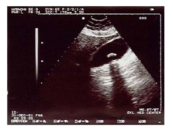
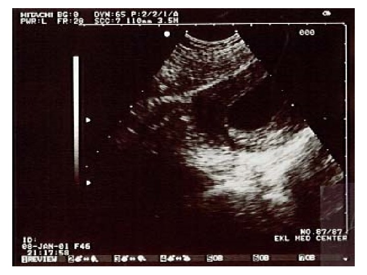
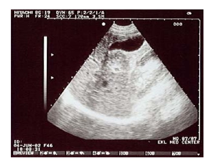
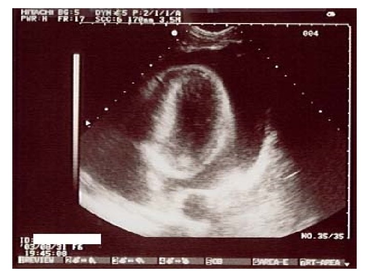
Ultrasound imaging: carotid artery • Doppler imaging looks at artery • Get image and trace of blood flow • This is a healthy artery. The flow is smooth and all in the same direction, like water in a large, slow river

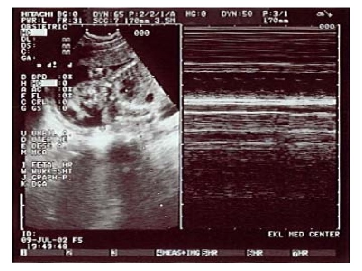
Ultrasound imaging: carotid artery • This is also a carotid artery. • The flow is not all in the same direction. It is turbulent, like rapids in a river. • This is usually due to a build-up of fatty deposits in the artery
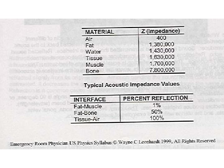

• Hyperechoic – Structure reflects most sound waves – Structure appears white on screen

• Anechoic – Structure allows most sound waves through – Structure appears black on screen

• Echogenic – Tissues in between – Allow some sound waves through and reflect others – Structures appear in various shades of gray depending on amount of reflection


• Homogeneous – Tissue has uniform texture

• Heterogeneous – Various degrees of echogenicity present

• Isoechoic – Two tissues with same of echogenicity


Fig. 16. Curvilinear probe and corresponding ultrasound image.

Fig. 17. Intracavitary probe and corresponding ultrasound image.

Fig. 18. Linear array transducer and corresponding ultrasound image.

Fig. 19. Phased array transducer and corresponding ultrasound image.
 Applications Curvilinear 2–")
Table 1 Transducer type and clinical use Probe Type Frequency (MHz) Applications Curvilinear 2– 5 FAST, renal, aorta, IVC, pelvic, bladder, bowel, appendicitis Linear 6– 15 Ocular, trachea, thyroid, thoracic, vascular access, DVT, MSK, soft tissue, appendicitis Intracavitary 8– 13 Peritonsillar abscess, pelvic Phased array/sector 1– 5 Cardiac, abdominal, renal, pediatric abdomen, bladder, bowel, IVC Abbreviations: DVT, deep venous thrombosis; FAST, focused assessment with sonography in trauma; IVC, inferior vena cava; MSK, musculoskeletal.
 control.")
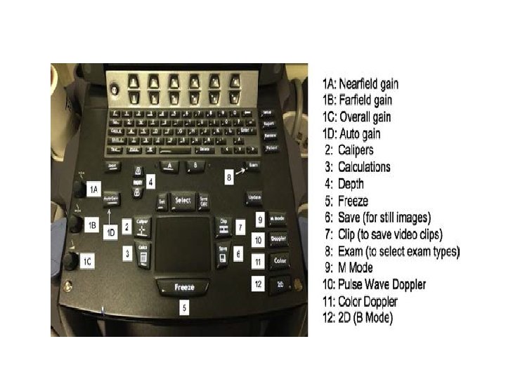
Fig. 23. Time gain compensation (TGC) control.

Fig. 24. Freeze, save, and video clip buttons on an ultrasound machine


- Slides: 53