Done by Supervised by OBJECTIVES Introduction Anatomy Disorders

Done by: Supervised by

OBJECTIVES • • • Introduction. Anatomy. Disorders of glands. Clinical approach. Surgical aspect.

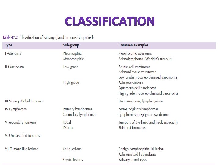
Salivary glands: are composed of 4 major glands, in addition to minor glands. Major: • 2 parotid glands. • 2 submand -ibular gland Minor: • Sublingual. • Multiple minor glands

1. PAROTID GLAND

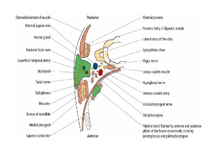
Important structure that run through the parotid gland: 1. Branch of facial nerve. 2. Terminal branch of external carotid artery that divided into maxillary & superficial temporal artery. 3. The retromandibular vein ( post. Facial ). 4. Intraparotid lymph node.
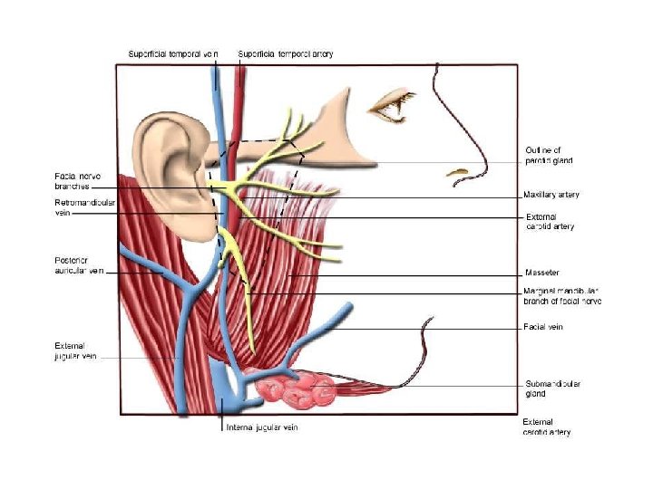

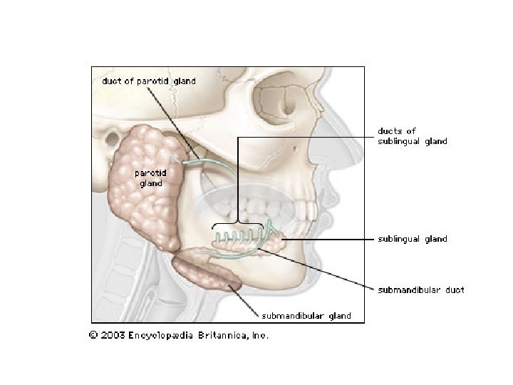
THE PAROTID DUCT: • Stensen’s duct is 5 cm long. • open opposite the second upper molar tooth

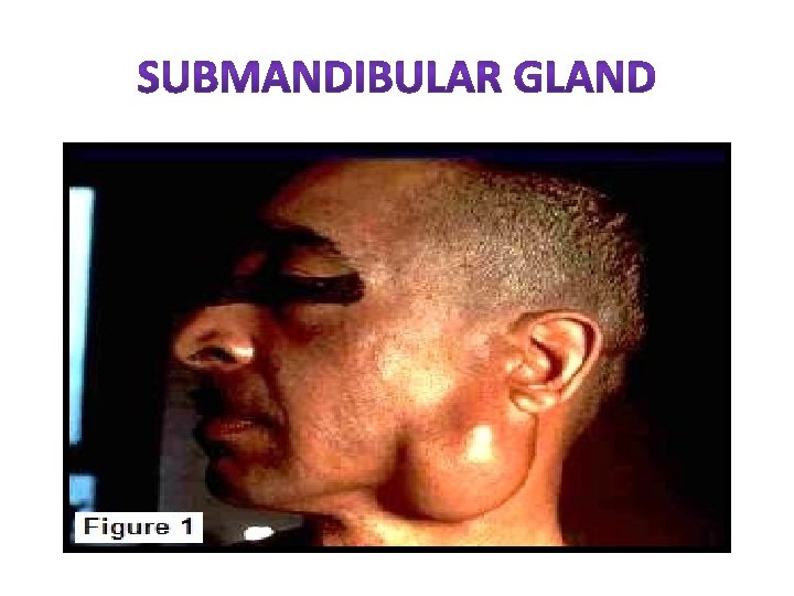
2. SUBMANDIBULAR GLAND • It’s paired of gland that lie below the mandible on either side. • Has 2 lobes, superficial & deep. • Warthon’s duct, drained submandibular gland that opens into anterior floor of mouth. Anatomical relationship: 1. Lingual nerve. 2. Hypoglossal nerve. 3. Anterior facial vein. 4. Facial artery. 5. Marginal mandibular branch of facial nerve.
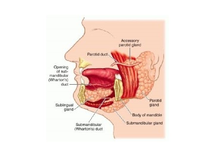

3. SUBLINGUAL GLAND • Lie on the superior surface of the mylohyoid muscle and are separated from the oral cavity by a thin layer of mucosa. • The ducts of the sublingual glands are called Bartholin’s ducts.

4. MINOR SALIVARY GLAND • About 450 lie under the mucosa • They are distirbuted in the mucosa of the lips, cheeks, palate, floor of mouth & retromolar area • Also appear in oropharyanx, larynx & trachea

It’s either: • Extravasation cyst result from trauma to overlying mucosa. • Mucous retention cyst in the floor of the mouth due to obstruction. • RANULA extravasation cyst that arises from sublingual gland.

• It is rare form of mucus retention cyst arise from both sublingual & submandibular. • The mucus collects around the gland &penetrates the mylohyoid diaphragm to enter the neck. Pt. presents with Dumbbell shaped swelling , soft, fluctuant & painless

• Tumors of minor & sublingual salivary gland are extremely rare. • 90% are malignant. • Most common site: upper lip, palate & retromolar region.

• The most common ectopic tissue is called stafne tissue… (what is it? ) • Presentation • Discovered by x-ray: • treatment

sialadenitis Acute chronic Acute on chronic

viral mumps Othe viral infections are extremly rare bacterial Most commonly due to obstruction… Treatment: antibiotics and surgically

• Most common cause is sialolithiasis which 80% happens in the submandibular gland… • Presentation: painful swelling in submandibular area • What would aggreveate it? • Clinical findings: tender, pus draining • investigations : x-ray • Treatment: surgical

• They are very rare in this gland 50% are benign… • Presentation • Investigations: CT and MRI… • Never do open biopsy but do FNA. . • Treatment is surgical…

• They extremly rare like agenesis, , duct atresia and congenital fistula formation…

A- viral infections: Mumps… Mode of infection Prodromal period Presentation Diagnosis Treatment is conservative Complications: Orchitis, oophoritis, pancreatitis, sensorineural deafness, nemimgoencephalitis but they are rare…

B- bacterial: Precipitating factors? ? ! Causative organisms Presentation Treatment : conservative and it might eed drainage…

• This occurs in 3 -6 years of age and the symptoms last for 3 -7 days accompanied with fever and malaise… • Diagnosis is made by HX and sialography showing a characteristic snowstorm appearance… • Treatment: -antibiotics -prophylactic antibiotics -parotidectomy. .
? - It is pathognomonic for HIV… • Presentation : very")
C- chronic parotitis (HIV)? - It is pathognomonic for HIV… • Presentation : very similar to sjogran’s syndrome… • Differentiated by negative autoantibody… • On investigation : CT and MRI show characteristic swiss cheese appearance of the cysts…

treatment: Surgery to improve the appearance although it’s painless

A- papillary obstruction: It less common than in submandibular gland… Most commonly due to trauma Presentation Treatment is papillotomy…

B- stone formation: As mentioned before it is 80% in submandibular but only 20 % in parotid Investigations: position… Treatment is surgical…

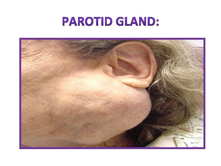
• The parotids are the commonest glands for tumors of salivary glands…

Slowly painless growing temor below the ear, or infront of it

Sometimes on the upper aspect of the neck:

• If it arised from the accessory lobe it will look like a presistant cheek swelling… • If it arises from the deep lobe it will present as parapharyngeal mass… • Symptoms: • Difficult swallowing • Snoring • Clinical examination…

• Investigations: CT AND MRI FNA OPEN BIOPSY IS CONTRAINDICATED… TREATMENT: SURGICAL…

1 -granulomatous sialadenitis: • Mycobacterial infection: • Sarcoidosis • Cat scratch disease • Toxoplasmosis • Syphilis • Deep mycosis • Wgner’s granulomatosis • Allergic sialdenitis due to radiotherapy of the head and neck…

• They are a group of diseases that are hard to diagnose and are not under any group of the other diseases: • Sialadenosis • Adenomatoid hyperplasia • Multifocal adenomporphic adenomatosis

• • Sjogran’s syndrome…: Benign lymphoepithilial lesions Xerostomia Sialorrhea

• It is an autoimmune condition causing progressive destruction of the salivary glands and the lacrimal glands…. . • Presentation is xerostomia and keratoconjunctivitis… • They also present with pain and asendng infection • . females more than males 10: 1 • Parotis is more common
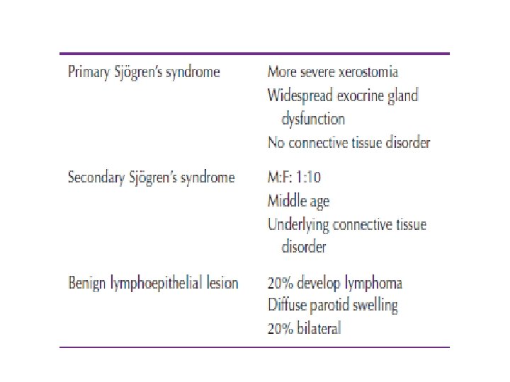

• The charachtaristic feature is progressive lymphocytic infiltration acinar cell destruction and prolifration of duct epithilium… • Diagnosis based on history… • Treatment remains symptomatic: Artificial tears… Salivary substitiuants or water… Floride to avoid dental carries… Complications are B cell lymphoma

• Normal salivary flow decreases with age… • Mostly in woman postmenopausal complaining of burning tongue of mouth. . • Causes: -chronic anxiety and depression. . -dehydration… -anticholinergic drugs… -sjogran’s syndrome… -radiotherapy of the neck and head

• Causes: some infections and drugs… • Drooling: In children that are mental handicap Also in cerebral palsy Management is surgical… Bilateral submandibular duct repositioning and sublingual duct excision… Bilateral submandibualr gland excision and repositioning of the parotis duct…

• History. • Clinical examination. • Investigation.

HISTORY • • • History of swellings / change over time? Trismus? Pain? Variation with meals? Bilateral? Dry mouth? Dry eyes? Recent exposure to sick contacts (mumps)? Radiation history? Current medications?
 • Diffuse or focal enlargement •")
CLINCAL EX. INSPECTION: • Asymmetry (glands, face, neck) • Diffuse or focal enlargement • Erythema extra-orally • Trismus • Medial displacement of structures intraorally? • Cranial nerve testing ( Facial , Hypoglossal nerve)

CLINCAL EX. PALPATION: • Palpate for cervical lymphadenopathy • Bimanual palpation of floor of mouth in a posterior to anterior direction – Have patient close mouth slightly & relax oral musculature to aid in detection – Examine for duct purulence • Bimanual palpation of the gland (firm or spongy/elastic).

INVESTIGATION 1. 2. 3. 4. 5. 6. Plain occlusal film. CT Scan. Ultrasound. Sialography. Radionuclide Studies. Diagnostic Sialendoscopy 2

1. PLAIN OCCLUSAL FILM • Effective for intraductal stones, while…. • intraglandular, radiolucent or small stones may be missed.

2. CT-SCAN • Large stones or small CT slices done. • Also used for inflammatory disorders 3. ULTRASOUND • Operator dependent, can detect small stones (>2 mm), inexpensive, non-invasive

4. SIALAOGRAPHY • Consists of opacification of the ducts by a retrograde injection of a water-soluble dye. • Provides image of stones and duct morphological structure • May be therapeutic, but success of therapeutic sialography never documented

4. SIALAOGRAPHY, CONT • Disadvantages: – – – Irradiation dose Pain with procedure Perforation Infection dye reaction Push stone further Contraindicated in active infection.

5. RADIONUCLIDE STUDY • is useful preoperatively to determine if gland is functional.

6. SIALENDOSCOPY • Allows complete exploration of the ductal system, direct visualization of duct pathology • Success rate of >95%2 • Disadvantage: technically challenging, trauma could result in stenosis, perforation

A-stone removal: -submandibular gland
")
1 -SUBMANDIBULAR GLAND -intracapsular dissection -extracapsular dissection…(suprehyoid neck dissection)

• So what are the indications of removal of the submandibular gland? ? ?

• • Structures to be preserved: Facial nerve marginal branch Platysma muscle fibers… Facial artery Hypoglossal nerve… Lingual nerve Anterior facial vein should be ligated

• • • Hematoma wound infection marginal mandibular nerve injury lingual nerve injusry hypoglossal nerve injury transection of the nerve to the myelohyoid muscle causing submental skin anesthesia…

• Superficial parotidectomy: • If the tumor lies in the superficial lobe a superficial peotidectomy should be performed with preserving the facial nerve… • It is the commonest procedure…

• 1 -the inferior portion of the cartilaginous canal called conley’s pointer the facial nerve lies 1 cm deep and inferior to it’s tip • 2 -the upper border of the posterior belly of the digastric muscle… • The facial nerve is superior to it… • A nerve stimulator might come in handy…

Whole gland is remover Facial nerve is transected Masseter muscle removed Neck dissection

• • Hematoma Infection Temporary facial nerve weakness. Transection of the facial nerve and permenant facial weakness. . Sialocele… Facial numbness. Permenant numbness of the ear lobe due to transection of the great auricular nerve… Frey’s syndrome

• • • Cause… Prevention… Treatment is incidence… Antiperspirants like ALCL Denervation by tympanic neurectomy Injection of botulinum toxin to the skin area

refrences • • Baily and love’s Schwart’s Browse Manual of clinical syrgery…

- Slides: 68