
Dominant Characteristics of Osteogenesis Imperfecta Brent Lehman Molecular
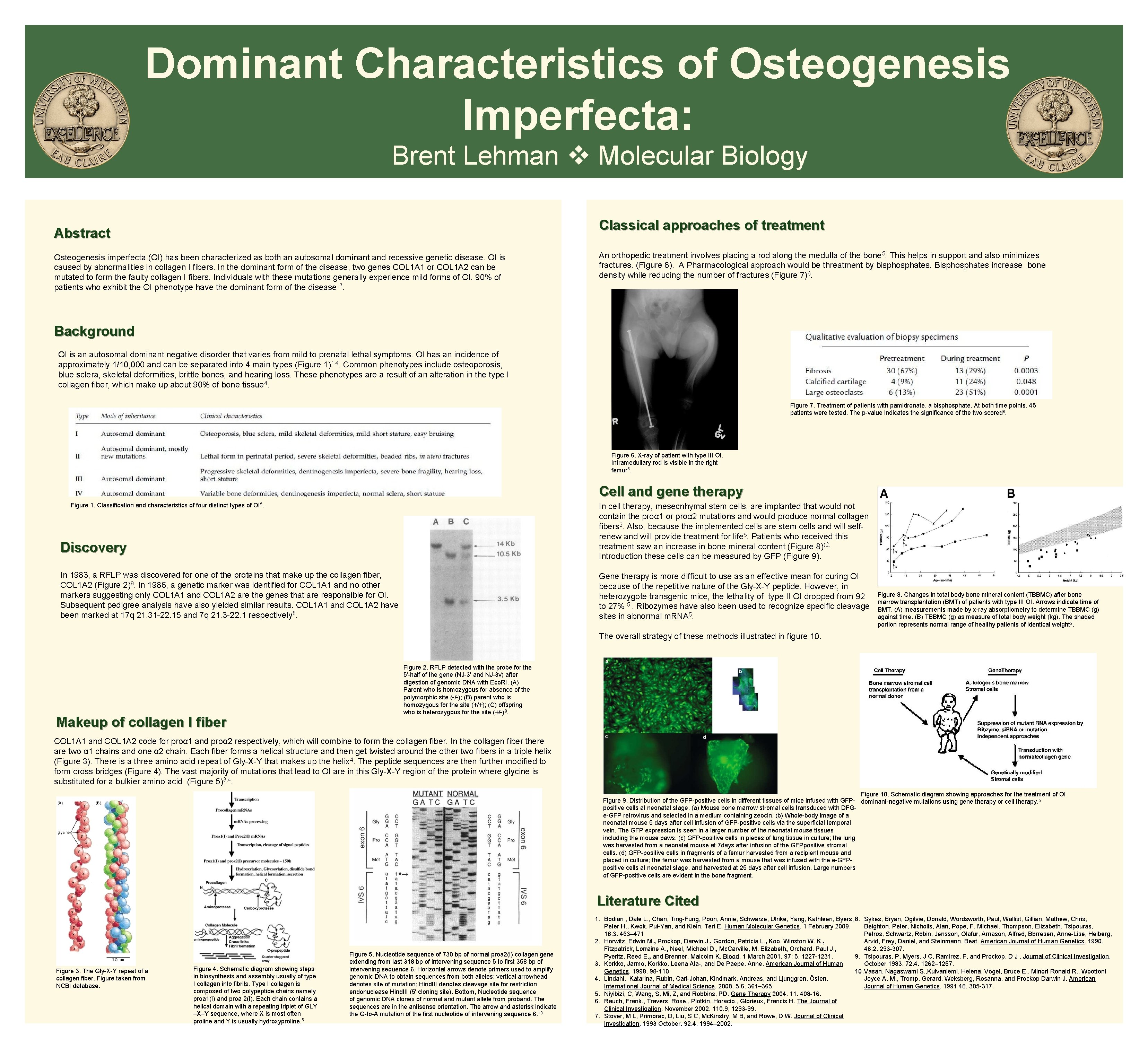
Dominant Characteristics of Osteogenesis Imperfecta: Brent Lehman Molecular Biology Classical approaches of treatment Abstract Osteogenesis imperfecta (OI) has been characterized as both an autosomal dominant and recessive genetic disease. OI is caused by abnormalities in collagen I fibers. In the dominant form of the disease, two genes COL 1 A 1 or COL 1 A 2 can be mutated to form the faulty collagen I fibers. Individuals with these mutations generally experience mild forms of OI. 90% of patients who exhibit the OI phenotype have the dominant form of the disease 7. An orthopedic treatment involves placing a rod along the medulla of the bone 5. This helps in support and also minimizes fractures. (Figure 6). A Pharmacological approach would be threatment by bisphosphates. Bisphosphates increase bone density while reducing the number of fractures (Figure 7)6. Background OI is an autosomal dominant negative disorder that varies from mild to prenatal lethal symptoms. OI has an incidence of approximately 1/10, 000 and can be separated into 4 main types (Figure 1)1, 4. Common phenotypes include osteoporosis, blue sclera, skeletal deformities, brittle bones, and hearing loss. These phenotypes are a result of an alteration in the type I collagen fiber, which make up about 90% of bone tissue 4. Figure 7. Treatment of patients with pamidronate, a bisphosphate. At both time points, 45 patients were tested. The p-value indicates the significance of the two scored 6. Figure 6. X-ray of patient with type III OI. Intramedullary rod is visible in the right femur 5. Cell and gene therapy Figure 1. Classification and characteristics of four distinct types of OI 5. In cell therapy, mesecnhymal stem cells, are implanted that would not contain the proα 1 or proα 2 mutations and would produce normal collagen fibers 2. Also, because the implemented cells are stem cells and will selfrenew and will provide treatment for life 5. Patients who received this treatment saw an increase in bone mineral content (Figure 8))2. Introduction these cells can be measured by GFP (Figure 9). Discovery In 1983, a RFLP was discovered for one of the proteins that make up the collagen fiber, COL 1 A 2 (Figure 2)9. In 1986, a genetic marker was identified for COL 1 A 1 and no other markers suggesting only COL 1 A 1 and COL 1 A 2 are the genes that are responsible for OI. Subsequent pedigree analysis have also yielded similar results. COL 1 A 1 and COL 1 A 2 have been marked at 17 q 21. 31 -22. 15 and 7 q 21. 3 -22. 1 respectively 8. Gene therapy is more difficult to use as an effective mean for curing OI because of the repetitive nature of the Gly-X-Y peptide. However, in heterozygote transgenic mice, the lethality of type II OI dropped from 92 to 27% 5. Ribozymes have also been used to recognize specific cleavage sites in abnormal m. RNA 5. Figure 8. Changes in total body bone mineral content (TBBMC) after bone marrow transplantation (BMT) of patients with type III OI. Arrows indicate time of BMT. (A) measurements made by x-ray absorptiometry to determine TBBMC (g) against time. (B) TBBMC (g) as measure of total body weight (kg). The shaded portion represents normal range of healthy patients of identical weight 2. The overall strategy of these methods illustrated in figure 10. Makeup of collagen I fiber Figure 2. RFLP detected with the probe for the 5'-half of the gene (NJ-3' and NJ-3 v) after digestion of genomic DNA with Eco. RI. (A) Parent who is homozygous for absence of the polymorphic site (-/-); (B) parent who is homozygous for the site (+/+); (C) offspring who is heterozygous for the site (+/-)9. COL 1 A 1 and COL 1 A 2 code for proα 1 and proα 2 respectively, which will combine to form the collagen fiber. In the collagen fiber there are two α 1 chains and one α 2 chain. Each fiber forms a helical structure and then get twisted around the other two fibers in a triple helix (Figure 3). There is a three amino acid repeat of Gly-X-Y that makes up the helix 4. The peptide sequences are then further modified to form cross bridges (Figure 4). The vast majority of mutations that lead to OI are in this Gly-X-Y region of the protein where glycine is substituted for a bulkier amino acid (Figure 5)3, 4. Figure 10. Schematic diagram showing approaches for the treatment of OI Figure 9. Distribution of the GFP-positive cells in different tissues of mice infused with GFP- dominant-negative mutations using gene therapy or cell therapy. 5 positive cells at neonatal stage. (a) Mouse bone marrow stromal cells transduced with DFGe-GFP retrovirus and selected in a medium containing zeocin. (b) Whole-body image of a neonatal mouse 5 days after cell infusion of GFP-positive cells via the superficial temporal vein. The GFP expression is seen in a larger number of the neonatal mouse tissues including the mouse paws. (c) GFP-positive cells in pieces of lung tissue in culture; the lung was harvested from a neonatal mouse at 7 days after infusion of the GFPpositive stromal cells. (d) GFP-positive cells in fragments of a femur harvested from a recipient mouse and placed in culture; the femur was harvested from a mouse that was infused with the e-GFPpositive cells at neonatal stage, and harvested at 25 days after cell infusion. Large numbers of GFP-positive cells are evident in the bone fragment. Literature Cited Figure 3. The Gly-X-Y repeat of a collagen fiber. Figure taken from NCBI database. Figure 4. Schematic diagram showing steps in biosynthesis and assembly usually of type I collagen into fibrils. Type I collagen is composed of two polypeptide chains namely proa 1(I) and proa 2(I). Each chain contains a helical domain with a repeating triplet of GLY –X–Y sequence, where X is most often proline and Y is usually hydroxyproline. 5 Figure 5. Nucleotide sequence of 730 bp of normal proa 2(I) collagen gene extending from last 318 bp of intervening sequence 5 to first 358 bp of intervening sequence 6. Horizontal arrows denote primers used to amplify genomic DNA to obtain sequences from both alleles; vertical arrowhead denotes site of mutation; Hind. III denotes cleavage site for restriction endonuclease Hind. III (5' cloning site). Bottom, Nucleotide sequence of genomic DNA clones of normal and mutant allele from proband. The sequences are in the antisense orientation. The arrow and asterisk indicate the G-to-A mutation of the first nucleotide of intervening sequence 6. 10 1. Bodian , Dale L. , Chan, Ting-Fung, Poon, Annie, Schwarze, Ulrike, Yang, Kathleen, Byers, 8. Sykes, Bryan, Ogilvie, Donald, Wordsworth, Paul, Wallist, Gillian, Mathew, Chris, Peter H. , Kwok, Pui-Yan, and Klein, Teri E. Human Molecular Genetics. 1 February 2009. Beighton, Peter, Nicholls, Alan, Pope, F. Michael, Thompson, Elizabeth, Tsipouras, 18. 3. 463– 471 Petros, Schwartz, Robin, Jensson, Olafur, Arnason, Alfred, Bbrresen, Anne-Lise, Heiberg, 2. Horwitz, Edwin M. , Prockop, Darwin J. , Gordon, Patricia L. , Koo, Winston W. K. , Arvid, Frey, Daniel, and Steinmann, Beat. American Journal of Human Genetics. 1990. Fitzpatrick, Lorraine A. , Neel, Michael D. , Mc. Carville, M. Elizabeth, Orchard, Paul J. , 46. 2. 293 -307. Pyeritz, Reed E. , and Brenner, Malcolm K. Blood. 1 March 2001, 97: 5, 1227 -1231. 9. Tsipouras, P, Myers, J C, Ramirez, F, and Prockop, D J. Journal of Clinical Investigation. 3. Korkko, Jarmo, Korkko, Leena Ala-, and De Paepe, Anne. American Journal of Human October 1983. 72. 4. 1262– 1267. Genetics. 1998. 98 -110 10. Vasan, Nagaswami S. , Kuivaniemi, Helena, Vogel, Bruce E. , Minort Ronald R. , Woottont 4. Lindahl, Katarina, Rubin, Carl-Johan, Kindmark, Andreas, and Ljunggren, Östen. Joyce A. M. , Tromp, Gerard, Weksberg, Rosanna, and Prockop Darwin J. American International Journal of Medical Science. 2008. 5. 6. 361– 365. Journal of Human Genetics. 1991 48. 305 -317. 5. Niyibizi, C, Wang, S, Mi, Z, and Robbins, PD. Gene Therapy 2004. 11. 408 -16. 6. Rauch, Frank. , Travers, Rose. , Plotkin, Horacio. , Glorieux, Francis H. The Journal of Clinical Investigation. November 2002. 110. 9, 1293 -99. 7. Stover, M L, Primorac, D, Liu, S C, Mc. Kinstry, M B, and Rowe, D W. Journal of Clinical Investigation. 1993 October. 92. 4. 1994– 2002.
- Slides: 1