DO NOW Turn in EndocrineNeurotransmitters handout Pick up


 the thalamus; directs several maintenance activities like eating,")


 ●storing long term memories")

 ●")


- Slides: 35
DO NOW Turn in Endocrine/Neurotransmitters handout Pick up brain graphic organizer Complete the NERVOUS NELLIE warm up and complete with your elbow partner.
The Brain – Studying & Structures Unit 3 – pg. 66 -79 Modified Power. Point from: Aneeq Ahmad -- Henderson State University. Worth Publishers © 2007
Learning Objectives ●Describe the nervous system and its subdivisions and functions: o major brain regions, lobes, and cortical areas; o brain lateralization and hemispheric specialization. ●Recount historic and contemporary research strategies and technologies that support research (e. g. , case studies, split-brain research, imaging techniques).
Brain Charts and Diagrams ●Have out the Brain Structure Chart- you'll fill in most of it with this Power. Point. What you don't get, you'll need to find in the book or from an app called "3 D Brain. " Download this on your phone
Techniques to Study the Brain lesion experimentally destroys brain tissue to study animal behaviors after such destruction. Usually lesions are done for scientific or medicinal purposes. Hubel (1990)
Clinical Observation Clinical observations have shed light on a number of brain disorders. Alterations in brain morphology due to neurological and psychiatric diseases are now being catalogued. Tom Landers/ Boston Globe
f. MRI Scan When the subject is in the scanner functional magnetic resonance imaging (f. MRI), the researchers will be able to communicate with him using an intercom system and a visual projection system. The image of the brain depicts, with colors of the rainbow, the amount of blood flow in each part of the brain, which indicates the amount of neural
STRUCTURES OF THE BRAIN – Diagram 1 Reticular Formation
STRUCTURES OF THE BRAIN – Diagram 2
Older Brain Structures Brainstem the oldest part of the brain, beginning where the spinal cord swells and enters the skull. Responsible for automatic survival functions.
Brain Stem Medulla: base of the brainstem, controls vital functions like heartbeat and breathing. Reticular Formation: a nerve network in the brainstem that plays an important role in controlling arousal & involved in attention and sleep (filtering out stimuli)
Brain Stem Thalamus: the brain’s sensory switchboard --directs messages to the sensory areas in the cortex and transmits replies to the cerebellum and medulla. Hal and Amos are traffic cops… Pons: bridge between the brain and spinal cord (especially dealing with motor messages) and/or sleeping (sleep cycle)
Cerebellum The “little brain” attached to the rear of the brainstem. It helps coordinate voluntary movements and balance.
The Limbic System a doughnut-shaped system of neural structures at the border of the brainstem and cerebrum, associated with emotions such as fear, aggression and drives for food and sex. It includes the hippocampus, amygdala, and hypothalamus.
Limbic System Amygdala: two almondshaped neural clusters linked to emotion of fear and anger. Hippocampus: two fingerlike structures attached to amygdala involved in processing (new) memories
Limbic System Hypothalamus: lies below (hypo) the thalamus; directs several maintenance activities like eating, drinking, body temperature, and sex. Helps govern the endocrine system via the pituitary gland. Ventromedial – “vomit” – tells you when to STOP eating Lateral – “let’s eat” – tell you when you are hungry
5 Minute Break—handout on cerebral cortex
Accounts for about 80% of brain’s total mass and does most of the sophisticated information processing in the brain
The Cerebral Cortex Corpus callosum: band of nerves that connect the two cortical hemispheres & carries messages between them
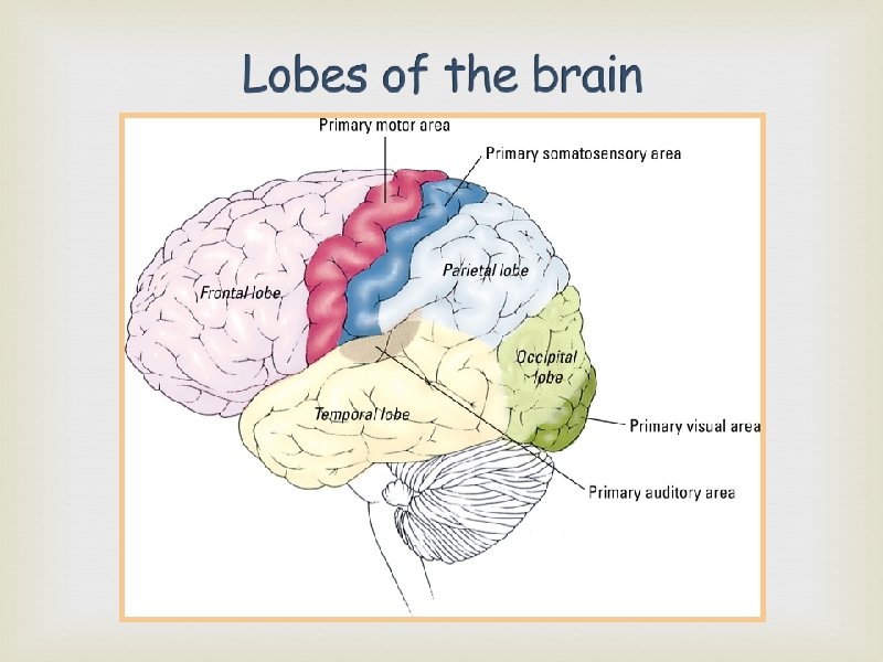
Each brain hemisphere is divided into four lobes, almost identical in each hemisphere, separated by prominent fissures. They are: ●frontal lobes (forehead) ●parietal lobes (top to rear head) ●occipital lobes (back head) ●temporal lobes (side of head)
Cerebral Cortex: FRONTAL LOBE The frontal lobe controls functions like: ●Judgment planning ●producing speech sounds temperament emotions (controlling them) personality ●movement (motor cortex)Works w/the motor cortex to make precise movements
PHINEAS GAGE In 1848 Gage was a respected, polite railroad foreman who was working to clear a path on the rails. As Gage was filling a hole with dynamite, it exploded sending a rod (3 ft. long) into Gage’s head (entering beneath his left eye and exiting through the top of his skull) He survived the accident -- no speech or motor difficulties and his memory was intact BUT… his personality was greatly changed – he became difficult to be around, short-tempered & often said inappropriate things o frontal lobed damaged…this prevented the censoring of thoughts and ideas.
PHINEAS GAGE
The temporal lobes control functions like: ●hearing (primary auditory cortex) ●storing long term memories ●speech and language Understanding “tempo” as your mnemonic device above your ear…where temporal lobe is located
The parietal lobes control functions like somatosensory cortex ●body position ●spatial reasoning like o Touch pressure o Temperature pain
The occipital lobes control functions like: ●all aspects of vision (primary visual cortex) ● each piece of visual cortex corresponds to a particular place on the retina…receiving only information from that place…the pieces are later put together to form the whole
Largely responsible for the voluntary movement of the parts of the body (located in the back of the frontal lobe) ● if a part of the motor cortex was electrically stimulated, it would cause that body part to move ● movements that are precise or delicate are controlled by considerably larger portions of the motor cortex Motor Homonculus
Largely responsible for perceiving touch and pressure on parts of the body (located in the front of parietal lobe) ● if a part of the sensory cortex was electrically stimulated, it would cause the person to “feel” pressure on that part of the body ● the more sensitive the area, the greater area of sensory cortex dedicated to it Sensory Homonculus
Uncommitted areas of the cortex that are involved in higher mental functioning – these areas integrate, interpret & act on information from the sensory/motor areas Broca’s Area: in left frontal lobe that directs muscle movements involved in speech Wernicke’s Area: in left temporal lobe that is involved in language comprehension and expression
Brain Caps Follow instructions and have FUN with it. You MUST complete the Brain Functions column. We’ll do the “mnemonic device” side together next class.
Orange Brain surgery: Group: Raisins (TOTAL of ~20, probably 2 1 LARGE ORANGE small boxes) 1 bag Sour patch watermelon slice 1 bag: Swedish fish 1 bag: Candy orange slice 1 bag: Life Saver gummy 1 box: Good N plenty or Mike n Ikes
Next class: VOCAB QUIZ— neurons/neurotransmitters nervous system Endocrine system NO Brain Questions next class: Reading Guide 3 D Brain caps—yup, we’re wearing em Brain Surgery materials
Orange Brain surgery: Materials provided by Materials needed: Mrs. Henry 1 Large Orange per group Raisins (2 total per group) Plastic knife Plain toothpicks Sour brite crawlers Sweet tart
Plasticity Divided Brain Right Brain, Left Brain Orange surgery