DNA Structure Function From DNA to Protein DNA


")


")
")
 t. RNA-binding sites Large")

")


 – Occurs in nucleus – RNA Polymerase uses 1 side")


- Slides: 51
DNA Structure & Function From DNA to Protein
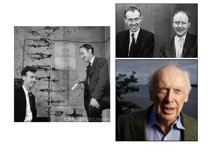
DNA • DNA makes up genes so it is called genetic material • Full name: deoxyribonucleic acid • Is one type of nucleic acid (the other is RNA – ribonucleic acid) • Structure was determined by Watson & Crick; they published the structure in 1953 • This opened the door to the whole new world of genetic engineering, cloning, etc.
DNA Structure • DNA is composed of nucleotides. • The structure of a DNA nucleotide is shown at the right. • Each one has 3 parts: – 5 -C sugar (deoxyribose) – Phosphate – Nitrogen base (1 of 4)
Nitrogen Bases in DNA: Thymine Cytosine Adenine Guanine
DNA Structure – cont’d • DNA nucleotides are joined together to make a strand. • Two strands of nucleotides compose a DNA molecule.
Simple Representations of DNA: (like a “ladder”)
Simply stated, DNA has the structure of a twisted “ladder. ”
DNA Structure & Replication Animation • http: //207. 4. 198/pub/flash/24/menu. swf
More realistic representation of DNA – note hydrogen bonds
DNA strands are complementary and antiparallel.
By the way. . . • Watson and Crick may not have been able to figure out the structure of DNA if they had not seen the work of Rosalind Franklin. • She used X-Ray Crystallography Technology to make pictures of DNA. • She died before Watson & Crick received the Nobel Prize; probably would have shared it with them had she lived.
Rosalind Franklin
DNA Replication occurs during S phase of Cell Cycle; allows each chromosome to duplicate itself • Occurs in a semi-conservative manner. • Each new strand is half “old” (1/2 of parental DNA molecule) and half “new”. • The parent DNA molecule is semi-conserved in each new DNA molecule produced by DNA Replication.
DNA Replication – cont’d
Eh. Enough notes for today.
How does a gene control a trait? Usually, by directing the synthesis of a protein. This involves 2 processes: – Transcription = using one side of the DNA of a gene as a template for making a complementary strand of m. RNA – Translation = a ribosome “reads” the coded information in the m. RNA and uses that to assemble a sequence of amino acids (a protein) with the help of t. RNA
To make a protein, what information is needed? – Need to know which amino acids to use and the order in which they need to be assembled • How can a gene have this information? – The information is written in 3 -letter CODE WORDS. Each code word is a sequence of 3 nitrogen bases in a gene. • ex: TAC or TTT, etc.
• Where are the genes located? – in the nucleus – (each chromosome is a sequence of genes) • Where are the proteins made? – in the cytoplasm (ribosomes) • Do you think that a gene can leave the nucleus? – NO
Questions to consider: 1. How can the CODE for making a particular protein get from the gene in the nucleus out to a ribosome? 2. Of what are ribosomes made? 3. How will the necessary amino acids to delivered to the ribosome?
The answers to these questions are found as we look at the 3 types of RNA.
Review: RNA Nucleotide Structure REMEMBER: RNA does not contain thymine. In its place, RNA has Uracil. The sugar is ribose instead of deoxyribose.
RNA • RNA molecules are single strands of nucleotides. • The 3 types of RNA are: – m. RNA = messenger RNA – r. RNA = ribosomal RNA – t. RNA = transfer RNA SO … BACK TO OUR QUESTIONS. .
1. How can the CODE for making a particular protein get from the gene in the nucleus out to a ribosome? • • The code is transferred from the nucleus to a ribosome in the form of a m. RNA (messenger RNA) molecule. This m. RNA molecule is made in the nucleus by an enzyme called RNA Polymerase in the process called TRANSCRIPTION.
• The sequence of nitrogen bases in m. RNA is determined by the sequence of nitrogen bases in the DNA of the gene being expressed. • Each 3 letter code word in m. RNA is called a codon (ex: AUG, AAA, etc. ). • The m. RNA leaves the nucleus and finds a ribosome.
Transcription (Tc)
2. Of what are ribosomes made? Ribosomes are made of r. RNA (ribosomal RNA) and protein. Each is composed of a small subunit and a large subunit. The small subunit binds m. RNA first to initiate TRANSLATION. Then the large subunit is added to form a complete ribosome. The large subunit has a “P” site and an “A” site.
Structure of a Ribosome (composed of r. RNA + protein) t. RNA-binding sites Large subunit m. RNA binding site Small subunit
3. How will the necessary amino acids to delivered to the ribosome? The necessary amino acids are delivered to the ribosome by t. RNA (transfer RNA) molecules.
Transfer RNA (t. RNA)
How will a specific t. RNA molecule “know” that the amino acid it carries is needed? Each t. RNA has a 3 -nitrogen base sequence called an ANTICODON. During TRANSLATION when a ribosome is reading a particular CODON, the t. RNA with the complementary ANTICODON locks on and delivers its amino acid.
Transcription DNA m. RNA Amino acid RNA polymerase 1 m. RNA is transcribed from a DNA template. Translation 2 Each amino acid attaches to its proper t. RNA with the help of a specific enzyme and ATP. Enzyme ATP t. RNA Anticodon Large ribosomal subunit Initiator t. RNA 3 Initiation of polypeptide synthesis The m. RNA, the first t. RNA, and the ribosomal sub-units come together. Start Codon m. RNA Small ribosomal subunit
New peptide bond forming Growing polypeptide 4 Elongation Codons A succession of t. RNAs add their amino acids to the polypeptide chain as the m. RNA is moved through the ribosome, one codon at a time. m. RNA Polypeptide 5 Termination Stop codon The ribosome recognizes a stop codon. The polypeptide is terminated and released.
What type of bond forms between the amino acids as they are brought in to form a protein? PEPTIDE BONDS
Protein Synthesis • http: //highered. mcgrawhill. com/sites/0072437316/student_view 0/ chapter 15/animations. html#
Review • Transcription (Tc) – Occurs in nucleus – RNA Polymerase uses 1 side of a gene as a template to make a complementary m. RNA. • Translation (Tl) – Occurs in cytoplasm – Ribosomes read codons; t. RNAs bring in the correct amino acids
PRACTICE: • Consider the following segment of a DNA molecule (only the nitrogen base pairs are shown; the sugar-phosphate backbone of each strand is understood to be present): – --A T G T T A C G A A A G A T G A-– -- • Pretend that this segment of DNA = a gene for a protein. Note: Only one side of the gene is used as a template to transcribe a m. RNA molecule. Let’s use the bottom half of our practice gene.
Transcribe this DNA base sequence into a m. RNA base sequence. TAC AAT GTG CTT TCT ACT DNA m. RNA codon t. RNA anticodon amino acid Fill in t. RNA anticodons. Use the CODONS & the genetic code chart on p. 222 to fill in the appropriate amino acids.
The Genetic Code The genetic code was broken in the early 1960’s. Note that: 1. in the genetic code, more than 1 codon can specify the same amino acid; the 3 rd position of each codon is called the “wobble” position; code is REDUNDANT 2. in the genetic code, no codon specifies more than one amino acid; code is not AMBIGUOUS 3. the genetic code contains some codons that are signals: -AUG is a start codon that specifies Met -UAA UAG UGA are all stop codons that signal for the ribosome to stop translation 4. the genetic code is universal – is shared by all organisms from the simplest bacteria to the most complex plants and animals.
How can we work with the Genetic Code?
Different formats for Genetic Code Chart:
Review of how a gene directs the synthesis of a protein:
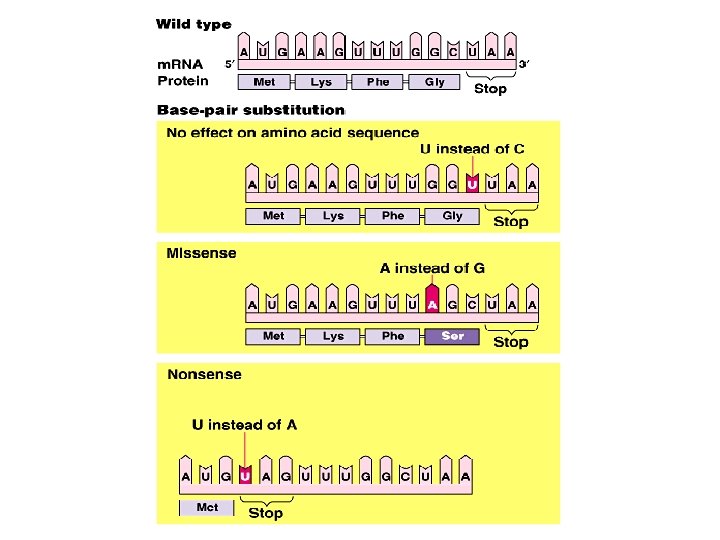
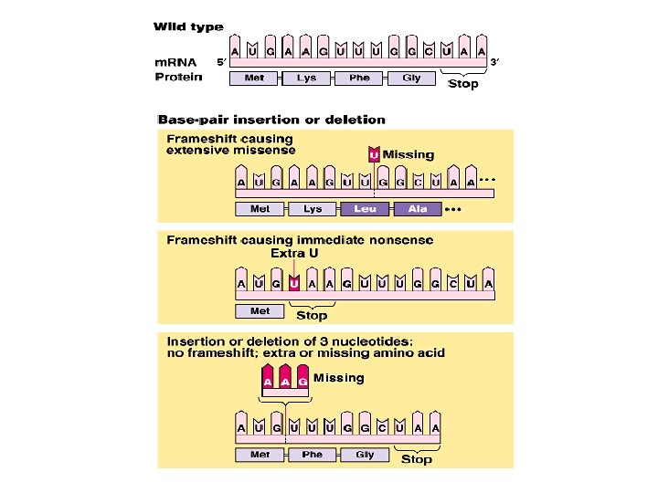
Mutations DFN = changes in genetic material 1. Chromosome Mutations • EX: Chromosome number • EX: Chromosome structure mutations – Duplications – Deletions – Inversions – translocations 2. Gene Mutations • Also called “point mutations” • EX: Base-pair substitution mutations – Missense – wrong amino acid put in – Nonsense – results in premature stop codon • EX: Insertion/Deletion of 1 or more base pairs – Cause translation (reading) “frameshift”
Chromosome Number Mutations can result from nondisjunction during Meiosis:
Chromosome Number Mutations – cont’d • Individuals have too many or too few chromosomes: • Having the wrong number of chromosomes causes a variety of syndromes: – Down Syndrome (trisomy 21) – Turner’s Syndrome (XO) – Klinefelter’s Syndrome (XXY)
Chromosome Structure Mutations:
Major Types of Gene Mutations: • The diagram at the right shows the basic idea for base substitution and base deletion mutations. • The next 3 slides show more detailed information concerning the consequences of these mutations.
Gene Mutation ex: missense due to base-pair substitution

 Dna and genes chapter 11
 Carrier vs channel proteins
 Protein-protein docking
 Dna polymerase function in dna replication
 Dna to protein steps
 Dna rna protein central dogma
 Rna and protein synthesis study guide
 Chapter 12 section 3 dna rna and protein
 Dna to protein steps
 Dna to protein steps
 Microarray analysis
 Dna rna protein
 Dna rna protein diagram
 Dna-protein interactions
 Dna rna protein trait
 Definition protein
 Nucleosome
 Protein function
 Protein function
 Protein function
 Protein function
 Protein function
 Biological role of proteins
 Function of plasma protein
 Protein function prediction via graph kernels
 Hbv protein function in the body
 Collagen fibrous protein
 Alpha helix and beta sheet
 Peptide bonds in primary structure of protein
 Super secondary structure of protein
 Primary secondary tertiary quaternary structure
 Secondary protein structure hydrogen bonds
 Quaternary structure of myoglobin
 Protein monomer
 Primary secondary and tertiary protein structure
 Adenine thymine cytosine and guanine
 Protein primary structure
 Quaternary structure of protein
 Protein tertiary structure bonds
 Protein structure
 Hierarchy of protein structure
 Anomalous
 Protein structure
 Super secondary structure of protein
 Super secondary structure of protein
 Hierarchy of protein structure
 Protein primary structure
 Leucine polar or nonpolar
 Misfolded
 Nnuuu
Nnuuu Primary secondary and tertiary structure of protein
 Protein structure


