DNA Structure A B Z forms DNA Discovery

DNA Structure: A, B & Z forms

DNA Discovery § DNA was first observed by a German biochemist named Frederich Miescher in 1869. But for many years, researchers did not realize the importance of this molecule. It was not until 1953 that James Watson, Francis Crick, Maurice Wilkins and Rosalind Franklin figured out the structure of DNA — a double helix — which they realized could carry biological information. § Watson, Crick and Wilkins were awarded the Nobel Prize in Medicine in 1962 "for their discoveries concerning the molecular structure of nucleic acids and its significance for information transfer in living material. " § Deoxyribonucleic acid or DNA is a molecule that contains the instructions an organism needs to develop, live and reproduce. These instructions are found inside every cell, and are passed down from parents to their children.

DNA Structure • DNA is made up of molecules called Nucleotides. • Each nucleotide contains a phosphate group, a sugar group and a nitrogen base. • The four types of nitrogen bases are adenine (A), thymine (T), guanine (G) and cytosine (C). • Nucleotides are attached together to form two long strands that spiral to create a structure called as double helix. • To fit inside cells, DNA is coiled tightly to form structures called as chromosomes. • Each chromosome contains a single DNA molecule. Humans have 23 pairs of chromosomes, which are found inside the cell's nucleus. PURINE: possess two rings Eg: Adenine & Guanine PYRIMIDINE: possess one ring Eg. Cytosine & Thymine

Antiparallel Orientation: Double-stranded DNA is an antiparallel molecule, meaning that it is composed of two strands that run alongside each other but point in opposite directions. In a double-stranded DNA molecule, the 5' end (phosphate-bearing end) of one strand aligns with the 3' end (hydroxyl-bearing end) of its partner, and vice versa.

Base Pairing The two strands of the DNA double helix are held together by hydrogen bonds between nitrogenous bases on opposite strands. Base pairing is very specific. If there is an A found on one strand, it must be paired with a T on the other (and vice versa). Similarly, G on one strand must always have a C for a partner on the opposite strand. These A-T and G-C associations are known as Complementary Base Pairs. Because a large purine (A or G) is always paired with a small pyrimidine (T or C), the diameter of the helix is uniform, coming in at about 2 nanometers. A-T pairs form two hydrogen bonds total, while GC pairs form three.

Right-handed helix In Watson and Crick's model, the two strands of DNA twist around each other to form a righthanded helix. All helices have a handedness, which is a property that describes how their grooves are oriented in space. The major groove is a wider gap that spirals up the length of the molecule, while the minor groove is a smaller gap that runs in parallel to the major groove. The base pairs are found in the center of the helix, while the sugar-phosphate backbones run along the outside. These grooves are important binding sites for proteins that maintain DNA and regulate gene activity. Image of a DNA double helix, illustrating its right-handed structure.

Various factors affect the conformation of the DNA in a cell leading to structural transitions into other DNA forms. Aform, B-form and Z-form of DNA are three generally found structural conformations of DNA.

A-DNA: A form of DNA is a structural conformation that any DNA molecule favours under dehydrating condition and also certain purine stretches favours the A-DNA forms. Discovered by Rosalind Franklin, the A-DNA helix conformation is wider when compared with the B-DNA conformations because of the base pairs stacking. The bases in the A-DNA are stacked little offcentre due to which a hollow region at the centre is visible when looked at the cross-section. Like B-DNA, A-DNA is the right-handed double helix structure with antiparallel strands held together by Watson-Crick base-pairing. Compared to B-DNA, it is more compact and shorter structure. Helical Parameters of A-DNA: Parameter A-form of DNA Helix sense Right-handed Diameter 23 Å (2. 3 nm) Helix rotation on per base 32. 7° pair Base pair tilt to axis +20° Helix pitch (rise per turn) 28. 6 Å (2. 86 nm) Helix rise per base pair 2. 6 Å (0. 26 nm) Base pairs per turn 11 Glycosyl bond angle Anti Sugar pucker C 3′-endo Major groove Narrow and Deep Minor groove Wide and Shallow

B-DNA The deoxyribonucleic acid conformation in the cells is the Watson–Crick form of the double helix. The right-handed helix B-DNA exists in relatively much higher hydrating conditions, unlike A-DNA. As under physiological conditions, the B form of DNA is predominant in the cells. Among the three different families of DNA helices, the B-DNA conformation is the most common and variable in structure. Helical Parameters of B-DNA: Parameter B-form of DNA Helix sense Right-handed Diameter 20 Å (2. 0 nm) Helix rotation on per base pair 34. 3° Base pair tilt to axis -6° Helix pitch (rise per turn) 34 Å (3. 4 nm) Helix rise per base pair 3. 4 Å (0. 34 nm) Base pairs per turn 10. 5 Glycosyl bond angle Anti Sugar pucker C 2′-endo Major groove Wide and Deep Minor groove Narrow and Deep

Z-DNA The Z-DNA is a left-handed double helical structure obtaining its name “Z-DNA” because of the irregular and zigzag shape of it backbone. The occurrence of Z-DNA is generally unfavourable, however, DNA sequences with alternating of purines and pyrimidines adopt left-handed Z-DNA conformation. A fascinating feature about Z-DNA, which makes it easily distinguishable from B-DNA that there is no major groove exists in ZDNA and a single narrow groove corresponding to the minor groove of the BDNA. The Z-DNA is thinner and more extended when compared to B-DNA. Helical parameters of Z-DNA : Parameter Z-form of DNA Helix sense Left-handed Diameter 18 Å (2. 0 nm) Helix rotation on per base pair 9° or 51° Base pair tilt to axis -7° Helix pitch (rise per turn) 44 Å (4. 4 nm) Helix rise per base pair 3. 8 Å (0. 38 nm) Base pairs per turn 12 Glycosyl bond angle Pyrimidine: anti, Purine: syn Sugar pucker C 2′-endo for pyrimidines C 3′-endo for purines Major groove Flat Minor groove Narrow and Deep
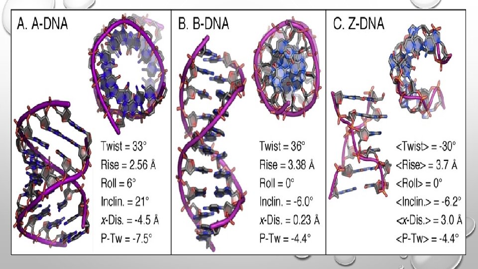

Side and Top view of A, B & Z DNA Conformations
- Slides: 12