DNA and Cell Cycle By Mckinley Meeks Jessica

DNA and Cell Cycle By: Mckinley Meeks & Jessica Zirkle

History of DNA Rosalind Franklin determined that DNA has a double stranded helix shape. Also, that DNA had phosphates on the outside in patterns that repeated multiple times throughout a sequence. Watson and Crick figured out that the sugars and phosphates were on the outside of the helix and the nitrogenous bases were making the rungs of the helix. They paired the bases to find that a purine and a pyrimidine fit perfectly together.

What is DNA? Deoxyribonucleic acid, a self-replicating material present in nearly all living organisms as the main constituent of chromosomes. It is the carrier of genetic information. ● Composed of nucleotides ● Forms a double helix ● Coils into chromosomes

DNA Structure and Replication Structure of a Nucleotide ● Phosphate group ● Sugar group ● Nitrogenous base

Sides of the Ladder ● Sugar-Phosphate backbone ● 3’ and 5’ ends ● Nucleotide line up across from its complimenting base pair

Hydrogen Bonding and Nitrogenous Bases

Double Helix ● formed using complementary base pairing and hydrogen bonds ● Nitrogenous bases from two single strands are joined using the complementary base pairing rule.

DNA Forming Chromosomes Structure in eukaryotes. ● the DNA is wrapped around proteins called histones forming nucleosomes. ● This forms a fiber known as chromatin. ● This forms a coil within a coil.

Replication of DNA
 and Frank Stahl (right) in 1958")
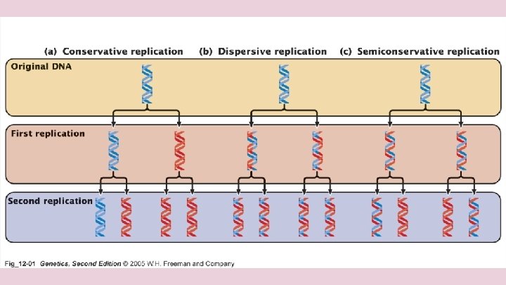
Experiment of Meselson and Stahl Matt Meselson (left) and Frank Stahl (right) in 1958 demonstrated that replication was semiconservative using radioactive nucleotides with dividing bacteria supporting Watson’s and Crick’s hypothesis

Prokaryotic DNA Replication The prokaryotic chromosome attaches to the plasma membrane. The DNA is then replicated in both directions.

Eukaryotic DNA Replication DNA replication occurs simultaneously in many locations along the very long eukaryotic chromosomes. Three replication bubbles are visible along the DNA within this cultured Chinese hamster cell. The arrows indicate the directions of DNA replication at the two ends of the bubble.

Helicases are enzymes responsible for the unwinding of the DNA molecule. They unwind the DNA in both directions

Releasing Stress in the DNA Molecule Since DNA is a double helix, there will be tension in the DNA strand that causes it to tangle as it is unwound by the helicase. The enzymes topoisomerase I and II are responsible for relieving that stress by clipping one or two strands of the DNA.

Adding Nucleotides as Triphosphates Nucleotides are always added on as triphosphates. When the nucleotides are added then two phosphates are cleaved off making a pyrophosphate.

Something to keep in mind. . . ● Synthesis ALWAYS occurs in the 5′ to 3′ direction! ● When a DNA molecule is being synthesized , the nucleotides are added as triphosphates, and two phosphates are removed. Nucleotides are always added to the 3′ end!

Primer- DNA Polymerase DNA polymerase must always attach the complementary nucleotide to a 3′ end of the deoxyribose sugar molecule. So, in the very beginning a small RNA primer must be laid down in order to start the process of DNA replication. Primase is the enzyme responsible for this.
 are being laid down by")
Putting Down a RNA Primer RNA nucleotides (red pentagons) are being laid down by primase before DNA polymerase begins DNA replication.

Lagging Strand ● Ligase ties the two strands together

Telomeres Once DNA has been replicated, there is one problem. The usual replication machinery provides no way to complete the 5′ ends after the RNA primer is removed, so repeated rounds of replication produce shorter and shorter DNA molecules. To compensate for this repeated shortening process, repetitive sequences of DNA are added. These are noncoding sequences and called telomeres.

Maintaining Telomere Length in Gametes and Germ Cells In gametes, the shortening of telomeres would cause serious problems. If chromosomes of gametes became shorter each time during replication, then essential genes would eventually be missing. An enzyme complex called telomerase catalyzes the lengthening of telomeres in gametes.

DNA Repair ● Errors in DNA replication occur about 1 in every 10, 000 base pairs. Not bad, but with 6 billion bases being replicated that amounts to 60, 000 mistakes every time a cell divides. ● DNA repair systems repair about 99% of these mistakes.

Cell Cycle G 1 phase: Organelles are forming and DNA is uncoiling during this time. Protein synthesis is also present during this phase. Most cells differentiate, if they do not then they will not reproduce and are said to be stuck in G 0. G 1 can last anywhere from seconds to years. S phase: DNA uncoils and replication occurs. Further organelle replication takes place. The S phase makes sure the daughter cell has the same genetic information as the mother cell.

Cell Cycle G 2 phase: cell prepares for mitosis, takes about 4 hours M phase: nucleus is replicated, cytoplasm divides to make identical daughter cells

Cyclin and Kinase Cyclins are a family of proteins that control the progression of the cell cycle A kinase is a type of enzyme that transfers phosphate groups to specific substrates

Chromosomes DNA is wrapped around histone proteins and coiled Chromosomes prevent DNA breakage and ensures that each cell gets one copy of each chromosome

Mitosis Unicellular: undergo mitosis to reproduce themselves Multicellular: cell division for growth and repair or to make a new organism

Prophase Centromere condenses in chromosomes Nucleoli disappears Spindles form Centrosomes move to opposite poles

Prometaphase Nucleolus no longer visible Centrosomes at opposite ends Microtubules extend through nuclear area Kinetochores form on the centromere on each chromatid Kinetochore microtubules attach to the kinetochores. Moving the chromosomes back and forth until they reach the middle of the cell.

Metaphase Chromosomes line up on the metaphase plate

Anaphase Cohesin proteins are split and sister chromatids separate Chromosomes pulled to opposite poles Kinetochore microtubules are disassembled Spindle poles move apart

Telophase Two daughter nuclei form Nuclear envelope forms Chromosomes unwind, forming chromatin

Cytokinesis In animals: Mitosis without cytokinesis results in multinucleated cells Animals pinch in the cell membrane Rings of acting form under cell membrane and pull to create a cleavage furrow In Algae: Inward growth of new cell wall and membrane In higher plants: Begins in the middle and moves towards the outside as membranous

Prokaryotes reproduce using binary fission: Chromosome attaches to the plasma membrane at the origin of replication DNA Replication occurs in both directions. The cytoplasm divides and pinches in forming two new cells.

Meiosis I Interphase: G 1, S, G 2 of the cell cycle Centrosomes with a pair of centrioles replicate

Meiosis I Prophase I: Chromosomes are visible as separated filaments Homologous chromosomes pair and align as long well-separated filaments. Recombination nodules attach and crossing over occurs.

Meiosis I Metaphase I The homologous chromosomes move to the metaphase plate

Meiosis I Anaphase I Centromeres do not separate Homologous chromosomes separate and move to opposite poles Spindle fibers force poles apart

Meiosis I Telophase I The chromosomes contain two hybrid chromatids The double-stranded chromosomes have migrated to the poles The number of chromosomes is the haploid number of chromosomes

Meiosis II Interkinesis This is the time between meiosis I and meiosis II Haploid daughter cells; no further DNA replication occurs during this time

Meiosis II Prophase II Centrosomes replicate and begin to move apart Chromosomes appear as long thin threads Nucleolus becomes less distinct Asters begin to form Twin chromatids become visible Kinetochore microtubules attach to the kinetochores Spindle forms Membrane begins to disappear.

Meiosis II Metaphase II Membrane has disappeared Double stranded chromosomes move to the metaphase plate Microtubules attach to both sides of the kinetochore The spindle fibers overlap one another

Meiosis II Anaphase II Centromeres have separated and move the single stranded chromosomes toward opposite poles Overlapping spindle microtubules from opposite poles interact and to pull the poles apart Cytokinesis may begin

Meiosis II Telophase II Nuclear membrane begins to form Chromosomes begin to uncoil, become thinner Nucleolus becomes less distinct Cytokinesis is nearly complete

Gametogenesis • Spermatogenesis: The four equal sized cells produced in meiosis will differentiate and become sperm cells. • Oogenesis: During cytokinesis, there is unequal division of the cytoplasm with usually one large cell and three smaller cells.

Advantages of Asexual reproduction Eliminates the need to find a mate May reproduce at a faster rate May produce larger number of offspring using less overall energy and resources Optimum in stable unchanging environments Uses mitosis

Types of asexual reproduction Budding - an offspring simply grows out of the body of the parent Gemmules - The parent releases a specialized mass of cells that can develop into an offspring. Fragmentation - occurs when the body of the parent breaks into distinct pieces, each of which can produce an offspring Regeneration- A piece of an organism is detached and develops into a completely new individual. Parthenogenesis – is a form of asexual reproduction in which growth and development of embryos occur without fertilization

Advantages of sexual reproduction Varied number phenotypic offspring. Better response to varying environment. Sexual reproduction involves the union of two haploid cells from different parents to produce a diploid zygote.

Diploid vs. Haploid

Human Somatic Cells Humans have 23 pairs of chromosomes in their somatic cells. These chromosomes have been stained and certain banding patterns appear. The two chromosomes in each pair are called homologous chromosomes, or homologs. Chromosomes in a homologous pair are the same length and carry genes controlling the same inherited characters.

Human Karyotype A karyotype is an ordered display of the pairs of chromosomes from a given cell arranged from longest to shortest. Autosomes are non-sex chromosomes. Notice that the sex chromosomes in this karyotype are not homologous.

Synapsing Homologous pairs form a protein structure between them called the synaptonemal complex. This keeps the homologs as a tetrad. Places where crossing over occurs is called a chiasma. In mitosis, kinetochore microtubules attach to both sides of the centromere, but in meiosis I they on attach to one side.

Crossing over

Review Game https: //quizizz. com/admin/quiz/5 aeaf 12 c 32 cfb 9001 b 469 e 89
- Slides: 55