Divisions of the Nervous System Unit 8 Human


 • Control center of the body • Relays")


 • Left and right hemispheres – Left brain – logic,")
 and grooves (sulci)")
 divide the cerebrum into four lobes 1. Occipital Lobe: Lobe visual")


 – Commonly called a stroke – The result of")

 Sensory Division Motor Division Somatic NS Autonomic NS Sympathetic Parasympathetic")
 • Peripheral Nervous System is made up of all")


, then")
 sensory neuron spinal cord motor neuron effector (muscle)")


 >")
")
- Slides: 38
Divisions of the Nervous System Unit 8: Human Body Systems Chapter 35 -3
Divisions of the Nervous System • 2 major divisions: Nervous System Central Nervous System Peripheral Nervous System Brain Spinal Cord Nerve cells
A. The Central Nervous System (CNS) • Control center of the body • Relays messages, processes & analyzes information • Made of two parts: – Brain – Spinal cord
• 1. The Brain – Impulses flow to and from the brain – 100 billion neurons, mainly interneurons
• 2. Spinal Cord – Main communication link between brain and the rest of the body – Processes information such as reflexes
1. Protection of the CNS • Brain and spinal cord are protected by: • a) Bone – skull and vertebrae • b) Meninges – layers of connective tissue that surround the organs • c) Cerebrospinal fluid – fluid found in between meninges and organ – Acts as a shock absorber – Continually circulates around the brain
• DISEASE: Meningitis – Inflammation of the meninges – Can be caused by an viral , bacterial or microorganism infection – Causes headaches, neck stiffness, confusion, sensitivity to light and sound – Can be life threatening • DISEASE: Hydrocephalus (“water on the brain”) – Accumulation of CSF in the brain – Exerts pressure on the brain causing brain damage
2. Regions of the Brain • Regions of the Brain – Cerebral hemispheres (Left & Right) – Diencephalon – Brain stem – Cerebellum
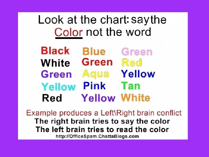
a. Cerebral Hemispheres (Cerebrum) • Left and right hemispheres – Left brain – logic, language, math – Right brain – creativity • Connected by the corpus callosum (communication link between right and left)
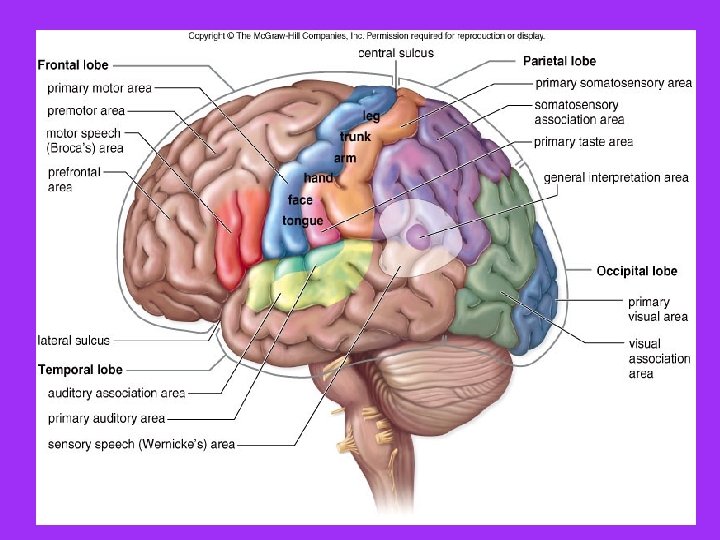
• The surface is made of ridges (gyri) and grooves (sulci)
Fissures (deep grooves) divide the cerebrum into four lobes 1. Occipital Lobe: Lobe visual integration 2. Parietal: Parietal spatial knowledge, math 3. Temporal: Temporal memories, auditory, language 4. Frontal: Frontal emotion, future planning, judgment, muscle movements, language
• Limbic System – involved in emotion, motivation, arousal, memory, and learning – Amygdala – fear – Hippocampus – memory formation
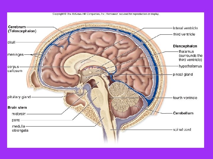
b. Diencephalon • Relay and control center • Sits on top of brain stem • Two main parts: – 1) Thalamus – relay between sensory areas and cerebrum – 2) Hypothalumus – regulates involuntary responses & hormone secretions of the pituitary gland
c. Brain Stem • Attaches brain to spinal cord • Parts of the brain stem – Midbrain – vision, hearing, motor control – Pons – breathing, sleep – Medulla oblongata involuntary activities (breathing, heart rate, blood pressure)
d. Cerebellum • “Little brain” inferior to and posterior to cerebral cortex • Coordination, posture, motor learning
3. Spinal Cord • Cylinder of nervous tissue that begins at base of brain • Protected by the vertebral column and meninges
• Spinal nerves extend from the cord through each vertebrae • Main communication link between brain and the body
4. Traumatic Brain Injuries & Diseases • Concussion – Slight or mild brain injury – Bleeding & tearing of nerve fibers happened – Recovery likely with some memory loss • Contusion – A more severe TBI – Nervous tissue destruction occurs – Nervous tissue does not regenerate • Cerebral edema – Swelling from the inflammatory response – May compress and kill brain tissue
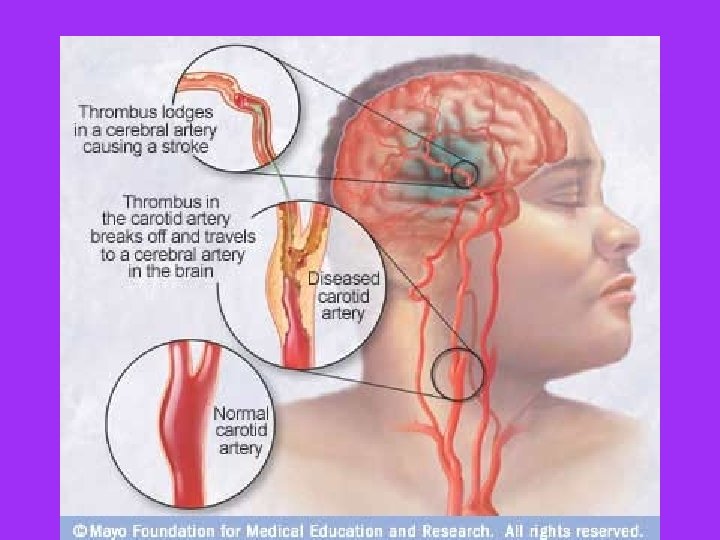
• Cerebrovascular Accident (CVA) – Commonly called a stroke – The result of a blocked or ruptured blood vessel supplying a region of the brain – Brain tissue supplied with oxygen from that blood source dies – Loss of some functions or death may result
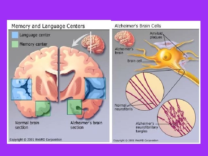
Alzheimer’s Disease • Progressive degenerative brain disease • Mostly seen in the elderly, but may begin in middle age • Structural changes in the brain include abnormal protein deposits and twisted fibers within neurons • Victims experience memory loss, irritability, confusion and ultimately, hallucinations and death
Peripheral Nervous System (PNS) Sensory Division Motor Division Somatic NS Autonomic NS Sympathetic Parasympathetic
B. Peripheral Nervous System (PNS) • Peripheral Nervous System is made up of all the nerves and ganglia (nerve cell bodies) that carry messages between the body and the CNS • Receives info from the environment • Transmits commands from CNS to organs and glands • Contains mostly motor and sensory neurons
Divisions of the PNS • Sensory division • Motor division – transmits impulses from sense organs to the central nervous system to the muscles or glands – Divided into 2 divisions • Somatic • Autonomic
Motor Division • Somatic N. S. • Autonomic N. S. • Regulates activities that are under conscious control • Example: movement of muscles (wiggle toe) • Involved in reflexes (quick, automatic response to stimulus) • Regulates activities that are automatic or involuntary • Example: heart rate • Consists of only motor nerves • Divided into two divisions – Sympathetic division – Parasympathetic division
Autonomic N. S. Division • Sympathetic • Parasympathetic • “Fight-or-Flight” • Takes over to increase activities • Remember as the “E” division = exercise, excitement, emergency, and embarrassment • “Rest and Digest • Conserves energy • Maintains daily necessary body functions • Remember as the “D” division = digestion, defecation, and diuresis (urination)
Peripheral Nervous System & Reflexes • The peripheral nervous system is also involved in reflexes. • A reflex is a quick and unconscious response to a stimulus • The brain is not involved with reflexes.
• The impulse travels up sensory neurons, to the spinal cord (interneuron), then immediately travels down motor neurons for a response. • The pathway the impulse travels is called the reflex arc
Reflex Arc • Receptor (sense organ) sensory neuron spinal cord motor neuron effector (muscle)
Internal Communication • Internal communication is critical to maintain homeostasis. • Sensory neurons are constantly sending information to the brain about the internal environment. • The brain responds by sending signals through the motor neurons to maintain homeostasis.
The Nervous System CNS Brain Spinal Cord PNS Somatic NS Voluntary Movement Autonomic NS Involuntary Movement Sympathetic Parasympathetic NS NS fight or flight rest & digest
Reflex Pathway • Sensory input > – Sensory Neuron > • Spine (Interneuron) > –Motor neuron » > Involuntary Reflex (response)
Conscious Response Pathway • Sensory Input > – Sensory Neuron > • Brain (Interneurons) > –Motor Neuron > » Voluntary Movement (Response)