Divisions of the nervous system CNS PNS EFFERENT


- Slides: 38
Divisions of the nervous system CNS PNS EFFERENT Somatic Skeletal muscle -voluntary ANS Cardiac & smooth muscles Glands -involuntary AFFERENT Somatic Visceral Cardiac & Skeletal muscle, tendons smooth muscles Glands joints, skin
PNS Terminology • Ganglia – neuron cell bodies • Peripheral nerves – neuronal axons • PNS neuroglia – Satellite cells • Enclose neuron cell bodies in ganglia – Schwann cells • Cover peripheral axons
Efferent Division of the PNS • the somatic nervous system and part of the autonomic nervous system • the somatic – control of skeletal muscle • the ANS – involuntary control over cardiac and smooth muscle + gland secretion
I - Olfactory II - Optic III - Oculomotor IV-Trochlear V - Trigeminal VI - Abducens VII - Facial VIII - Acoustic IX - Glossopharyngeal X - Vagus XI - Accessory XII - Hypoglossal -cranial nerves – 12 pairs -considered part of the peripheral nervous system (PNS) -olfactory & optic contain only sensory axons = sensory nerves -remaining are motor or mixed nerves (both motor and sensory axons)
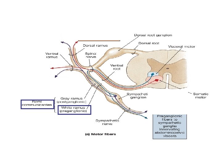
Spinal Nerve • after passing through intervertebral foramina the spinal nerve branches = ramus/rami • Dorsal ramus Sensory/motor innervation to skin and muscles of back • Ventral ramus -Ventrolateral body surface, body wall structures, muscles of the upper and lower limbs • in addition to these rami - the spinal nerves also give off a meningeal branch reenters the vertebral canal and supplies the vertebrae, vertebral ligaments and meninges
• rami communicantes = branches from the spinal nerve -defined as a connection between a spinal nerve and the sympathetic trunk of the ANS sympathetic trunk
Nerve Plexuses • Four major plexuses – Cervical plexus – Brachial plexus – Lumbar plexus – Sacral plexus • Joining of ventral rami of spinal nerves to form nerve networks or plexuses • Found in neck, arm, low back & sacral regions • No plexus in thoracic region
Somatic Nervous System • considered the voluntary aspect of the PNS – but the muscles of posture and balance are controlled involuntarily by the lower brain centers (brain stem, cerebellum) • cell bodies located in the ventral gray horn of the spinal cord • the axon of a somatic motor neuron extends from the CNS continuously to its skeletal muscle target • terminals release acetylcholine – contraction • can only stimulate its target
Somatic Nervous System - somatic/motor axons emerge from the ventral gray horn and travel into the spinal nerve - they then travel through either the: – dorsal ramus to end up at the muscles of the back – OR the ventral ramus to end up at the muscles of the limbs and body wall (chest/abs/pelvis)
Somatic Nervous System • somatic motor neurons that originate in the ventral gray horn (or the brain stem) receive incoming information from many converging presynaptic neurons – both excitatory and inhibitory on these motor neurons – the neurons that synapse with these motor neurons are: – 1. reflex neurons originating in the spinal cord – 2. neurons from motor areas of the brain – form the descending white matter tracts – these neurons synapse with the somatic motor neurons and regulate their activity • activation – impulse sent to muscles • inhibition – no impulse, no contraction
Somatic Nervous System • no matter what motor pathway you learn – they eventually control the somatic motor neuron in the ventral gray horn or the brain stem • therefore the somatic motor neuron is considered the final common pathway – considered the only way any other part of the nervous system can influence muscle activity
Somatic Motor pathways • all excitatory and inhibitory signals that control skeletal muscle movement converge on the somatic motor neurons • these somatic motor neurons originate in one of two places – 1. Brain Stem – 2. Ventral Gray Horn of Spinal Cord • these motor neurons extend from the brain stem and SC to innervate the skeletal muscles – also called lower motor neurons (LMNs) – their axons extend through the cranial and spinal nerves to skeletal muscle – only LMNs provide output from the CNS to skeletal muscle fibers
Somatic Motor pathways • neurons in four distinct circuits control movement by providing input to these LMNs – – 1. local circuit neurons 2. upper motor neurons (UMNs) 3. basal ganglial neurons 4. cerebellar neurons
Somatic Motor pathways • 1. local circuit – input arrives at LMNs from nearby interneurons called local circuit neurons – receive input from somatic sensory receptors and higher centers of the brain – help coordinate rhythmic activities in muscle groups
• 2. UMNs: Upper Motor Neurons – provide input to the local circuit and LMNs – essential for planning, initiating and directing sequences of voluntary movements – extend from the brain to the LMNs via two types of somatic motor pathways • 1. direct motor pathways: nerve impulses for voluntary movement – lateral corticospinal, anterior corticospinal and corticobulbar (brain stem) – UMNs originate in the motor cortex and travel down the spinal cord as the corticospinal tracts to synapse with the LMN – OR – UMNs exit the brain stem – the LMN emerges as spinal nerves or through the brain stem and out as cranial nerves
• 2. indirect motor pathways: or extrapyramidal pathways – nerve impulses follow complicated circuits that involve the cortex, basal ganglia, thalamus and brain stem – descending axons/tracts pass outside the pyramids of the medulla – 1. rubrospinal – 2. reticulospinal – 3. vestibulospinal
• 3. Basal ganglia pathways – assist movement by providing input to the UMNs – “okays” the motor pathways that emerge from the motor cortex – also suppresses unwanted movements and initiates and terminates movement – the production of dopamine by the substantia nigra also effects muscle tone by modifying this path – caudate nucleus and putamen receive sensory input from several areas of the brain – to know what muscles are doing Motor Cortex Basal Ganglia Thalamus
Somatic Motor pathways • 4. Cerebellar – function involves four activities • 1. monitoring intentions for movement • 2. monitoring actual movement • 3. comparing the command (intention and movement) with sensory information • 4. correction – to UMNs – travels via the thalamus to the UMNs in the cerebral cortex – or can go directly to the UMNs in the brain stem Cerebellum Thalamus Motor Cortex Midbrain
The Neuromuscular Junction • end of the lower motor neuron (synaptic terminal or axon bulb) in very close association with a muscle fiber/cell • distance between the bulb and the folded sarcolemma = synaptic cleft • nerve impulse leads to release of a specific neurotransmitter (acetylcholine) • this release will result in activation of the muscle cell and contraction • therefore the NMJ is ALWAYS excitatory • the only way inhibition can take place is through the inhibition of the neuron “connecting” with the muscle –i. e. upper motor neurons http: //www. blackwellpublishing. com/matthews/neurotrans. html
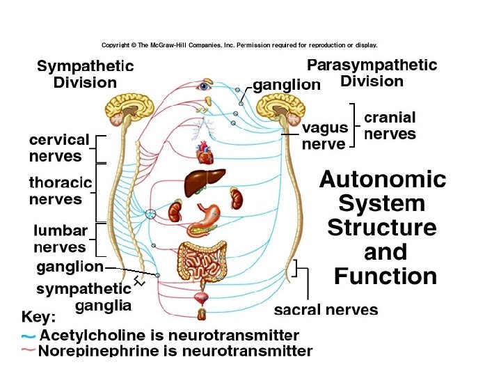
ANS • two divisions that innervate the same organs • efferent branch regulates “visceral” activities (motor commands, involuntary, organs) • also has an afferent branch that receives sensory information from these areas • generally divided into the: – A. Parasympathetic – B. Sympathetic
ANS • involuntary motor commands and sensory information • supplies cardiac and smooth muscle, glands (i. e. viscera) • comprised on two neurons – preganglionic and postganglionic – preganglionic synapses with the cell body of the postganglionic within the ganglion – the pregang and postgang neurotransmitters can differ – the postganglionic neuron is unmyelinated – glands are innervated by the preganglionic neuron – e. g adrenal gland which then releases epinephrine or norepinephrine in response
Parasympathetic Division • cell bodies of the pre. G neurons are located in the brain stem – axons form the four cranial nerves III, VII, IX and X – known as cranial parasympathetic outflow • also found in the lateral gray horns of the sacral spinal nerves 2 through 4 – emerge as part of the cranial or spinal nerve – known as sacral parasympathetic outflow
Parasympathetic Division • parasympathetic ganglia are located near or in the target • called terminal ganglia – the pre. G fibers are very long because they must extend from the CNS to an organ – synapse with post. G within the terminal ganglia – four major terminal ganglia are located close to the organ they innervate – 1. otic (parotid gland) – 2. submandibular (submandibular and sublingual glands) – 3. pterygopalatine (lacrimal gland) – 4. ciliary (pupils)
Sympathetic Division • • • for visceral motor commands cell bodies of the pre. G neurons are located in the lateral gray horns of T 1 to L 2 sympathetic ganglia: – site of the synapse between the pre. G and post. G neurons – short pre. G lead into these ganglia – long post. G axons lead out to target – two groups: 1. sympathetic trunk ganglia: or paravertebral chain ganglia -vertical row lateral to the vertebral column -3 cervical, 11 or 12 thoracic, 4 or 5 lumbar and 4 or 5 sacral 2. prevertebral ganglia: or the collateral ganglia -post. G neurons innervate the abdominal organs -anterior to the vertebral column and are near the large abdominal arteries -three major prevertebral ganglia: celiac, superior mesenteric and inferior mesenteric celiac ganglion
Sympathetic Division • axons exit the lateral gray horn through the ventral root of the spinal cord • axons form part of the spinal nerves T 1 to L 2 • form part of the spinal nerve along with somatic motor nerve axons and parasympathetic pre. G axons • BUT the axons then enter the rami communicantes and pass to the nearest sympathetic trunk ganglion – synapse with the post. G neuron celiac ganglion *** whether it is sympathetic or parasympathetic – the pre. G neurons release Ac. H
Sympathetic Dominance • fight or flight • protective response • elevated heart rate, blood pressure, respiration rate • increase blood flow to skeletal muscles, lungs, heart, brain • decrease blood flow to digestive, reproductive and urinary organs
Parasympathetic Dominance • “rest and digest” response • dominates in quiet, stress-free situations • resets the system after sympathetic stimulation – e. g. slow the heart rate and lower blood pressure
ANS Neurotransmitters • specific neurons release specific NTs – have distinct names • cholinergic neurons –release of Ac. H – all pre. G neurons from sympathetic and parasympathetic neurons – all parasympathetic post. G neurons – two types of receptors • 1. nicotinic • 2. muscarinic • adrenergic neurons – release of NE – most sympathetic post. G are adrenergic – two types of receptors • 1. alpha – a 1 and a 2 • 2. beta – b 1 and b 2 and b 3
ANS receptors • the NTs released by the ANS can either stimulate or inhibit its target – depends on the receptors located in the target 1. Cholinergic receptors – respond to Ac. H • a. nicotinic – named because they are also activated by nicotine – found on the cell bodies of the post. G neurons within the ganglia of the symp. and parasymp. division – i. e. respond to Ac. H release from symp and parasympathetic pre. G fibers – binding of Ac. H opens channels for the movement of multiple ions including Na and K – if more positive ions (e. g. Na) enter the post. G neuron within the ganglion – depolarization and initiation of an AP by the post. G neurons adrenergic R nicotinic R muscarinic R
• b. muscarinic receptors • can bind either Ach or muscarene (Amanita muscaria mushroom) • called metabotropic receptors - specific for one kind of ion – e. g. ligand-gated Na channel • expressed on tissues “downstream” of post-ganglionic neurons – at the target tissue – e. g. neuromuscular junction (ligand-gated sodium channel) adrenergic R nicotinic R muscarinic R
2. Adrenergic receptors – respond to NE/Epi • alpha and beta classes – a 1, a 2, b 1, b 2, b 3 • distributed in a specific pattern in target tissues and respond to either NE or Epi or both – epinephrine made by the adrenal glands and NOT by neurons • respond to activation by activating G proteins -> second messengers (c. AMP or Ca) • therefore they are called G protein coupled receptors adrenergic R nicotinic R muscarinic R
Reflexes Reflex arc • Neural “wiring” of reflex • Requires 5 functional components: 1. sensory receptor, 2. sensory neuron, 3. integrating center (SC or BS), 4. motor neuron & 5. effector
• By development – Innate, acquired • Where information is processed – Spinal, cranial • Motor response – Somatic, visceral • Complexity of neural circuit – Monosynaptic Classification of Reflexes
• Stretch reflex: causes contraction in response to stretch • regulates skeletal muscle length and tone • monosynaptic - only one synapse in the CNS - between and single sensory and motor neuron • all monosynaptic reflexes are ipsilateral reflexes - input and output on same side • sensory receptors are found in muscle spindles – activated when stretched – e. g. Patellar reflex – hit with a mallet stretches the quadriceps and its tendon - results in contraction – muscle spindles in the quadriceps muscles are activated – reflex results Spinal Reflexes
• Tendon reflexes: controls muscle tension by causing muscle relaxation before muscle contraction rips tendons • generally polysynaptic - more than one CNS synapse involved between more than two different neurons • sensory receptor synapses with 2 interneurons • 1. an inhibitory IN synapses with motor neurons and causes inhibition and relaxation of one set of muscles • 2. a stimulatory IN synapses with motor neurons and causes contraction of the antagonistic muscle Spinal Reflexes
Postural Reflex withdrawl crossed extensor