
Disorders of Melanocytes Melanoma DR AMIT GUPTA ASSOCIATE





- Slides: 37
Disorders of Melanocytes Melanoma DR AMIT GUPTA ASSOCIATE PROFESSOR DEPARTMENT OF SURGERY
Nevus Histopathology: Main cell is nevus cell These are large ovoid cells with vesiculated nuclei, and pale cytoplasm They are derived from neural crest cells
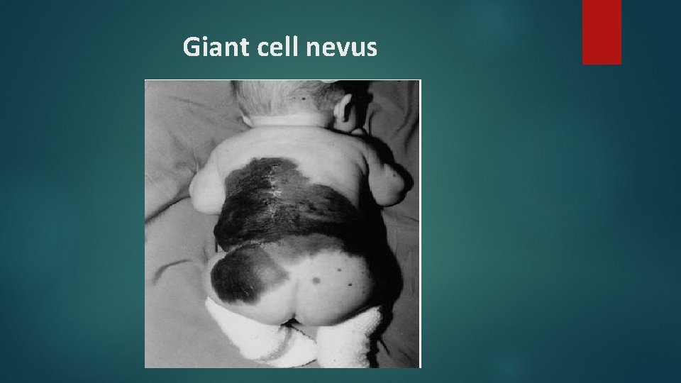
Congenital nevi Rarer , 1% of neonates larger and may contain hair Congenital giant lesions (giant hairy nevus) most often occur in a bathing trunk distribution or on the chest and back Develop malignant melanoma in 1 to 5% of the cases Excision of the nevus is the treatment of choice
Giant cell nevus
Acquired melanocytic nevi Classified as junctional, compound, or dermal, depending on the location of the nevus cells. Nevus cells accumulate in the epidermis (junctional), migrate partially into the dermis (compound), and finally rest completely in the dermis (dermal). Eventually most lesions undergo involution.
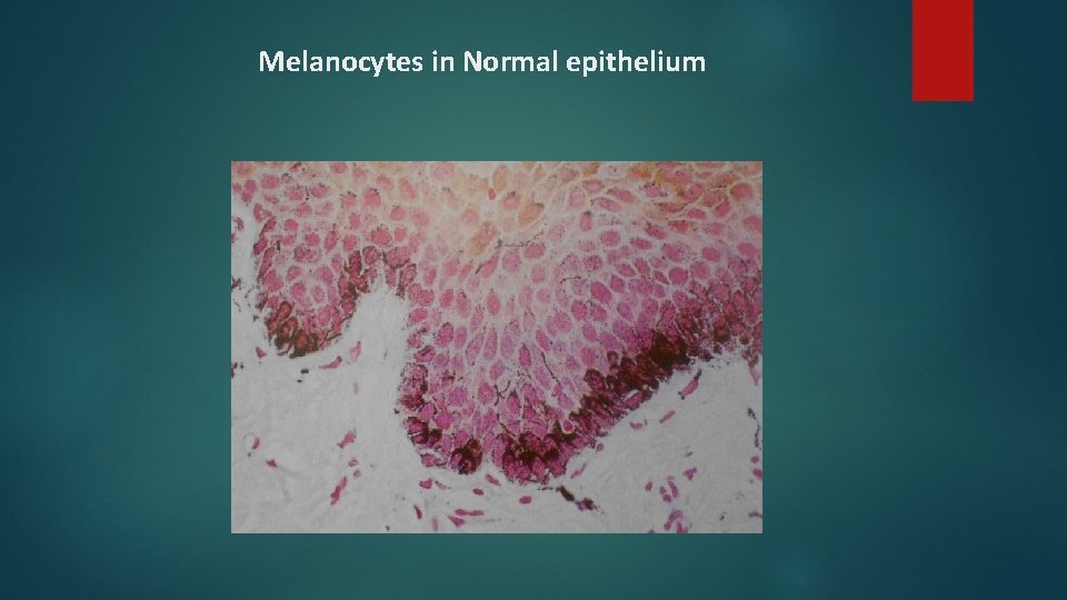
Melanocytes in Normal epithelium
Compound Nevus - Shows junctional activity & nests of nevus cells in connective tissue.
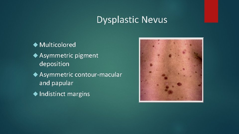
Dysplastic Nevus Multicolored Asymmetric pigment deposition Asymmetric contour-macular and papular Indistinct margins
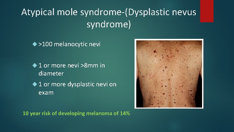
Atypical mole syndrome-(Dysplastic nevus syndrome) >100 melanocytic nevi 1 or more nevi >8 mm in diameter 1 or more dysplastic nevi on exam 10 year risk of developing melanoma of 14%
Management Close monitoring- full body exams every 6 months Dermoscopy of all atypical appearing nevi The magnified visualization of pigmented skin lesions beyond what would be visible by the physician Increases diagnostic accuracy by 10 -20% Excision of any changing or markedly atypical nevi
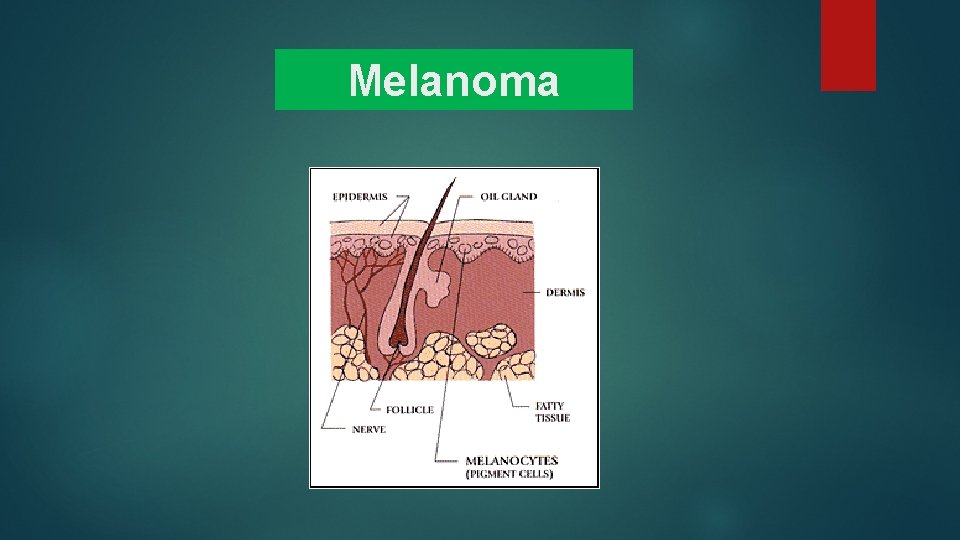
Melanoma
Melanoma: Pathogenesis § Cell of origin: melanocyte § Etiology: • Cumulative and prolonged UVB and/or UVA exposure • UVA exposure from tanning beds increases risk for melanoma
Risk Factors § Individual risk factors for development of melanoma • Increasing age • Fair skin; blue eyes, red or blond hair; freckling • Greater than 100 acquired nevi • Atypical nevi • Immunosuppression • Personal or family history of melanoma (two or more 1 st degree relatives) • Ultraviolet exposure: Risk directly related to # of severe blistering sunburns before puberty; tanning booth use • Genetic syndromes
Heredity of Melanoma § 10 % of melanomas are familial and have a genetic basis § The genes CDKN 2 A and CDK 4 together make up 50% of all inherited familial cases § Other identified genes include p 53, BRCA 2 § 50% of familial melanoma patients have no identified mutation – i. e. , their genes have not been identified yet
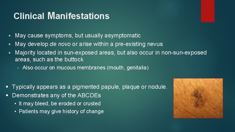
Clinical Manifestations May cause symptoms, but usually asymptomatic § May develop de novo or arise within a pre-existing nevus § Majority located in sun-exposed areas, but also occur in non-sun-exposed areas, such as the buttock § • Also occur on mucous membranes (mouth, genitalia) § Typically appears as a pigmented papule, plaque or nodule. § Demonstrates any of the ABCDEs • It may bleed, be eroded or crusted • Patients may give history of change
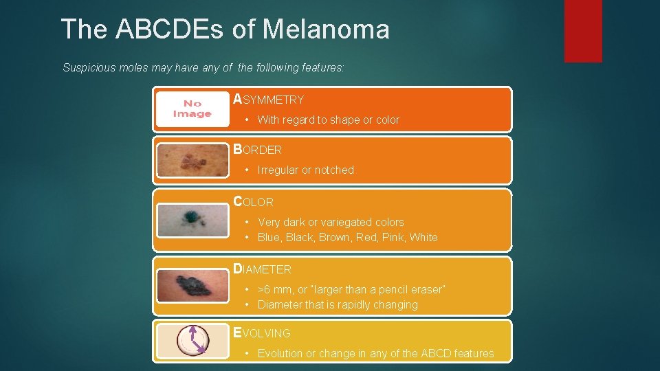
The ABCDEs of Melanoma Suspicious moles may have any of the following features: ASYMMETRY • With regard to shape or color BORDER • Irregular or notched COLOR • Very dark or variegated colors • Blue, Black, Brown, Red, Pink, White DIAMETER • >6 mm, or “larger than a pencil eraser” • Diameter that is rapidly changing EVOLVING • Evolution or change in any of the ABCD features
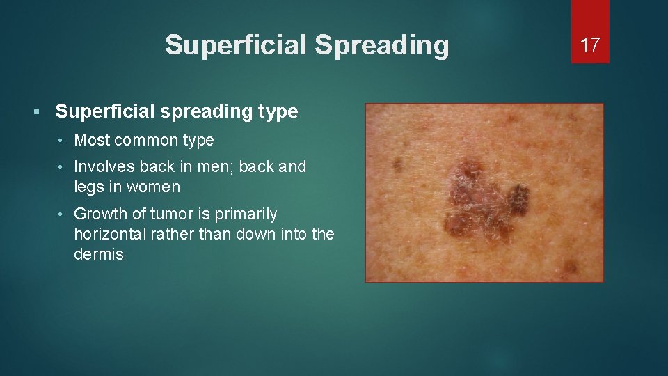
Superficial Spreading § Superficial spreading type • Most common type • Involves back in men; back and legs in women • Growth of tumor is primarily horizontal rather than down into the dermis 17
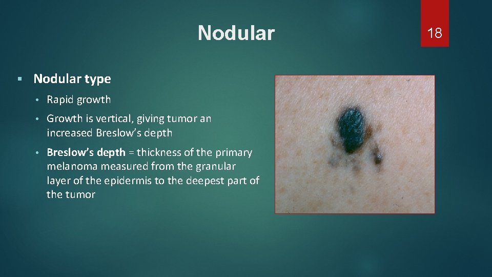
Nodular § Nodular type • Rapid growth • Growth is vertical, giving tumor an increased Breslow’s depth • Breslow’s depth = thickness of the primary melanoma measured from the granular layer of the epidermis to the deepest part of the tumor 18
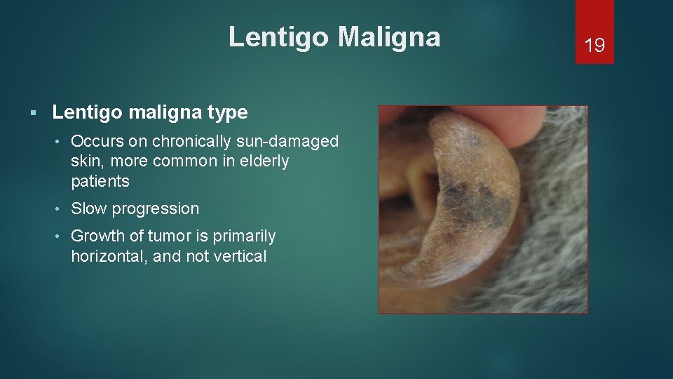
Lentigo Maligna § Lentigo maligna type • Occurs on chronically sun-damaged skin, more common in elderly patients • Slow progression • Growth of tumor is primarily horizontal, and not vertical 19
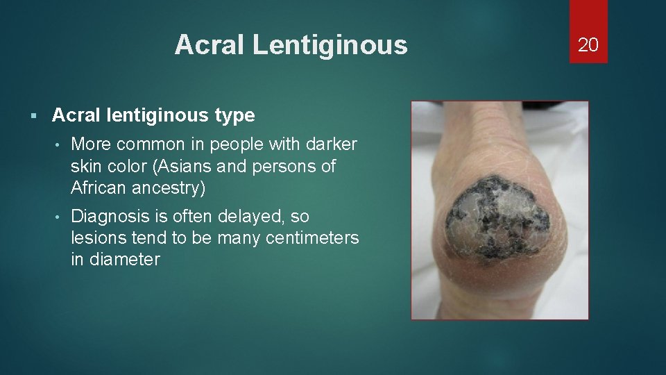
Acral Lentiginous § Acral lentiginous type • More common in people with darker skin color (Asians and persons of African ancestry) • Diagnosis is often delayed, so lesions tend to be many centimeters in diameter 20
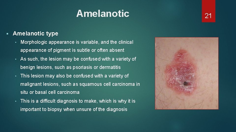
Amelanotic § Amelanotic type • Morphologic appearance is variable, and the clinical appearance of pigment is subtle or often absent • As such, the lesion may be confused with a variety of benign lesions, such as psoriasis or dermatitis • This lesion may also be confused with a variety of malignant lesions, such as squamous cell carcinoma in situ or basal cell carcinoma • This is a difficult diagnosis to make, which is why it is important to biopsy when unsure of the diagnosis 21
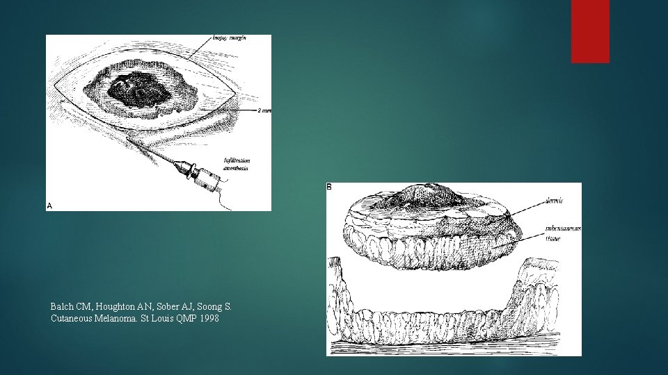
Diagnosis Biopsy Excision (Golden standard) *İncision biopsy *Punch biopsy Partial thickness or shaving biopsies are contraindicated *All dermis layers should be removed
Balch CM, Houghton AN, Sober AJ, Soong S. Cutaneous Melanoma. St Louis QMP 1998
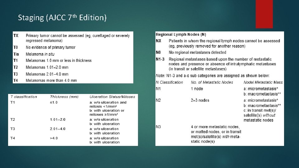
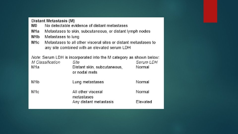
Staging (AJCC 7 th Edition)
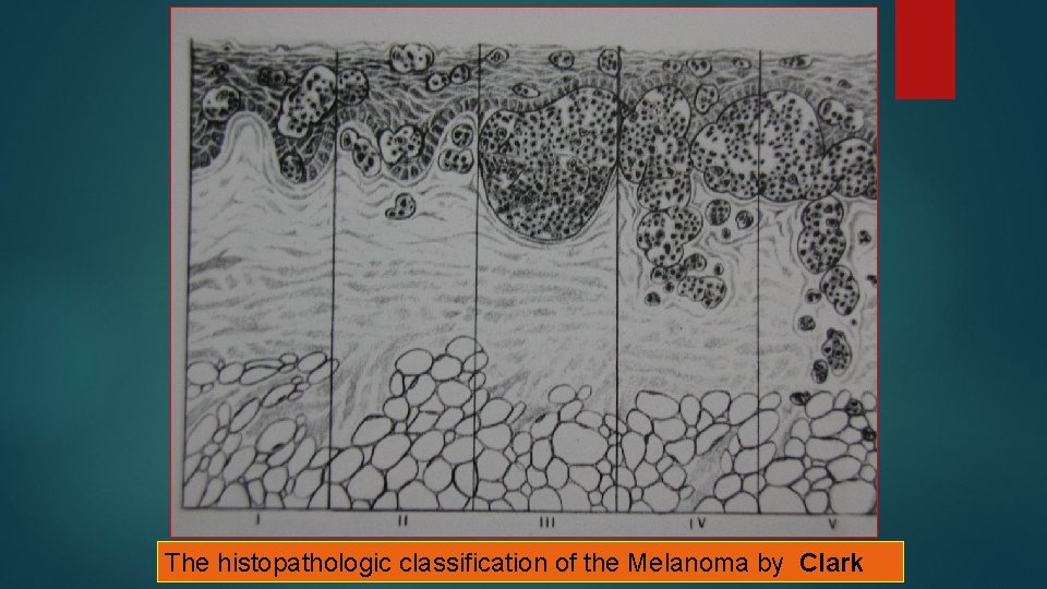
Clark Classification Level I — the atypical melanocytes are confined to the epidermis (in situ melanoma); Level II — the atypical melanocytes have extended into the papillary dermis but have not reached the reticular dermis; Level III — the atypical melanocytes have penetrated to the interface between the papillary dermis and the reticular dermis but do not extend into the reticular dermis; Level IV — the atypical melanocytes have extended into the reticular dermis; Level V — the atypical melanocytes have reached into the subcutaneous fat.
The histopathologic classification of the Melanoma by Clark
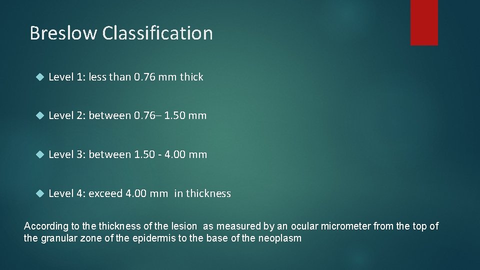
Breslow Classification Level 1: less than 0. 76 mm thick Level 2: between 0. 76– 1. 50 mm Level 3: between 1. 50 - 4. 00 mm Level 4: exceed 4. 00 mm in thickness According to the thickness of the lesion as measured by an ocular micrometer from the top of the granular zone of the epidermis to the base of the neoplasm
Prognostic Factors n n n Breslow thickness (most important) Clark invasion level Ulceration Age, sex, location Size and surgical margins Others (Mitotic index, growth phase, regression. . . )
Surgical Treatment n Biopsy n Wide Local Excision n Staging with Sentinel Lymph Node biopsy n Therapeutic Lymph Node Dissection n Treatment of Distant Metastasis
Wide Surgical Excision Suggested surgical margins: (according to breslow thickness) In-situ MM: 0. 5 -1 cm Breslow thickness < 1 mm : 1 cm Breslow thickness 1 -4 mm: 2 cm Breslow thickness >4 mm: > 3 cm
Sentinel Lymphadenectomy
Sentinel Lymphadenectomy Sentinel lymph node shows the regional node status If sentinel lymph node negative, others lymph nodes in the basin are also negative If sentinel lymph node contains tumor cells, It means disease spread to the regional nodal basin
Sentinel Lymphadenectomy Sentinel node negative no additional treatment, follow the patient Sentinel lymph node positive Therapeutic lymph node dissection
Advantages of Sentinel Lymphadenectomy n Provides staging n Prevention of Elective Lymph node dissection morbidity
Sites of Distant Metastasis n n n n Skin Subcutaneous Tissue Distant Lymph Nodes Pulmonary Liver Brain Bone Intestine
Chemotherapeutic agents • Melphalan • Interferon • Interleukin-2 • Dacarbazine