Disorders of Bones Joints and Skeletal Muscle Nrsg

Disorders of Bones, Joints, and Skeletal Muscle Nrsg 407

Normal Skeleton

Bones 3 Functions: mechanical, metabolic, and hematopoietic Composed of lamellar bone Other type is woven bone present in the fetus and during healing from fractures and bone disease Lamella bone is organized by 2 structural bone Compact bone-hard outer shell Spongy bone-central marrow cavity

Types of Bone Cells Osteoprogenitor cells-found in bone marrow and periosteum which differentiate into osteoblasts and osteoclasts Osteoblasts: bone-forming cells Osteoclasts: bone-dissolving cells

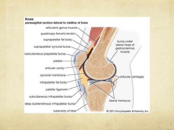
Joints Busiest, hardest working parts of the body Place where two bones meet Classified according to movement they allow

Joints cont’d Fibrous joints are synarthroses by joining bones by fibrous tissue Includes bones of the skull Synovial joints-diarthroses, join bones by ligaments, have a space and allow range of motion Knee, elbow, shoulder, wrist, hip Cartilaginous joints-amphiarthroses, join bones by cartilage, have no space, allow for limited movement Joints between vertebral bodies and wrists
 Composed")
Skeletal Muscle Motor unit: Lower motor neuron Motor end plate Skeletal muscle cell(s) Composed of two fiber types Type I-red/slow twitch Type II-white/fast twitch

Osteoporosis Decreased mineralization of bones, increased bone porousness, and decreased bone mass Bone tissue no longer provides proper mechanical support and strength Primary-most common Post-menopausal women Secondary-due to another condition (hyperparathyroidism)

Primary Osteoporosis Postmenopausal women Less tall – compression of spine curvature of spine, drooped posture Weak abdominal muscles – protruding abdomen Compression of lungs from posture – resp insufficiency

Fractures Broken bone, defined as a discontinuity of the anatomy of a bone Risk factors: 80+ years old Weight less than 130 lb Long-term use of benzodiazepines Lack of walking/running for exercise Poor vision Brain disease affecting physical stability/mental capacity

Types of Fractures

Types of Fractures Avulsion – ligament or tendon attached to bone pulls away Comminuted – many small fragments (> 2 pieces) Displaced – displacement of fracture fragments, can be axially displaced, angulated or rotated Greenstick – incomplete fracture in which the bone bends Impacted – one broken end driven and wedged into the other – commonly seen with comminuted fxs Interarticular – related to joints Longitudinal – lengthwise along bone Oblique – across the shaft of the bone, combo of bending and twisting Pathologic – related to disease making bones brittle Spiral – fracture line spirals around the shaft of the bone Stress – bone subjected to repeated stress, AKA fatigue fx

How Fractures Heal Inflammatory phase: Hematoma Reparative phase: Granulation Callus formation Consolidation Remodeling phase

Treatments for Fractures Closed reduction Open reduction internal fixation Pins, plates, screws, nails, grafts, implants

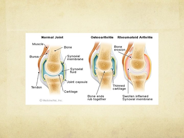
Arthritis Disease of painful joint condition contributing to joint abnormalities Most common: osteoarthritis Progressive, noninflammatory erosion of joint cartilage Primary-not attributed to specific circumstance Secondary-abnormal stress on a joint Obesity Harsh physical activity (knees of professional athletes) Physical malformation creating abnormal joint stress Joints of individuals with peripheral nerve disease

Rheumatoid Arthritis Systemic, chronic autoimmune disease involving synovial joints Exact cause unknown but viruses and genetics suspected Autoimmune reaction due to T lymphocytes B lymphocytes also play a role Inflammatory reaction stimulates growth of blood vessels and fibrous tissue into the synovium and joint cartilage Highly vascular inflammatory membrane covers cartilage surface and releases destructive enzymes to dissolve cartilaginous plate

RA Signs and Symptoms 2 main signs: Deviation of bones in hand toward radial side of arm while the fingers deviate towards the opposite side (Z deformity) Rheumatoid nodules-painless 1 -2 cm subcutaneous inflammatory nodules Diagnosis depends on clinical signs (not laboratory tests) Detection of RF in blood can confirm but not exclude if not found

Carpel Tunnel Syndrome

Carpal Tunnel Syndrome Condition of the tendons and tendon sheaths of ventral wrist Repetitive finger and wrist motions

Fibromyalgia Pain syndrome with no objective abnormalities Clinical syndrome of fatigue, muscle, tendon, and ligament pain, tenderness, and stiffness NOT associated with any objective signs of disease Diagnosis depends on ruling out all other potential causes of patient’s symptoms
- Slides: 22