Diseases of the Small IntestineI Diarrhea Diarrhea is


- Slides: 15
Diseases of the Small Intestine-I
Diarrhea • Diarrhea is an increase in fecal mass caused by an increase in fecal water and/or solid content. It is typically accompanied by an increase in frequency, fluidity, and/or volume of feces • The mech- anistic approach to the genesis of diarrhea is simple: permeability diarrhea is due to inflammation or neoplastic infiltration causing exudation, and secretory diarrhea is caused by chemi- cal toxins and bacterial toxins
• However, most SI diseases have a component of osmotic diarrhea, and even in a situation as simple as lactase deficiency, mixed mechanisms occur • Bacterial fermentation of unabsorbed solutes is often a complicating factor in malabsorption. The fecal p. H is often low because of the production of volatile fatty acids, and some products (hydroxylated fatty acids, unconju- gated bile acids) cause colonic secretion; signs of large- intestinal diarrhea frequently accompany prolonged SI disease.
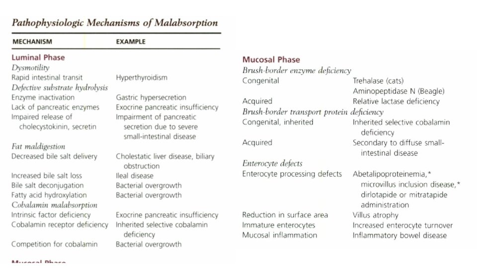
Malabsorption • Failure of food assimilation is sometimes classified as primary failure to digest (maldigestion) or primary failure to absorb (malabsorption). • However, such a classification is misleading, because failure of absorption is an inevitable consequence of failure to digest. The preferred use of the term malabsorption is to describe defective absorption of a dietary constituent resulting from interference with the digestive and/or absorptive processing of that molecule.
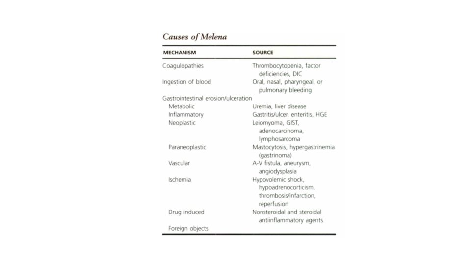
Melena • The presence of dark, tarry, oxidized blood in feces, a condi- tion called melena, reflects either swallowed blood or generalized or localized GI bleeding proximal to the large intestine • The general approach to melena is to rule out bleeding diatheses; ingestion of blood from other lesions, such as oral masses; toxicity, as with nonsteroidal antiinflammatory drugs (NSAIDs); or underlying metabolic disorders, such as hypoad- renocorticism, before pursuing primary GI causes.
Borborygmi and Flatulence • Borborygmi is the abdominal rumbling noise caused by the propulsion of gas through the Gl tract. Swallowed air and bacterial fermentation of ingesta are the main causes of borborygmi and flatulence and often result in an offensive odor. • Feeding a diet that is highly digestible with a low fiber content leaves little material for bacterial fermentation and can effect a cure in some cases. • If borborygmi or flatulence continue despite dietary modification or addition of adsorbents, the animal may be excessively aerophagic or may have malabsorption.
Weight Loss or Failure to Thrive • Weight loss or failure to thrive accompanied by diarrhea is a feature of malabsorption, and the diagnostic approach is the same as for chronic diarrhea. However, malabsorption causing weight loss does not invariably cause diarrhea.
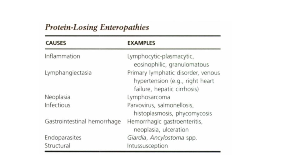
Protein-Losing Enteropathy • When SI disease is severe enough for protein leakage into the gut lumen to exceed plasma protein synthesis, hypoproteinemia develops. • Chronic diarrhea associated with panhypoprteinemia usually requires intestinal biopsy to define the cause of the PLE • Hypoproteinemia associated with GI disease is much less common in cats, and most often accompanies Gllymphoma, but infrequently results in ascites.
• Dog breeds predisposed to PLE • Hasenji, Lunde- hund, • Soft-Coated Wheaten Terrier, • Yorkshire Terrier, • Shar-Pei.
DIAGNOSIS • Serum concentrations of both albumin and glohulin are reduced in most patients with PLE. Exceptions are hyperglohulinemia found in histoplasmosis and Basenji enteropathy • Survey abdominal radiographs often are unhelpful in patients with PLE because of the loss of contrast, • Radiographs may show pleural effusion, metastatic neoplasia, or changes consistent with histoplasmosis. Although intestinal function tests may confirm the presence of malabsorption. they rarely provide a definitive diagnosis, and intestinal biopsy is more appropriate. Because many intestinal causes of PLE arc diffuse. endoscopy is the safer way to obtain biopsies, but surgical biopsy may be required for definitive diagnosis for transmural lymphoma and lymphangiectasia