Diseases of the Salivary Glands Diseases of the


 or the parotid gland (rarely) because")


 Almost exclusive to the parotid gland, it causes a")


 While not due to disease of the salivary glands, children or")

- Slides: 25
Diseases of the Salivary Glands
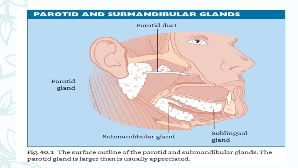
Diseases of the Salivary Glands The salivary glands consist of: 1 the parotid glands; 2 the submandibular glands; 3 minor salivary glands throughout the mouth and upper air passages. (The sublingual collection is included in this group. ) Parotid gland The parotid gland lies on the side of the face in close relationship to the ear, the angle of the mandible and styloid muscles. The facial nerve enters the posterior pole of the parotid gland divides within its substance into its various branches, which exit at the anterior margin of the gland. It is the presence of the facial nerve within the parotid that makes surgery of this gland so difficult. Its duct opens opposite the second upper molar tooth, where it forms a small visible papilla
Diseases of the Salivary Glands Its secretomotor nerve supply comes from the glossopharyngeal nerve via the tympanic plexus in the middle ear. The saliva produced is entirely serous. The surface outline of the gland is shown in Fig. 40. 1. The submandibular salivary gland The submandibular gland lies in the floor of the mouth below and medial to the mandible and its greater part is external to the mylohyoid muscle. The deep part of the gland curves around the back of the mylohyoid and the duct runs forwards to open at the sublingual papilla. The deep part of the gland lies on the lingual nerve, from which it receives its secretomotor supply derived from the facial nerve via the chorda tympani in the middle ear. The saliva from the submandibular gland is both serous and mucous.
Diseases of the Salivary Glands The minor salivary glands can be seen and felt in the lips, cheeks, palate and upper air passages. They produce mainly mucous saliva (remember the noun is mucus) and are responsible for a large proportion of the total saliva secreted. They are subject to many of the diseases that affect the major salivary glands.
Protection of the tracheobronchial tube HISTORY-TAKING — SALIENT POINTS In asking the history, enquire particularly about pain and swelling of the glands in relation to eating. If the duct is obstructed, the whole gland will become tense and painful and enlarge visibly during saliva production, and will resolve slowly over about an hour. If a lump is present, ask about variation in size and whether it is related to food. Tumours do not enlarge during salivation, but do tend to get bigger with the passage of time. Ask about dryness of the mouth, remembering that obstruction of even two major glands produces little apparent change. Persistent dryness suggests diffuse salivary gland disease. Ask about recent contact with mumps.
EXAMINATION OF THE SALIVARY GLANDS First, inspect the salivary glands externally, noting any swellings or asymmetry. The function of the facial nerve should be tested in all its divisions. The parotid and submandibular ducts should be inspected to assess saliva flow, redness and the presence of pus or an obvious stone. The mouth should be inspected to see if it is excessively dry. After inspection, the glands should be palpated carefully by bimanual exploration. It is only in this way that a proper assessment can be made. The ducts should be felt carefully for calculi and then massaged gently towards the opening to express any pus present. The patient can be given an acid-drop to suck and any enlargement on salivation assessed. The ears should be inspected to make sure that there is no salivary fistula or tumour extension through the anterior meatal wall. Gentle probing of the duct may produce a gush of turbid saliva.
INVESTIGATION 1 Plain X-ray, including occlusal views, will show a radio-opaque calculus. 2 Ultrasonic scanning of the gland is quick, non-invasive and exposes the patient to no radiation. It will identify masses, cysts and calculi but is only comprehensible to the radiologist! 3 Sialography will outline the duct system. Contrast medium is injected into the gland after cannulation of the duct, and will show radiolucent stones or strictures. A solid tumour will not fill with contrast, but an area of sialectasis will be seen as droplets in the dilated ducts. Sialography contributes little to tumour diagnosis and is not usually performed in such cases. 4 If there is a large parotid tumour with parapharyngeal extension, an MR scan will outline its extent.
Acute inflammation Mumps is the commonest acute inflammatory condition of salivary glands. It affects mainly the parotid glands, which become uniformly swollen and painful, but the submandibular glands may also be involved. Its incidence had fallen to very low levels as a result of immunization, but is now rising alarmingly as some parents decline to have their children immunized. Acute suppurative parotitis is uncommon and usually occurs in debilitated patients. Treatment is with antibiotics, rehydration and oral hygiene. If an abscess develops, surgical incision is required.
Acute sialadenitis may affect the submandibular gland (commonly) or the parotid gland (rarely) because of the presence of a duct calculus. The affected gland is painful and swollen and is made worse by eating. The patient is usually unwell with a pyrexia. Removal of the stone provides dramatic relief in most cases. Recurrent acute inflammation of the major salivary glands presents a problem of management if no stone is present. If there is a duct stricture, gentle dilatation may be curative. In childhood, recurrent episodes of acute inflammation will usually subside by puberty and should be treated conservatively.
Chronic inflammation of the parotid or submandibular gland is usually due to sialectasis (duct dilatation leading to stasis and infection) and will not usually respond to conservative measures. The gland is thickened with episodic pain and infection, and can be felt easily on bimanual examination. TREATMENT The submandibular gland so affected can be excised; chronic sialectasis of the parotid poses a difficult problem. Excision has a high risk of facial nerve damage and long-term antibiotics should be tried before resorting to parotidectomy.
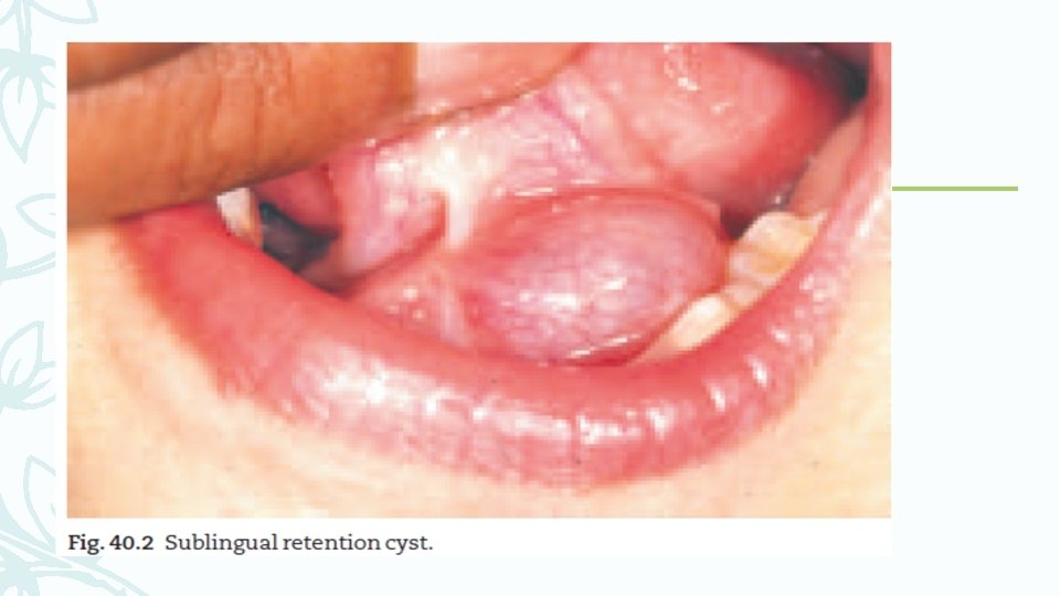
Sjögren’s syndrome is an auto-immune systemic disorder affecting the salivary and lacrimal glands. There is enlargement of the glands and loss of secretion, leading to dryness of the eyes and mouth. In many cases, biopsy of the lip mucosa will show minor salivary glands heavily infiltrated by lymphocytes. Symptomatic relief can be obtained by the use of artificial saliva or glycerin and warm-water mouthwash. Salivary retention cysts Salivary cysts occur most commonly in the floor of the mouth, where they may become very large and expand the loose tissues (Fig. 40. 2). The name ‘ranula’ is often applied. Less commonly, such retention cysts occur on the mucosal aspect of the lips.
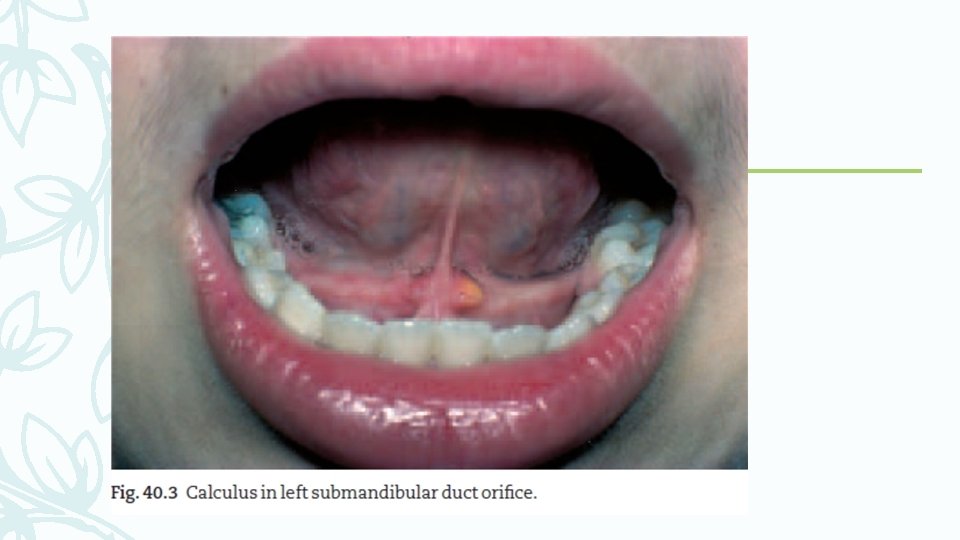
Salivary calculi Most salivary calculi occur in the submandibular gland because of the mucoid nature of its saliva, which can become inspissated (Fig. 40. 3). However, calculi do also occur in the parotid gland. CLINICAL FEATURES The flow of saliva from the affected gland becomes obstructed, causing the gland to swell during salivation. Such swelling is painful and its size may be alarming. The swelling will usually resolve over about an hour. The calculus can be seen if it presents at the duct opening, or felt within the duct or gland. INVESTIGATION Most, but not all, calculi are radio-opaque, and X-rays should be performed as described above.
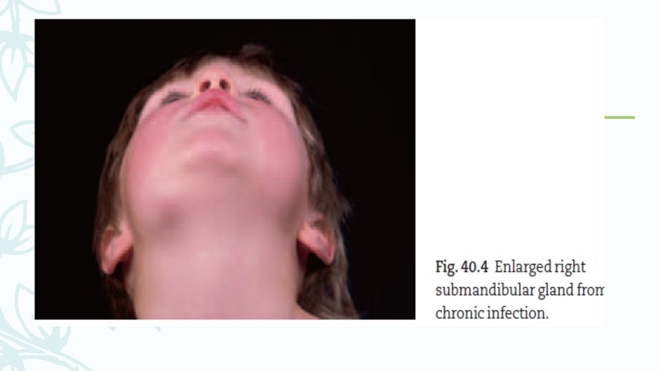
TREATMENT 1 Intraductal calculi can be removed from the duct under local anaesthetic. A suture should first be passed around the duct proximal to the stone to prevent its movement back into the gland. Removal of such a stone may be more difficult than you might expect. 2 If the stone is within the substance of the salivary gland, excision of the gland will have to be considered. The submandibular gland presents no problem as it is straightforward to excise, but parotidectomy for calculus requires a high degree of skill. Salivary gland tumours Salivary glands, because they contain lymph nodes within their structure, may be the site of metastases from a non-salivary primary site or from blood dyscrasias such as leukaemia (Fig. 40. 4). Salivary gland disease is uncommon in childhood, but a solid parotid tumour presenting under the age of 16 is more likely (60: 40) than not to be malignant.
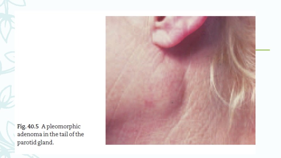
It is not usually possible to detect clinically whether a tumour in a salivary gland is benign or malignant. Fine needle aspiration cytology may be helpful in predicting the type of tumour present. All such tumours should be treated as malignant until the diagnosis is confirmed by histology. The same tumours occur in minor salivary glands as in the major glands but malignant tumours in minor glands have a more aggressive course. PATHOLOGICAL CLASSIFICATION Benign tumours Pleomorphic salivary adenoma (mixed salivary tumour, PSA) (Fig. 40. 5) Occurs most frequently in the parotid gland. Propensity for recurrence if not removed with surrounding cuff of tissue. PSA accounts for about 90% of parotid tumours in adults.
Warthin’s tumour (cystic lymphoepithelial lesion) Almost exclusive to the parotid gland, it causes a smooth swelling in the tail of the gland that may feel cystic. Haemangioma A rare tumour, usually congenital or occurring in early childhood; occurs most commonly in the parotid gland. A haemangioma is also often present on the skin of the face or in the mouth.
Malignant tumours Adenoid cystic carcinoma The commonest malignant tumour of salivary glands. With early perineural invasion, the long-term prognosis is poor but survival for many years is usual. Muco-epidermoid tumours Can arise in any of the salivary glands and have a variable degree of malignancy; the majority will behave as benign tumours but a small proportion are aggressively malignant. Acinic cell tumours Are usually of low-grade malignancy and occur almost exclusively in the parotid gland. Malignant pleomorphic adenomata May arise in an existing adenoma. The malignant change is made apparent by a rapid increase in size and, in the case of parotid tumours, the development of facial weakness. A benign tumour causes no such weakness.
Squamous carcinoma of the submandibular and parotid glands is uncommon and has a very poor prognosis. Radical excision followed by radiotherapy is the only treatment that offers any chance of cure. Lymphoma can occur in any of the salivary glands, major or minor. Surgery has no part in treatment other than for biopsy, but radiotherapy and/or chemotherapy may be curative. The lymphoma arises from lymphoid tissue within the salivary gland.
Salivary incontinence (drooling) While not due to disease of the salivary glands, children or adults with, for example, cerebral palsy or stroke may be unable to control the saliva produced, particularly from the sublingual and submandibular ducts. This results in much distress and discomfort to patient and relatives. It can often be relieved by surgical relocation of the submandibular ducts to a posterior position near the tonsil combined with excision of the sublingual gland.
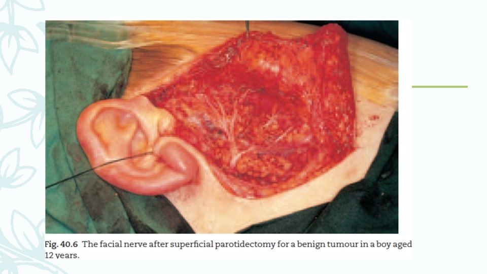
Surgery of the salivary glands SUBMANDIBULAR GLAND EXCISION This is usually performed for tumour removal but may be made necessary by calculus or by chronic infection. The gland is approached externally and care is taken not to damage the marginal mandibular branch of the facial nerve or the lingual nerve. PAROTIDECTOMY Again, usually undertaken for removal of tumour (Fig. 40. 6). The facial nerve is identified at an early stage in the operation and its branches followed carefully. Usually the tumour lies superficial to the nerve but if it is deep, the nerve will need to be mobilized. All patients having parotidectomy must be warned of the risk of facial nerve damage.