DISEASES OF THE SALIVARY GLANDS 1 Sialolithiasis Nonspecific


- Slides: 47

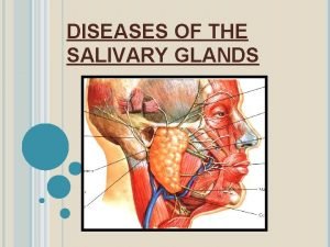
DISEASES OF THE SALIVARY GLANDS 1

Sialolithiasis & Nonspecific sialadenitis • Submandibular • Stones nidus - impacted food debris - dehydration with decreased secretion • Stone → duct obstruction • Nonspecific interstitial inflammation • Or suppuration (abscess) – Staph. Strep 2

Sialolithiasis & Nonspecific sialadenitis • • Unilateral Painful – during meals Purulent ductal discharge + Recurrent 3

MUCOCELE • • Trauma Lower lip Fluctuant swelling Blockage / rupture of salivary duct → leakage of saliva into the surrounding stroma • RANULA – mucocele of the sublingual 4

MUCOCELE 5

Sjogren’s syndrome • Immune destruction • Salivary – xerostomia (dry mouth) • Lacrimal – keratoconjunctivitis sicca (dry eyes) 6

TUMOURS • • Approx. 30 in number 2% of all tumours Benign – 5 th – 7 th decade Malignant – later than above 7

BENIGN TUMOURS • • • PLEOMORPHIC ADENOMA – 50% Warthin’s tumour Oncocytoma Basal cell adenoma Canalicular adenoma Ductal papilloma 8

PLEOMORPHIC ADENOMA • Benign tumour of glands – ADENOMA • Remarkable histologic diversity PLEOMORPHIC • Commonest • Also called Mixed tumour – epithelial and connective tissue components 9

PLEOMORPHIC ADENOMA • Cell of origin → EPITHELIAL epithelial-myoepithelial / ductal reserve cells 10

PLEOMORPHIC ADENOMA Gross • < 6 cms • Bosselated • Well-demarcated / encapsulated / tongue-like protrusions • Cut surface: gray-white / variegated / myxoid / bluish translucent (chondroid) 11

PLEOMORPHIC ADENOMA 12

PLEOMORPHIC ADENOMA 13

PLEOMORPHIC ADENOMA Microscopy • HETEROGENITY ! → PLEOMORPHIC • EPITHELIAL : epithelial & myoepithelial cells arranged in ducts, acini, irregular tubules, strands, sheets NO DYSPLASIA • MESENCHYMAL : myxoid, hyaline, chondroid, osseous 14

PLEOMORPHIC ADENOMA 15

PLEOMORPHIC ADENOMA 16

PLEOMORPHIC ADENOMA CLINICAL ASPECTS • Slow growing • Painless • Parotid – swelling below & in front of the year • RECURRENT – tongue-like protrusions Enucleation – 25% with Parotidectomy – 4% 17

CARCINOMA ex PLEOMORHIC ADENOMA OR MALIGNANT MIXED TUMOUR Adenocarcinoma 18

WARTHIN’S TUMOUR Papillary cystadenoma lymphomatosum Adenolymphoma 19

WARTHIN’S TUMOUR Gross • Encapsulated • Cut surface : pale-grey narrow cystic / cleft-like / convoluted spaces contain serous / mucinous fluid 20

WARTHIN’S TUMOUR 21

WARTHIN’S TUMOUR Microscopy Cystic spaces - Narrow / cleft-like CYSTADENOMA Lining – double-layered epithelium – infoldings PAPILLARY • Inner (lumen) – palisade of columnar cells with abundant finely granular eosinophilic cytoplasm (oncocytic) • Outer (below the first) – cuboidal / polygonal Stroma - dense lymphoid tissue (germinal centres + ) LYMPHOMATOSUM 22

WARTHIN’S TUMOUR 23

24

WARTHIN’S TUMOUR 25

WARTHIN’S TUMOUR • HISTOGENESIS ? Aberrant inclusions of lymphoid tissue in salivary glands 26

WARTHIN’S TUMOUR CLINICAL ASPECTS • Benign • Very rarely recurs 27

MALIGNANT TUMOURS • • Mucoepidermoid ca Acinic cell ca Adenoid cystic ca Malignant mixed tumour 28

Malignant tumours • Old age • Max (70 -90%) – sublingual • Malignant tumours more in smaller glands 29

MUCO-EPIDERMOID CARCINOMA • 15% • 60 -70% in the parotid, also in others • MOST COMMON MALIGNANT • Most common radiation-induced 30

MUCO-EPIDERMOID CARCINOMA Gross • < 8 cms. • Apparently circumscribed / infiltrative • Cut surface : pale grey-white small mucin-filled cysts 31

MUCO-EPIDERMOID CARCINOMA Microscopy • Mixtures of three cell types arranged in cords, sheets, cystic 1. mucus-secreting cells 2. intermediate cells (hybrid) squamoid features with small to large mucusfilled vacuoles 3. squamous cells 32

MUCO-EPIDERMOID CARCINOMA 33

MUCO-EPIDERMOID CARCINOMA • LOW GRADE – Mucus cells predominant • HIGH GRADE – squamous cells predominant 34

MUCO-EPIDERMOID CARCINOMA CLINICAL COURSE Low grade Spread Local invasion Recurrence 15% 5 -yr survival >90% High grade Metastasis(30%) 25 -30% 50% 35

ADENOID CYSTIC CARCINOMA • Mouth – minor, rare in parotid • Nose, paranasal sinuses • Upper airways 36

ADENOID CYSTIC CARCINOMA Gross • Small • Poorly encapsulated, infiltrative • Gray-pink 37

ADENOID CYSTIC CARCINOMA Microscopy • Small cells with dark compact nuclei, scanty cytoplasm • Arranged in tubules, solid, cribriform • SPACES BETWEEN CELLS FILLED WITH HYALINE MATERIAL – basement membrane material • Perineural invasion - pain 38

ADENOID CYSTIC CARCINOMA 39

40

ADENOID CYSTIC CARCINOMA 41

ADENOID CYSTIC CARCINOMA CLINICAL ASPECTS • • Malignant Slow growing Stubbornly recurrent Painful – perineural invasion 42

Acinic cell carcinoma (tumour) • Most PAROTID – composed of serous cells 43

Acinic cell carcinoma Gross • Small • Encapsulated 44

Acinic cell carcinoma Microscopy • Look like serous cells • Clear cytoplasm • Solid sheets, microcystic, glandular, follicular, papillary • Little anaplasia 45

Acinic cell carcinoma 46

Acinic cell carcinoma CLINICAL ASPECTS • Good prognosis • Recurrence rare 47
 Ts of liver
 Intercalated duct histology
 Youtube
 Submandibular gland excision
 Dysphagia terminology
 Salivary gland swelling
 Function of salivary glands
 Sialography
 Sialography
 Phadebas paper
 Weber salivary gland
 Colon
Colon Minor salivary glands
 Modes of glandular secretion
 Specific vs nonspecific defense
 романенко
 Th and tc cells
 V/q scan pulmonary embolism
 J point
 Nonspecific vs specific immunity
 Nonspecific host defense mechanism
 Salivary amylase digestion of starch
 Mucous acini
 Pleomorphic adenoma
 Warthin's tumor
 Function of bile
 Fosdick calcium dissolution test
 Oncocytoma salivary gland
 Cylindroma slideshare
 Salivary gland
 Salivary gland tumor
 Lactobacillus count test
 Oncocytoma salivary gland
 Oncocytoma salivary gland
 Salivary gland pathology
 Simple branched alveolar gland
 Salivary gland disorders classification
 Oxyntic glands
 Major endocrine glands male and female
 What glands are flask shaped with short ducts
 Layer of dermis
 Multicellular exocrine glands can be classified
 The only example of unicellular exocrine glands are
 Pineal and pancreas glands
 Gonads glands
 Serous vs mucous
 Coiled gland
 Adrenal glands

