
Diseases of the Pleura DR Alaa Rashad Diseases












- Slides: 76
Diseases of the Pleura DR/ Alaa Rashad
� � ﺍﻟﻠﻪ ﺍﻟ�ﺣﻤ� ﺍﻟ�ﺣﻴ ﺑﺴ Diseases of the Pleura • Pleura is two Layers, Visceral Pleura (VP) which covers the surface of the lung and lines the interlobar fissures is continous at the hilum with the parietal pleura (PP), which lines the hemithorax.
Pleural space • A potential space with a very small amount of lymph fluid (3 -15 ml) • It is under a dynamic state of secretion and re-absorption. • Pleural fluid act as a lubricant to allow free motion of the lungs in the Pleural space. • Pleural spaces (Rt & Lt) are not connected together and separated by mediastinum.
Nerve supply • Pain fibers • Phrenic nerve in parietal pleura central part of diaphragm Blood supply • Visceral pleura bronchial art& branches of pulmonary art • parietal pleura intercostal arteries
Types of pleural diseases • • Dry pleurisy Pleural effusions Pneumothorax Pleural tumours
Dry or fibrinous pleurisy (DP) Causes : 1. Traumatic 2. Underlying lung disease. Pneumonia Pulmonary infarction Branchial cancer. T. B Rheumatoid arthritis. S. L. E. Uraemia. Epidemic Myalgia (Bornholm's disease) due to coxakie B virus is sometimes with dry pleurisy.
The cardinal symptom of DP is : Pdeuritic chest pain : which is sharp localized may radiate to the ipsilateral chest wall or shoulder. Increased by respiratory efforts cough and sneezing. Stapping
On auscultation it is called pleural rub : Friction sound. Continons in inspiration and expiration. Localized. Best heard early in the disease and after resolution. Increased on pressing the stethoscope. Together with signs of the underlying disease.
• • - Differential diagnosis of (DP): Intercostal myalgia. Herps zoster (before rash) Rib fracture. Costo chondritis Treatment of DP : Treat the cause. Symptomatic treatment for pain.
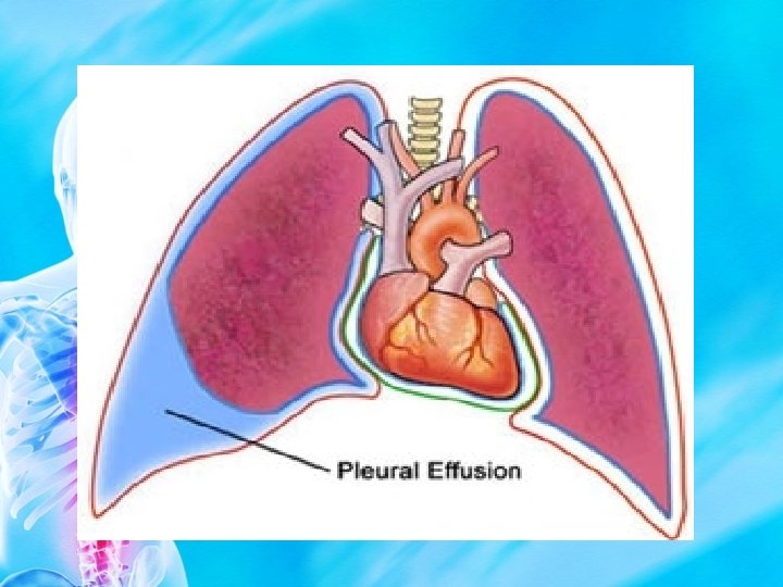
Pleural effusion (PE) Definition : Abnormal accumulation of fluid in the pleural space. PE is a sign of disease and not a diagnosis in itself.
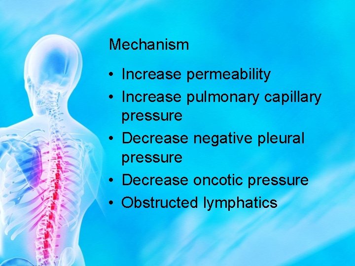
Mechanism • Increase permeability • Increase pulmonary capillary pressure • Decrease negative pleural pressure • Decrease oncotic pressure • Obstructed lymphatics
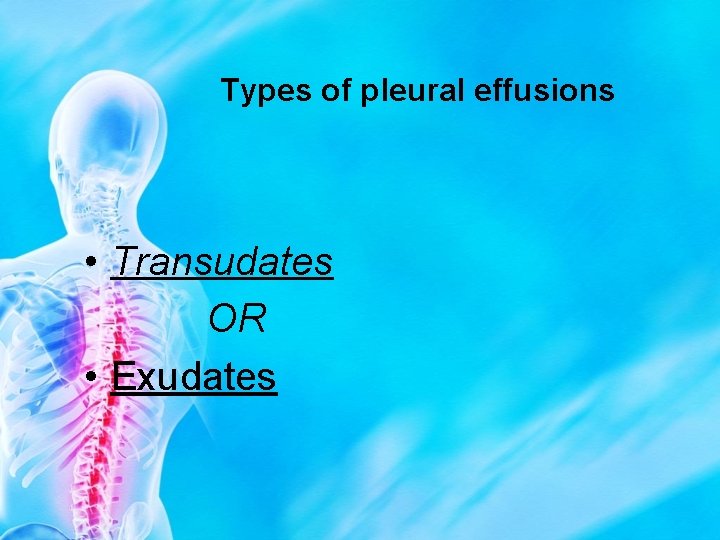
Types of pleural effusions • Transudates OR • Exudates
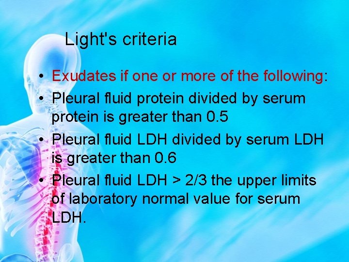
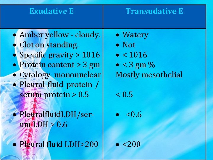
Light's criteria • Exudates if one or more of the following: • Pleural fluid protein divided by serum protein is greater than 0. 5 • Pleural fluid LDH divided by serum LDH is greater than 0. 6 • Pleural fluid LDH > 2/3 the upper limits of laboratory normal value for serum LDH.
Causes of pleural effusion Transudates • Very Common causes • Heart failure • Liver cirrhosis
Transudates • • • Less Common causes Hypoalbuminaemia Peritoneal dialysis Hypothyroidism Nephrotic syndrome Mitral Stenosis
Causes of pleural exudates • • • Common causes Malignancy Parapneumonic effusions Tuberculosis Pulmonary embolism
Exudates • Less Common causes • Rheumatoid arthritis and other autoimmune pleuritis • Benign Asbestos effusion • Pancreatitis • Post-myocardial infarction • Post CABG
Exudates • Rare causes • Yellow nail syndrome (and other lymphatic disorders • Drugs • Fungal infections
• (II) Purulent effusion (empyema) = Pus causes of acute empyema : 1. 2 ry to lung disease: pneumonia – lung abscess – brochiectasis. 2. 2 ry to chest wall dieases : mediastinits – T. B glands – septic lymphadenitis. 3. 2 ry to Mediastinal disease : Mediastinits – T. B glands septic lymphadenitis. 4. 2 ry to infradiaphragmatic conditions : Subphrenic abscess – liver abscess. 5. 2 ry to pyogenic infections in the neck. 6. Hematogenous in cases of pyemia and septicemia. 7. 2 ry infection in serous effusion hemothorax or chylothorax. 8. Trauma.
• (III) Hemorrhagic effusion : • Causes : (1)Malignant pleural effusion 1 ry pleural neoplasm is rare but 2 ry malignat effusion is common from : lung cancer. Metastasis from breast, stomach, pancreas and uterus. Lymphatic spread. Lymphomas. (2)Trauma (hemothorax) (3)Pulmonary thrombo – embolism. (4)Hemorrhagic diseases. (5)Severe fulminating infections as in T. B.
• (IV) Chylous effusion (chylothorax) • Arises from : (1)Surgical trauma to a main lymph channel during intrathoracic operations. (2)Malignant involvement of the thoracic duct. (3)Compression injuries of the trunk.
Diagnosis of Pleural effusion (PE) Symptoms : The clinical manifestations of PE are variable and often are related to the underlying disease. The most common symptoms are : (1)Progressive dyspnea : Is the most common symptom. Indicates a large effusion (not < 500 ml). Other factors (e. g. underlying lung disease, cardiac dysfunction or anemia) may cause dysnea.
(2) Chest pain : Mild or severe is described as sharp or stabbing, is increased with deep inspiration and is pleuritic pain. May be localized to the chest wall or referred to the ipsilateral shoulder or upper abdomen because of involvement of diaphragmatic pleura. It often diminishes in intensity as the effusion increases in size.
Chest pain signifies pleural irritation, which can aid in diagnosis of the cause of effusion, since most transudative effusions do not cause direct pleural irritation. Other symptoms are related to the underlying disease e. g: - Edema of lower limbs, orthopnea and paroxysmal nocturnal dysnea in C. H. F. - Night sweats, fever, hemoptysis and weight loss with T. B. - High grade fever, purulent sputum and pleuritic chest pain in bacterial pneumonia. - If the patient is a previously healthy young adult conside T. B.
- Abdominal symptoms or signs as pain pancreatitis , ascitis. - Recent abdominal surgery subphrenic abscess, liver cirrhosis. - If there any reason for hypoproteinemia (Malnutrition, malabsorption and nephrotic syndrome). - Is there evidence of malignancy ? - History of recent trauma. - Occupational exposure to Asbestos? - joint symptoms rheumatoid arthritis.
Signs • • • Decrease expansion Dull percusion node Decrease vocal resonance Decrease air entry Signs of associated disease (for example : chronic liver disease-CCF-nephrotic syndrome -SLE-RA-Ca lung)
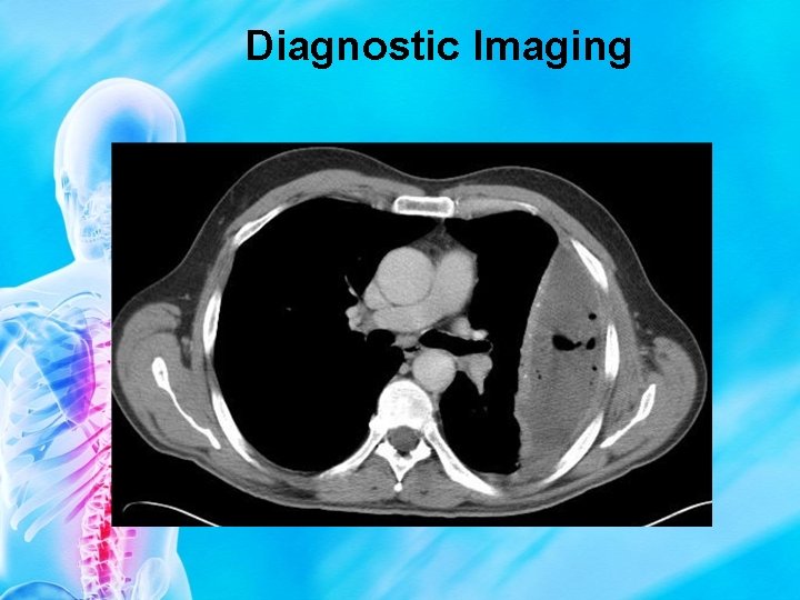
DIAGNOSIS • • CXR PLEURAL ASPIRATION PLEURAL BIOPSY Medical thoracoscopy CT scan VAT Bronchoscopy
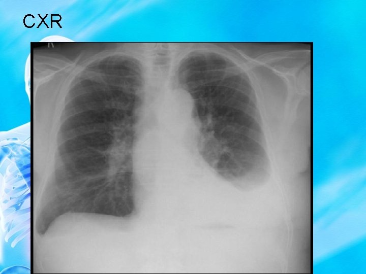
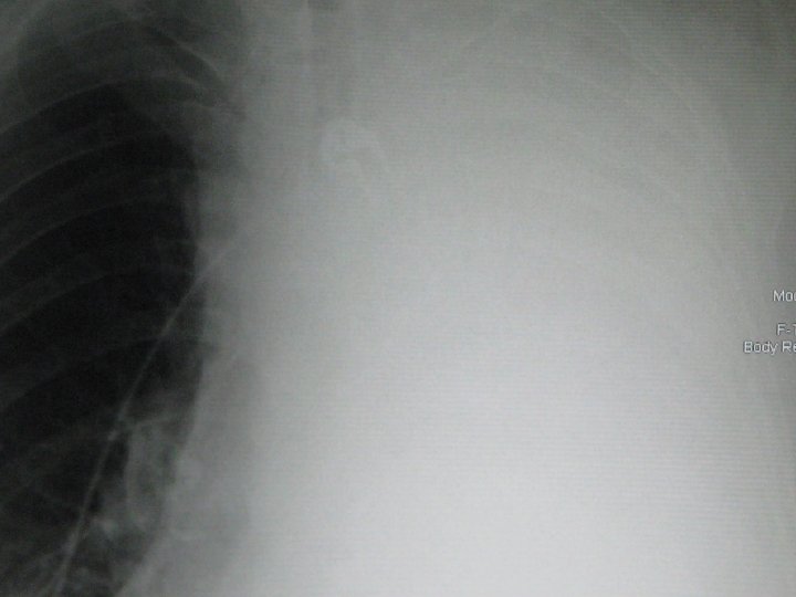
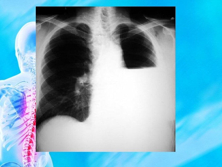
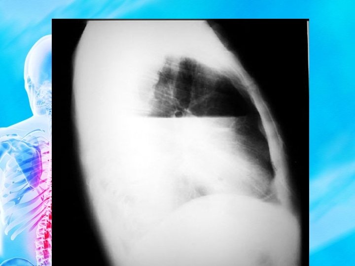
CXR
Diagnostic Imaging
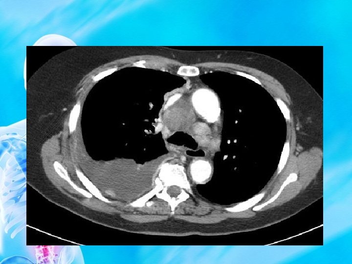
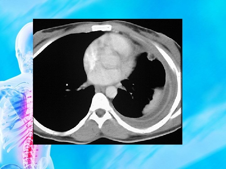
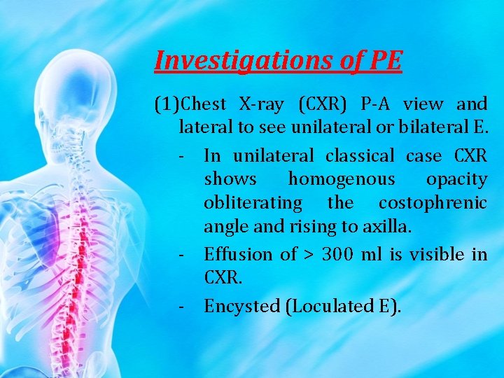
Investigations of PE (1)Chest X-ray (CXR) P-A view and lateral to see unilateral or bilateral E. - In unilateral classical case CXR shows homogenous opacity obliterating the costophrenic angle and rising to axilla. - Effusion of > 300 ml is visible in CXR. - Encysted (Loculated E).
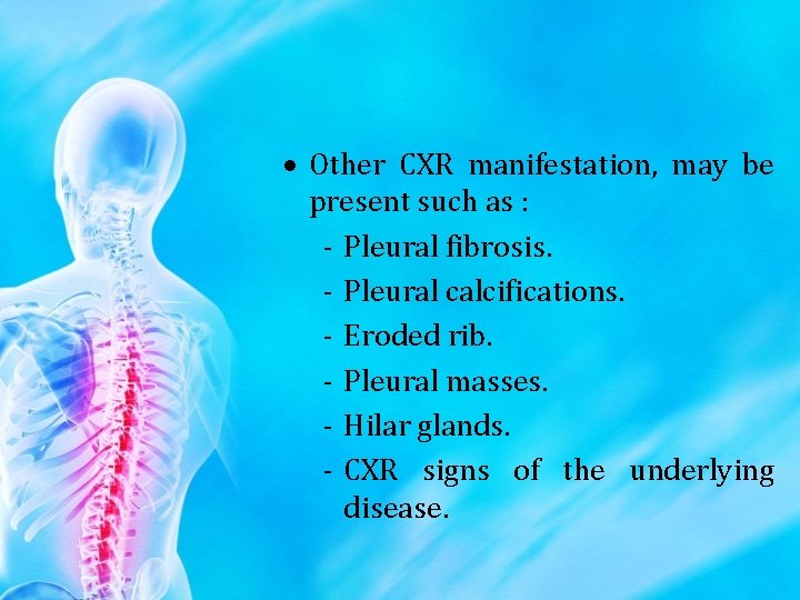
Other CXR manifestation, may be present such as : - Pleural fibrosis. - Pleural calcifications. - Eroded rib. - Pleural masses. - Hilar glands. - CXR signs of the underlying disease.
• • Pleural (fluid aspiration) = Thoracentesis = thoracocentesis Diagnostic aspiration to obtain fluid to see the gross picture and for laboratory studies for microscopic, bacteriological, chemical and cytological. • Therapeutic in some cases of massive E to relieve dysnea.
Exudative E Amber yellow - cloudy. Clot on standing. Specific gravity > 1016 Protein content > 3 gm Cytology mononuclear Pleural fluid protein / serum protein > 0. 5 Transudative E Watery Not < 1016 < 3 gm % Mostly mesothelial < 0. 5 Pleuralfluid. LDH/serum LDH > 0. 6 <0. 6 Pleural fluid LDH>200 <200
(1)Percutaneaus pleura (Biopsy) blind pleural biopsy from parietal pleura using cope or Abram's needle used for undiagnosed cases. (2)Thoracoscopy or pleuroscopy. Under vision. Invasive. Considered when less invasive tests have failed to give a diagnosis.
(1)Bronchoscopy in cases with underlying collapse or in patients with marked bronchitic symptoms or hemoplysis. (2)Ultrasound to determine the presence and location of fluid in the pleural space. Helpful in located or subpulmonic E. (3)Other tests may be useful CT, tuberculin test, CBC and ESR and sputum studies in some cases.
Treatment • Effusion is a sign of a disease and not disease in itself. (1)Treat the underlying cause. (2)Symptomatic treatment for cough. Fever and chest pain. (3)Therapeutic aspiration. To relieve dysnea in cases with massive effusion especially in malignant effusion may be the main line of treatment. Aspiration until dryness of the pleural sac in postpneumonic effusion because it is potiential empyema.
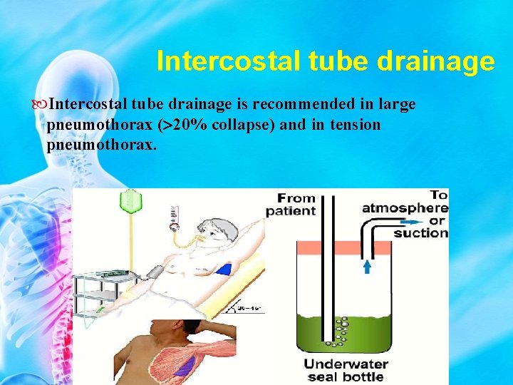
Aspiration in T. B. effusion if no adequate improvement obtained after 4 weeks from starting anti T. B. drugs. (1)Intercostal tube drainage with under water seal. for: Empyema or pyopneumothorax. Hemothorax. Malignant E.
(1)Pleurodesis using (tetracycline or talc powder) to obliterate the pleural space by induction of adhesions (e. g. in malignant effusion as it is massive and rapidly accumulating). (2)Ligation of thoracic duct in chylothorax. (3)Surgical closure of bronchopleural fistula.
Pneumothorax Definition : It is collection of air in the pleural cavity with secondary lung collapse
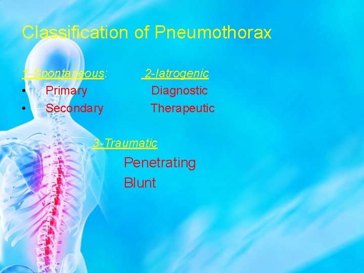
Classification of Pneumothorax 1 -Spontaneous: • Primary • Secondary 2 -Iatrogenic Diagnostic Therapeutic 3 -Traumatic Penetrating Blunt
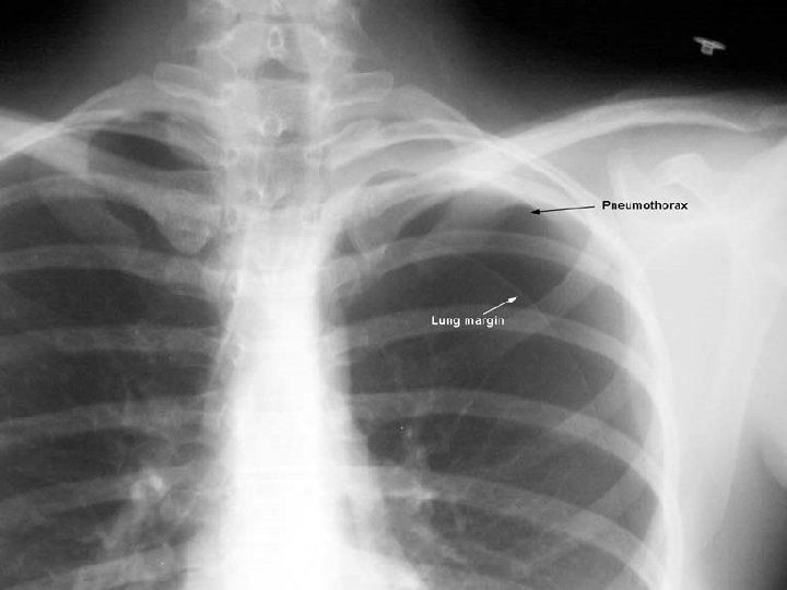
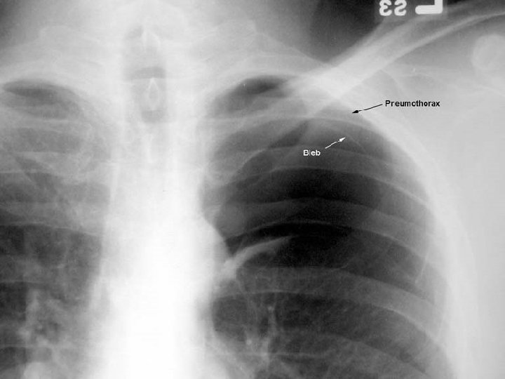
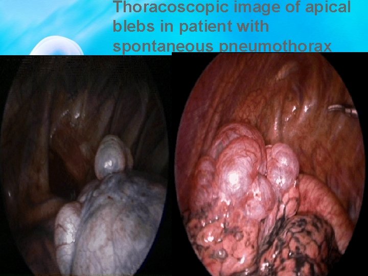
Primary Spontaneous Pneumothorax Occurs in people without any underlying lung disease or any trauma Predominantly disease of young adults AGE: 20 – 40 yrs CAUSES: Apical blebs Stature Familial Smoking
Thoracoscopic image of apical blebs in patient with spontaneous pneumothorax
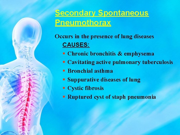
Secondary Spontaneous Pneumothorax Occurs in the presence of lung diseases CAUSES: Chronic bronchitis & emphysema Cavitating active pulmonary tuberculosis Bronchial asthma Suppurative diseases of lung Cystic fibrosis Ruptured cyst of staph pneumonia
IATROGENIC PNEUMOTHORAX Transthoracic needle aspiration (24%) Subclavian vessel puncture (22%). Thoracocentesis (22%). Pleural biopsy(8%). Mechanical ventilation (7%).
Traumatic Pneumothorax Penetrating : • Stab wound • Gunshot Blunt: • Motor car accident
Types of Pneumothorax • Open pneumothorax : The pleura is connected with the atmosphere. results when a penetrating chest wound enables air to rush in and cause the lungs to collapse. • Closed pneumothorax: due to tear in the pleura, results when the chest wall is punctured or air leaks from a ruptured bronchus (or a perforated esophagus)
• Tension pneumothorax : resulting from a wound in the chest wall which acts as a valve that permits air to enter the pleural cavity but prevents its escape. The accumulation of air under pressure in the pleural space, it trapped during expiration so the air pressure within the thorax higher than atmospheric pressure, It compresses the lung, and cause cardiopulmonary impairment. Also called pressure pneumothorax.
• Symptoms: Clinical presentation 1 -Acute pleuretic chest pain. 3 -Acute dyspnea 2 -Cough 4 -Shock 5 -Death(untreated severe tension pneumothorax)
Signs • General: Anxious, irritable. Tachypnea, tachycardia with weak pulse. Cyanosis. Inspection: mobility, bulging Palpation: mediastinal shift, TVF Percussion: hyper-resonant Auscultation: air entry, amphoric breathing in tension type
Complications • • Recurrence Tension pneumothorax Re-expansion pulmonary edema Respiratory failure Heart failure Hemothorax Pyo-pneumothorax
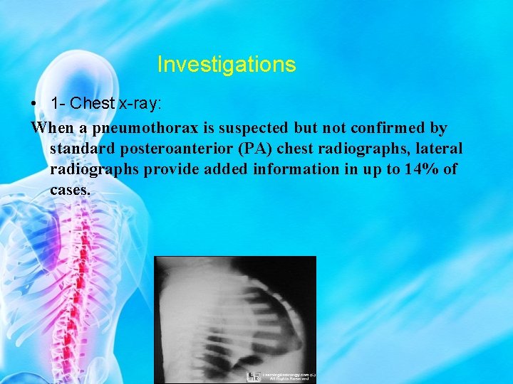
Investigations • 1 - Chest x-ray: When a pneumothorax is suspected but not confirmed by standard posteroanterior (PA) chest radiographs, lateral radiographs provide added information in up to 14% of cases.
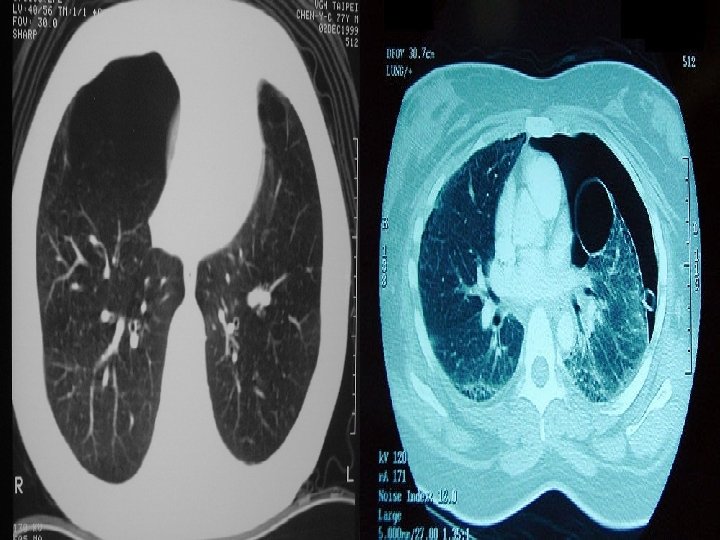
CT scanning is recommended When differentiating a pneumothorax from complex bullous lung disease. When aberrant tube placement is suspected When the plain chest radiograph is obscured by surgical emphysema.
TREATMENT OPTIONS FOR SPONTANEOUS PNEUMOTHORAX
Observation should be the treatment of choice for small closed pneumothorax (<20% collapse) without significant breathlessness. Observation alone is only recommended in patients with small secondary pneumothorax of < 1 cm depth or isolated apical pneumothorax in asymptomatic patients. If a patient with a pneumothorax is admitted overnight for observation, high flow (10 l/min) oxygen should be administered, with appropriate caution in patients with COPD who may be sensitive to higher concentrations of oxygen.
Intercostal tube drainage is recommended in large pneumothorax ( 20% collapse) and in tension pneumothorax.
Chemical pleurodesis can control difficult or recurrent pneumothorax but should only be attempted if the patient is either unwilling or unable to undergo surgery. Ø Substance used : • Tetracycline hydrochloride • 50% glucose solution • Autologus blood • Iodized talc or kaolin
Referral to thoracic surgeons In cases of persistent air leak or failure of the lung to reexpand, the managing respiratory specialist should seek an early (3– 5 days) thoracic surgical opinion. • Open thoracotomy and pleurectomy remains the procedure with the lowest recurrence rate for difficult or recurrent pneumothorax. Minimally invasive procedures, thoracoscopy (VATS), pleural abrasion, and surgical talc pleurodesis are all effective alternative strategies.
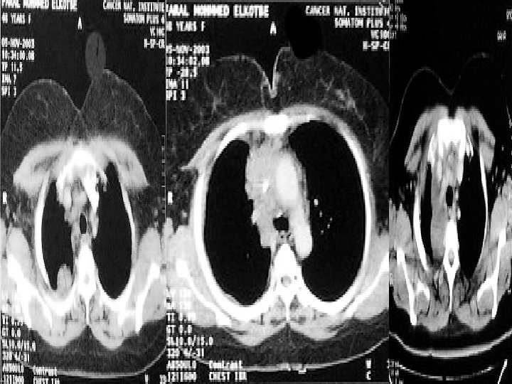
Pleural Tumors • Benign: it is localized as fibroma • Malignant: either primary as mesothelioma or secondary.
Pleural Diseases: Malignant Mesothelioma • Neoplasm of pleural serosa • Linked to asbestos exposure • Coalescence of pleural plaques • May be restricted to parietal pleura but can involve visceral pleura • Can lead to contracture of all structures in affected hemithorax
Types of mesothelioma • Mesothelioma can be divided into three different types according to the pathological biopsy: • Epithelioid: which has the best prognosis out of the three types. • Sarcomatoid: which has about a 7 – 20% survival rate. • Mixed/biphasic: which has about a 20 – 35% survival rate
• Mesothelioma is most frequently not diagnosed until it is in an advanced stage. • Because symptoms of mesothelioma, are non-specific, which can include pain in the lower back and shortness of breath, are often mistaken for other problems,
• Chest x-ray Diagnosis • Chest Sonography • Chest CT • Thoracoscope • MRI
Treatments • Treatment options for all three types of mesothelioma are the same. • Until recently there was no effective treatment for mesothelioma. • A new chemotherapy drug, Alimta, was recently approved by the FDA. • Surgical removal is possible in some cases but in many cases removal of the tumor alone is not enough. • Radiotherapy: is difficult and risky
Prognosis • Mesothelioma patients prognosis in large part depends on which type of mesthelioma they have. • With the new chemotherapy, as well as surgery mesothelioma patients are beginning to live longer than they could have hoped for in the past.
Contact me • • Dr/ Alaa Rashad Chest department, South Vally University, Tel; Clinic; 01021991127 E-mail; alaasma 27@yahoo. com Facebook; Dr. Alaarashad Kena Chest center Prof Alaa Rashad Beside Kena Chest Hospital