Diseases of the Nervous System Kun Mu MD
 MD, Ph. D Department of Pathology School")

 Sulci(脑沟) Parietal lobe(顶叶) Occipital")
 Cell bodies-- grey matter Cell processes– axons--White")

 Cell bodies-- grey matter Cell processes– axons--White")


 Cell bodies-- grey matter Cell processes– axons--White")

 Red neuron")
 正常神经细胞 中央型尼氏体溶解")
---Coagulation necrosis")
 o 2. Red")
 Negri body-rabies(狂犬病)")
 Neurofibrillary")
 n")
 o 2. Reactive astrogliosis(反应性胶质化)")

 o 2. Reactive astrogliosis(反应性胶质化)")
")
 正常室管膜细胞 颗粒状室管膜炎")
 o 2. Foamy cell(gitter")

")
 o 2. Hernia(脑疝) o 3.")
 : increased water content within the brain")


: Abnormal protrusion of brain tissue through an opening. Causes")
 o Within the intact skull, there are 3")
 o Tentorial hernia(小脑天幕疝,海马沟回疝) o Foramen magnum hernia(枕骨大孔疝,小 脑扁桃体疝)")

 refers to the accumulation of excessive CSF within the ventricular")
 Obstructive hydrocephalus o 2. communicating hydrocyphalus(交通性脑积水) Overproduction")


 o")
 o")
 Lymphs")

 o Cranial nerve paralysis(颅神经麻痹) o Cerebral ischemia and infarct(脑缺血和梗死)")


 Blood Stream Brief Viremia")


 Diffuse (grade 2) Anaplastic")

 o Histologic type: fibrillary(纤维型), protoplasmic(原 浆型), gemistocytic(肥胖细胞型). o Gross: a poorly")


 o Older adults o Median survival: 12 months o Highly malignant o")
 o o o Common in childhood Most slow growing of")
 毛细胞型星形细胞瘤年龄分布")


 o Origin: primitive neuroectodermal cells o Age: 1 st-2 nd decade of life.")
 o Origin: primitive neuroectodermal cells o Age: 1 st-2 nd decade of life.")

 o Origin: primitive neuroectodermal cells o Age: 1 st-2 nd decade of life.")


 Neurofibroma(神经纤维瘤) Perineurioma (神经束膜瘤) Malignant Peripheral")
 o Common Site A. 左侧小脑桥脑角听神经瘤 B. 脊髓神经鞘瘤")


 o Type I (common): (17 q) n Plexiform & solitary neurofibromas n Optic")
 o Type I (common): (17 q) n Plexiform & solitary neurofibromas n Optic")
 Café-au-lait spots Multiple neurofibromas")


- Slides: 117
Diseases of the Nervous System Kun Mu(牟坤) MD, Ph. D Department of Pathology School of Medicine, Shandong University
Contents u Structure of the NS and basic pathologic changes u Common complications of CNS u Infectious diseases of NS u Neoplasms of NS
I. Structure of the CNS and basic pathologic changes Gyri(脑回) Sulci(脑沟) Parietal lobe(顶叶) Occipital lobe(枕叶) Cerebellar peduncles( 小脑脚) Cerebellum(小脑) Fourth ventricle(四脑室) Pons(桥脑) Spinal Cord (脊髓) Frontal lobe(额叶)
Microscopic structure of the CNS o Neuron(神经元) Cell bodies-- grey matter Cell processes– axons--White matter bundles
Staining of neurones in the cerebral cortex
Microscopic structure of the CNS o Neuron(神经元) Cell bodies-- grey matter Cell processes– axons--White matter bundles o Glia(胶质细胞) Astrocytes Oligodendrocytes Ependymal cells
Staining of astrocyte in the cerebral cortex
Glia-oligodendrocytes
Ependymal cells
Microscopic structure of the CNS o Neuron(神经元) Cell bodies-- grey matter Cell processes– axons--White matter bundles o Glia(胶质细胞) Astrocytes Oligodendrocytes Ependymal cells o Microglia (小胶质细胞) o Meningocyte(脑膜细胞) o Vessel (血管)
I. Basic pathology changes of CNS o Neuron o Astrocytes o Oligodendrocytes o Ependymal cells o Microglia
1. Basic pathology changes of the Neuron o o o Chromatolysis (尼氏体溶解) Red neuron (红色神经元) Simple neuronal atrophy(单纯性神经元萎缩) Cellular inclusion(包含体形成) Cell Structure protein change(细胞结构蛋白异常) Waller degeneration( Waller变性)
1. Basic pathology changes of the Neuron o 1. Chromatolysis (尼氏体溶解) 正常神经细胞 中央型尼氏体溶解
1. Basic pathology changes of the Neuron o 2. Red neuron (红色神经元)---Coagulation necrosis
1. Basic pathology changes of the Neuron o 1. Chromatolysis (尼氏体溶解) o 2. Red neuron (红色神经元) o 3. Simple neuronal atrophy(单纯性神经元萎缩) Common in some chronic degenerative diseases
1. Basic pathology changes of the Neuron o 4. Cellular inclusion(包含体形成) Negri body-rabies(狂犬病)
1. Basic pathology changes of the Neuron o 5. Cell Structure protein change(细胞结构蛋白异常) Neurofibrillary Tangles-AZ Lewy bodies-PD
1. Basic pathology changes of the Neuron o 6. Waller degeneration ( Waller�性) n Degeneration n Demyelination(脱髓鞘) n Regeneration Trichrome染色:髓鞘染 成红色,胶原纤维染成青色, 可见髓鞘缺失、崩解。
2. Basic pathology changes of the astrocytes o 1. Swelling(肿胀) o 2. Reactive astrogliosis(反应性胶质化)
2. Basic pathology changes of the astrocytes o 2. Reactive astrogliosis
2. Basic pathology changes of the astrocytes o 1. Swelling(肿胀) o 2. Reactive astrogliosis(反应性胶质化) o 3. Amyloid body(淀粉样小体)
3. Basic pathology changes of the oligodendrocyte o Satellitosis(卫星现象)
4. Basic pathology changes of the ependymal cell o Ependymal granulation(颗粒状室管膜炎) 正常室管膜细胞 颗粒状室管膜炎
5. Basic pathology changes of the microglia o 1. Neuronophgia(噬神经细胞现象) o 2. Foamy cell(gitter cell)(格子细胞形成) o 3. Microglial nodules(胶质结节)
5. Basic pathology changes of the microglia 噬神经细胞现象 neuronophagia 格子细胞 gitter cell
5. Basic pathology changes of the microglial nodules (胶质结节)
II. Common Complications of CNS o 1. Brain edema(脑水肿) o 2. Hernia(脑疝) o 3. Hydrocephalus( 脑积水)
1. Brain edema o Brain edema (脑水肿) : increased water content within the brain parenchyma o Vasogenic edema: A state of increased extracellular fluid volume; Brain tumor, abscess, infarct, hemorrhage o Cytotoxic edema: The swelling of cellular elements; Hypoxia, toxication
1. Brain edema
1. Brain edema 脑 水 肿
2. Herniation o Herniation (脑疝形成): Abnormal protrusion of brain tissue through an opening. Causes : Raised ICP (Intracranial pressure) o Intracranial expanding lesions – tumor, haematoma, abscess
2. Herniation o Intracranial pressure (ICP) o Within the intact skull, there are 3 major components of intracranial pressure: the brain, the CSF and blood. o Any increase in volume of one of these 3 components will produce an increase in ICP o Normal ICP is 0. 6 -1. 8 k. Pa
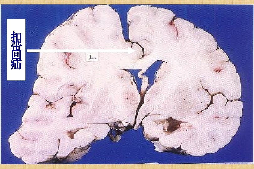
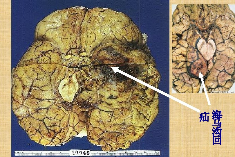
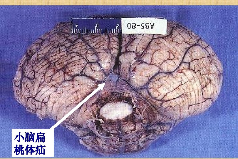
2. Herniation-Types o Supracallosal hernia(扣带回疝,大脑镰下疝) o Tentorial hernia(小脑天幕疝,海马沟回疝) o Foramen magnum hernia(枕骨大孔疝,小 脑扁桃体疝)
2. Herniation
3. Hydrocephalus o Hydrocephalus(脑积水) refers to the accumulation of excessive CSF within the ventricular system
3. Hydrocephalus-Types o 1. noncommunicating hydrocyphalus (非交通性脑积水) Obstructive hydrocephalus o 2. communicating hydrocyphalus(交通性脑积水) Overproduction of CSF Defective absorption of CSF
3. Hydrocephalus-Types Causes: o Increased secration of CSF :脉络丛肿瘤 o Obstraction to the flow of CSF:肿瘤、粘 连性阻塞 o failure of absorption of CSF:蛛网膜粒的 闭塞
3. Hydrocephalus-Types Dilated lateral ventricles seen in a coronal section
III. Infectious diseases of CNS o Epidemic meningitis o Epidemic Encephalitis B
Routes of infection o Hematogenous spread o Direct implantation Trauma, Nasal or paranasal sinuses inflammation o Invasion via the peripheral nerves
Structure of menginges
Meningitis o Acute suppurative meningitis: bacteria o Acute lymphocytic meningitis: virus o Chronic meningitis: TB
1. Epidemic meningitis o Group A meningococcus accounts for an estimated 80– 85% of all cases in the meningitis belt, with epidemics occurring at intervals of 7– 14 years. o Suppurative immflamation
1. Epidemic meningitis. Transmission o The bacteria are transmitted from personto-person through droplets of respiratory or throat secretions from carriers o The bacteria overwhelm the body's defenses allowing infection to spread through the bloodstream to the brain.
1. Epidemic meningitis-Gross o There abundant creamy, purulent exudate, most prominently over the superior surface of the cerebrum. o The exudation full in sulci. o The gyri are swollen. o The vessels are congested.
1. Epidemic meningitis-Gross
1. Epidemic meningitis-Micro o Neutrophilic exudate, dilated vessels, edema in the subarachnoid space
1. Epidemic meningitis-Clinical signs o Signs of infection (fever, malaise, skin rashes…. ) o Sign of high intracranial pressure a. headache b. vomiting o Signs of meningeal irritation a. neck stiffness, episthiotonus(角弓反张) b. Kernig positive
1. Epidemic meningitis-Clinical signs o Signs of infection (fever, malaise, skin rashes…. ) o Sign of high intracranial pressure a. headache b. vomiting o Signs of meningeal irritation a. neck stiffness, episthiotonus b. Kernig positive o CSF changes: a. cloudy purulent b. abundant neutrophils c. high protein level and reduced glucose level
1. Epidemic meningitis-CSF values Cells Acute bacterial PMNs Acute viral Lymphs Chronic (TB) Lymphs Protein Glucose Normal Pressure
1. Epidemic meningitis-CSF values monocyte neutrophil Bacteria
1. Epidemic Complications o Hydrocephalus(脑积水) o Cranial nerve paralysis(颅神经麻痹) o Cerebral ischemia and infarct(脑缺血和梗死)
Fulminant meningococcal septicemia 暴发性脑膜炎球菌败血症 o an extreme form of endotoxin-induced sepsis and coagulopathy. The condition is diagnosed clinically by the appearance of hemorrhagic skin lesions and compromised circulation
Fulminant meningococcal septicemia 暴发性脑膜炎球菌败血症
2. Epidemic Encephalitis B o Epidemic encephalitis B is acute infectious disease caused by encephalitis B virus o Usually occurs in summer & fall o The virus is transmitted by mosquito
2. Epidemic Encephalitis BTransmission Virus Replication in Mononuclear-phagocyte System (MPS) Blood Stream Brief Viremia Blood Brain Barrier CNS
Blood Brain Barrier-血脑屏障
2. Epidemic Encephalitis B-Gross o Pathologic location: cerebral parenchyma
2. Epidemic Encephalitis B-Micro o Congestion and inflammation: endothelial cells swelling, perivascular cuffing o Degeneration and necrosis of neurons: softing focus, neruonophagia, satellitosis o Proliferation of microglial cells: microglial nodule
2. Epidemic Encephalitis B-Micro o Congestion and inflammation: endothelial cells swelling, perivascular cuffing
2. Epidemic Encephalitis B-Micro o Degeneration and necrosis of neurons: softing focus, neruonophagia, satellitosis
2. Epidemic Encephalitis B-Micro o Proliferation of microglial cells: microglial nodule
2. Epidemic Encephalitis BClinical feature o High fever o Headache o Vomiting o Sleepiness o Convulsion抽搐
V. Tumors of Nerous System o Tumors of CNS o Peripheral Nerve Tumors
Tumors of CNS o Comprise: 10% of all tumors o Peak incidence 20 -50 yr o Common childhood tumor o Supratentorial tumors in adults o Infratentorial tumors in childhood o Location determines prognosis o Rare extra neural metastasis
WHO Grade I Ø Slow growing Ø Non-malignant tumors Ø Patients have long-term survival Grade II Ø Relatively slow growing Ø Sometimes recur as higher grade tumors Ø May be non-malignant or malignant. Grade III Ø Malignant tumors Ø Often recur as higher grade tumors Grade IV Ø Highly malignant and aggressive
Tumors of CNS o Gliomas o Medulloblastoma o Meningiomas
Tumors of CNS o Gliomas---derived from glial cells, include Astrocytoma Oligodendrogliomas Ependymoma
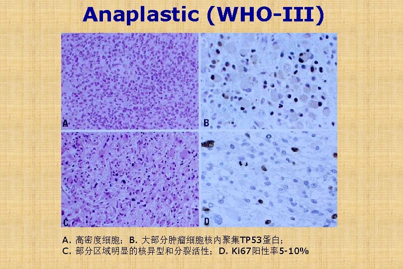
Histologic Classification of Glial Tumors Astrocytic Tumors Pilocytic (grade 1) Diffuse (grade 2) Anaplastic (grade 3) Glioblastoma (grade 4) Oligodendroglial tumors and mixed variants Oligodendroglioma, well differentiated (grade 2) Anaplastic oligodendroglioma (grade 3) Mixed oligodendroglioma/astrocytoma (grade 2) Mixed anaplastic oligodendroglioma/astrocytoma (grade 3) Ependymal Tumors Myxopapillary ependymoma (grade 1) Ependymoma (grade 2) Anaplastic ependymoma (grade 3)
Astrocytoma o 4 th to 6 th decade, Cerebrum. o Grading system based on n n Anaplasia Mitotic activity Necrosis Endothelial proliferation o Diffuse (WHO-II), anaplastic (WHO-III) & Glioblastoma (WHO-IV) o Molecular markers: GFAP, Ki 67, TP 53, EGFR, IDH 1, MGMT…
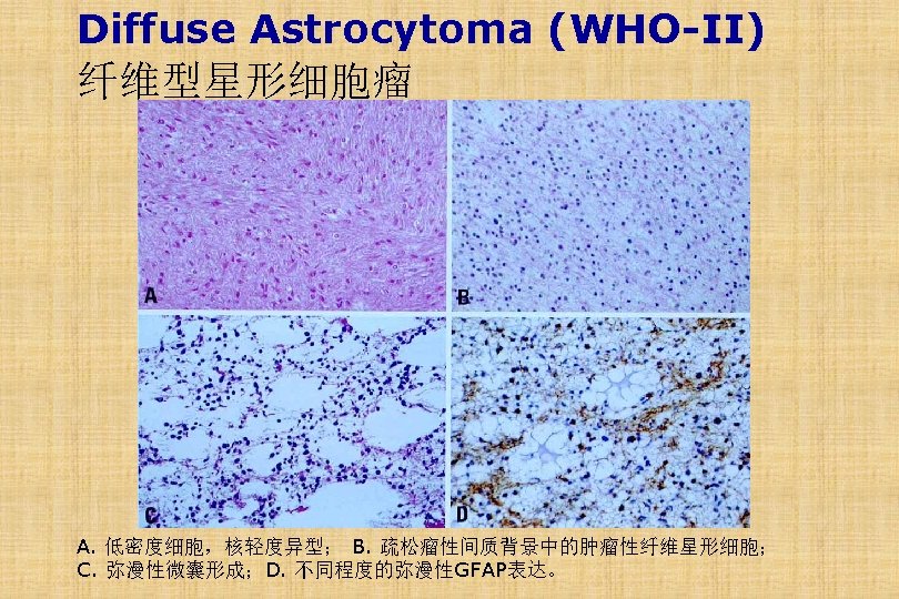
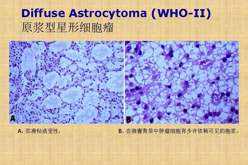
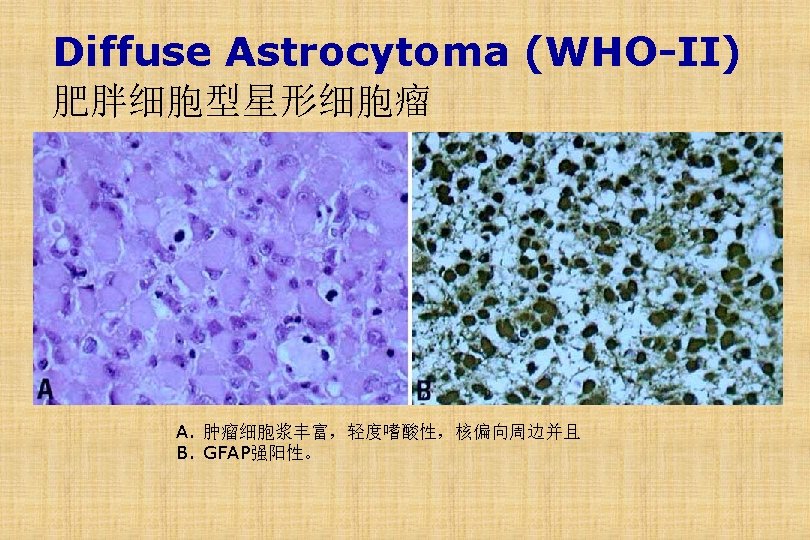
Diffuse Astrocytoma (WHO-II) o Histologic type: fibrillary(纤维型), protoplasmic(原 浆型), gemistocytic(肥胖细胞型). o Gross: a poorly defined, gray, infiltrative tumor that expands and distorts the invaded brain. The cut surface of the tumor is either firm, or soft and gelatinous; cystic degeneration may be seen. o Micro: a mild to moderate increase in the number of glial cell nuclei, somewhat variable nuclear pleomorphism, and tumor cells can be seen infiltrating normal tissue.
Glioma Cerebrum
Glioma Cerebrum
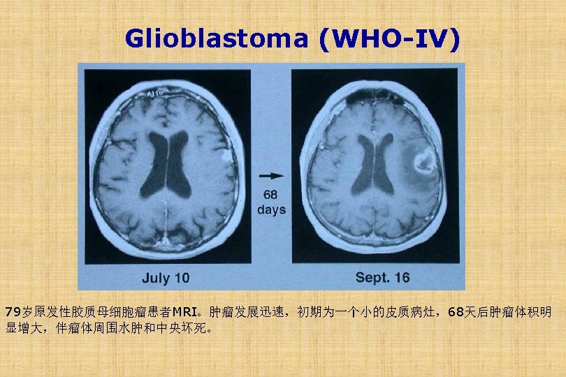
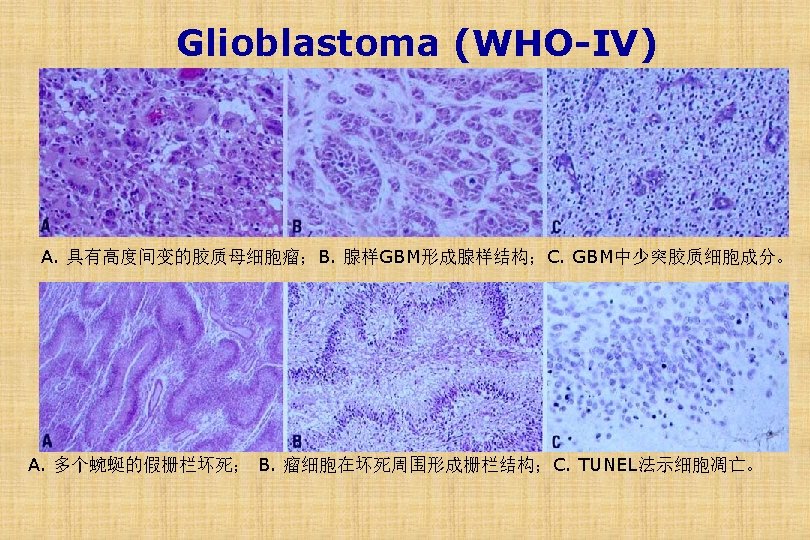
Glioblastoma (WHO-IV) o Older adults o Median survival: 12 months o Highly malignant o Diffuse infiltrative tumors
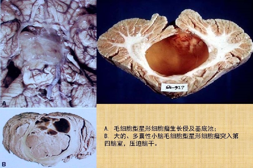
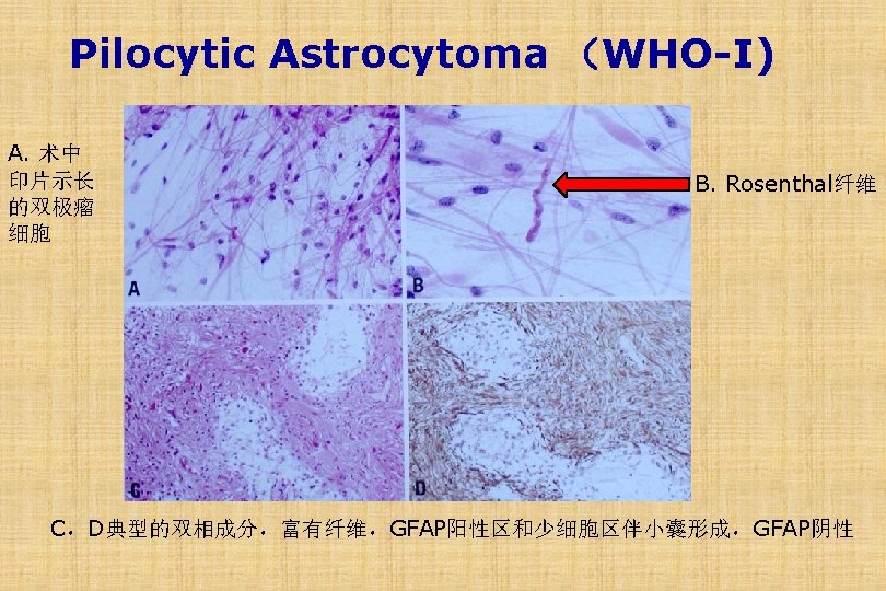
Pilocytic Astrocytoma ( WHO-I) o o o Common in childhood Most slow growing of the gliomas Sites: cerebellum, around III IV V. , optic nerve Grossly cystic with mural nodule Microscopic n elongated hair-like (pilo) cells n Rosenthal fibers
Pilocytic Astrocytoma (WHO-I) 毛细胞型星形细胞瘤年龄分布
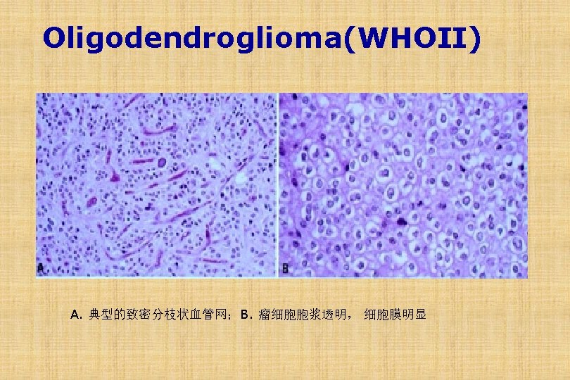
Oligodendroglioma o Cells of origin: Oligodendrocytes o Common in cerebral hemispheres o Calcifications common o Grades: Low grade (WHO-II) Anaplastic (WHO-III)
Tumors of CNS o Gliomas o Medulloblastoma
Medulloblastoma(髓母细胞瘤) o Origin: primitive neuroectodermal cells o Age: 1 st-2 nd decade of life. Most common brain tumor at this age.
Medulloblastoma(髓母细胞瘤) o Origin: primitive neuroectodermal cells o Age: 1 st-2 nd decade of life. Most common brain tumor at this age. o Site: vermis of cerebellum, may project into the V ventricule
Cerebellum Medulloblastoma
Medulloblastoma(髓母细胞瘤) o Origin: primitive neuroectodermal cells o Age: 1 st-2 nd decade of life. Most common brain tumor at this age. o Site: vermis of cerebellum, may project into the V ventricule o Subarachnoid dissemination o May cause hydrocephalus
Medulloblastoma-Subarachnoid dissemination A. 肿瘤转移至硬脑膜内表面;B. 肿瘤转移至马尾。
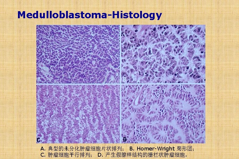
Medulloblastoma-Histology Homer Wright Rosettes
Tumors of CNS o Gliomas o Medulloblastoma o Meningiomas
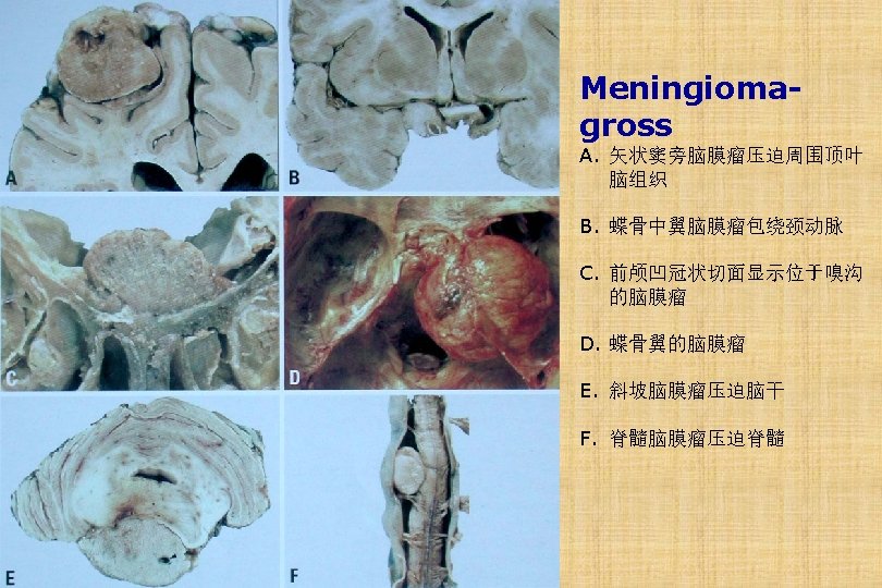
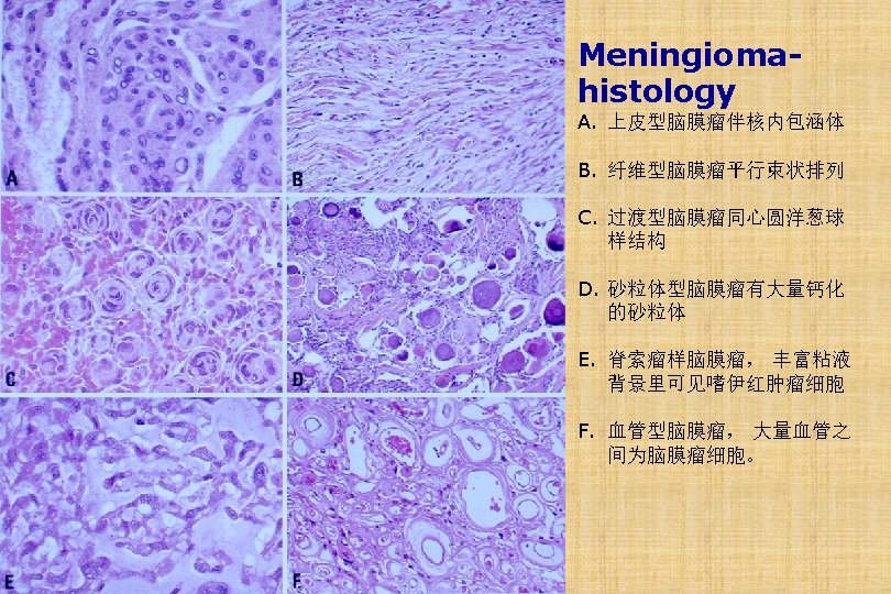
Meningioma o Meningiomas are predominantly benign tumors of adults o Usually attached to the dura, that arise from the meningothelial cell of the arachnoid o More common in women
Meningioma o Gross:Meningiomas are usually rounded masses with a well-defined dural base that compress underlying brain but are easily separated from it.
V. Tumors of Nerous System o Tumors of CNS o Peripheral Nerve Tumors
Peripheral Nerve Tumors o Nerve Sheath Tumors Neurilemmoma (神经鞘瘤) Neurofibroma(神经纤维瘤) Perineurioma (神经束膜瘤) Malignant Peripheral Nrve Sheath Tumor (恶性外周神经鞘瘤) o Neural cell tumor Neuroblastoma (神经母细胞瘤) Ganglioneuroma (节细胞神经瘤)
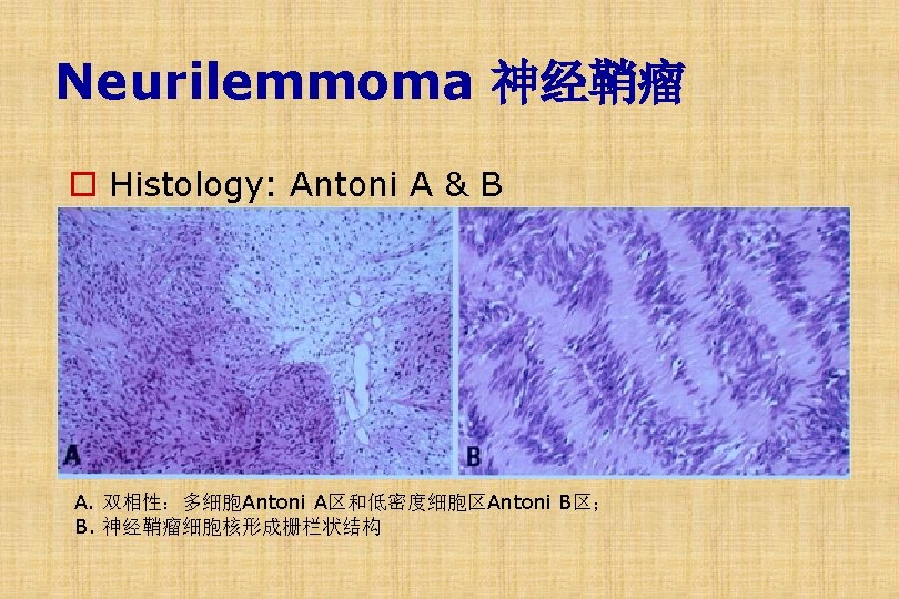
Neurilemmoma 神经鞘瘤 (Schwannoma) o Common Site A. 左侧小脑桥脑角听神经瘤 B. 脊髓神经鞘瘤
Neurilemmoma 神经鞘瘤 o Gross
Neurilemmoma 神经鞘瘤 Antony A Antony B
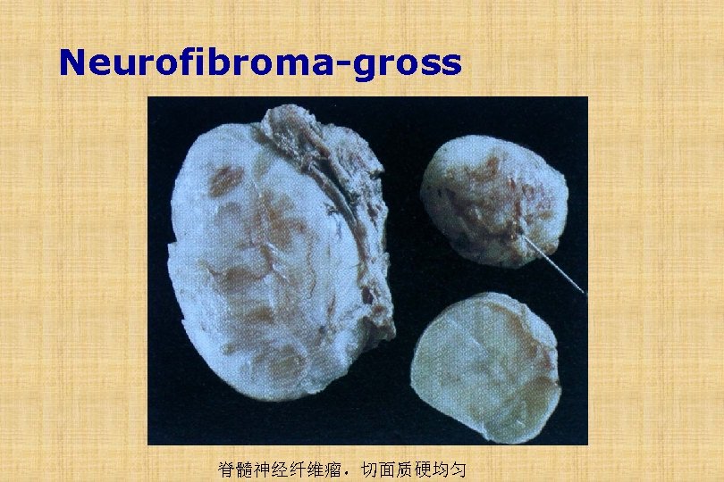
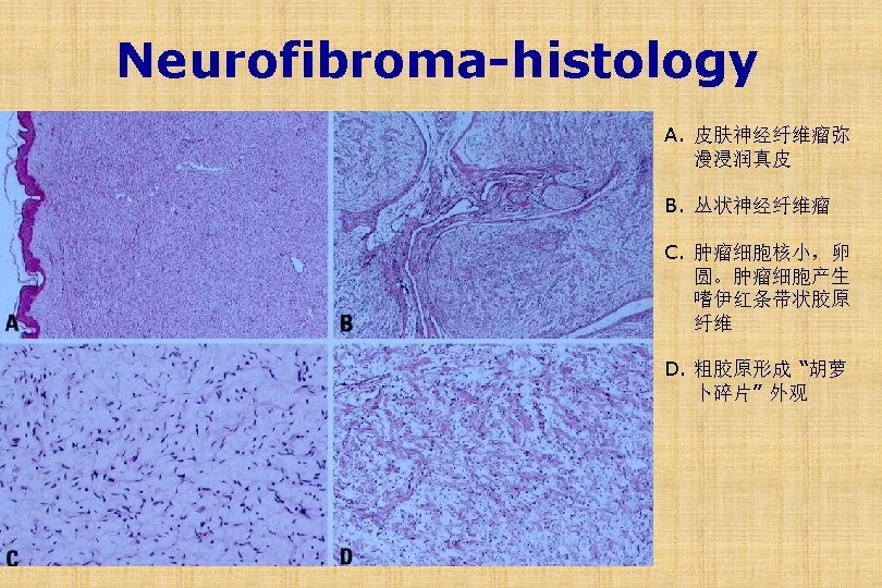
Neurofibroma神经纤维瘤 o Schwann cell, perineurial cells & fibroblasts o Two types: o Classic form - Cutaneous / nerve - Solitary collagen matrix, spindle cells o Plexiform (丛状的)- Multiple, infiltrative, myxoid
Neurofibromatosis(神经纤维瘤病) o Type I (common): (17 q) n Plexiform & solitary neurofibromas n Optic nerve gliomas, Lisch nodules(虹膜色素缺陷 瘤), Café au lait spots(咖啡牛奶斑).
Neurofibromatosis(神经纤维瘤病) o Type I (common): (17 q) n Plexiform & solitary neurofibromas n Optic nerve gliomas, Lisch nodules(虹膜色素缺陷 瘤), Café au lait spots(咖啡牛奶斑). o Type II (rare): (22 q) n Bilateral acoustic schwannoma/osis n Multiple meningioma/osis, ependymoma of spinal cord
Neurofibromatosis(神经纤维瘤病) Café-au-lait spots Multiple neurofibromas
Case Study o 22 -year-old female, presented with a history of headache and convulsion of four months duration. o Brain MRI : Right frontal lobe lesion with an irregular outline, mass-effect, peripheral contrast uptake, and central necrosis.
Thank You