
Diseases of the gastrointestinal tract Dr Nesreen Bataineh
Diseases of the gastrointestinal tract Dr. Nesreen Bataineh MD, FRCPath
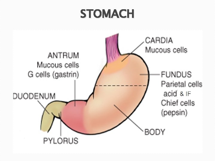
STOMACH
Gastric disorders �Congenital disorders q. Gastric heterotopia q. Diaphragmatic hernia q. Pyloric stenosis �Gastritis �Gastric ulceration �Tumors
Gastritis � Definition: “Inflammation of the gastric mucosa”. q Acute gastritis q Chronic gastritis (majority of cases).
Acute Gastritis � Definition: “An acute inflammation of gastric mucosa”. of a transient nature. � May be accompanied by hemorrhage and erosion UGIT hemorrhage. � Usually
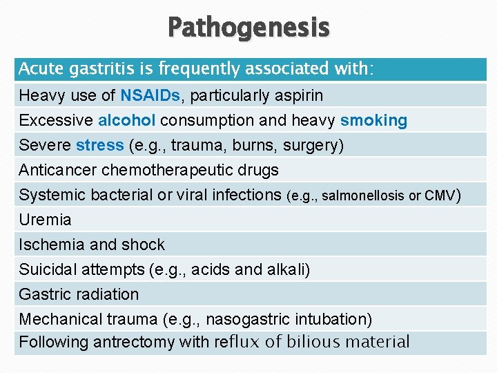
Pathogenesis Acute gastritis is frequently associated with: Heavy use of NSAIDs, particularly aspirin Excessive alcohol consumption and heavy smoking Severe stress (e. g. , trauma, burns, surgery) Anticancer chemotherapeutic drugs Systemic bacterial or viral infections (e. g. , salmonellosis or CMV) Uremia Ischemia and shock Suicidal attempts (e. g. , acids and alkali) Gastric radiation Mechanical trauma (e. g. , nasogastric intubation) Following antrectomy with reflux of bilious material
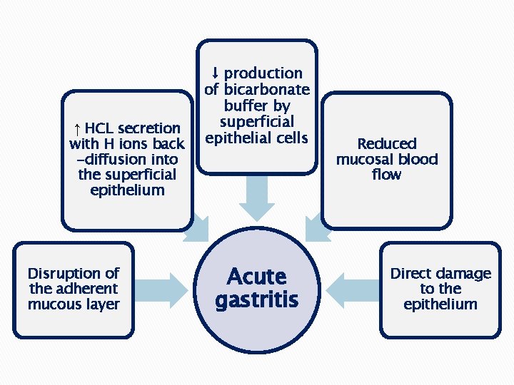
↑ HCL secretion with H ions back -diffusion into the superficial epithelium Disruption of the adherent mucous layer ↓ production of bicarbonate buffer by superficial epithelial cells Acute gastritis Reduced mucosal blood flow Direct damage to the epithelium
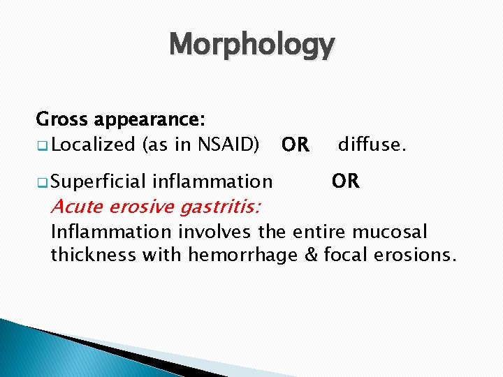
Morphology Gross appearance: q Localized (as in NSAID) q Superficial inflammation Acute erosive gastritis: OR diffuse. OR Inflammation involves the entire mucosal thickness with hemorrhage & focal erosions.
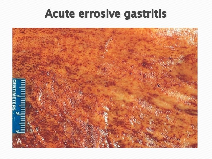
Acute errosive gastritis
Morphology Microscopic appearance: � Mucosal edema � Inflammatory infiltrate rich in neutrophils � + Erosions, hemorrhage & regeneration of epithelial cells � Removal of the cause may lead to complete restitution of the normal mucosa.
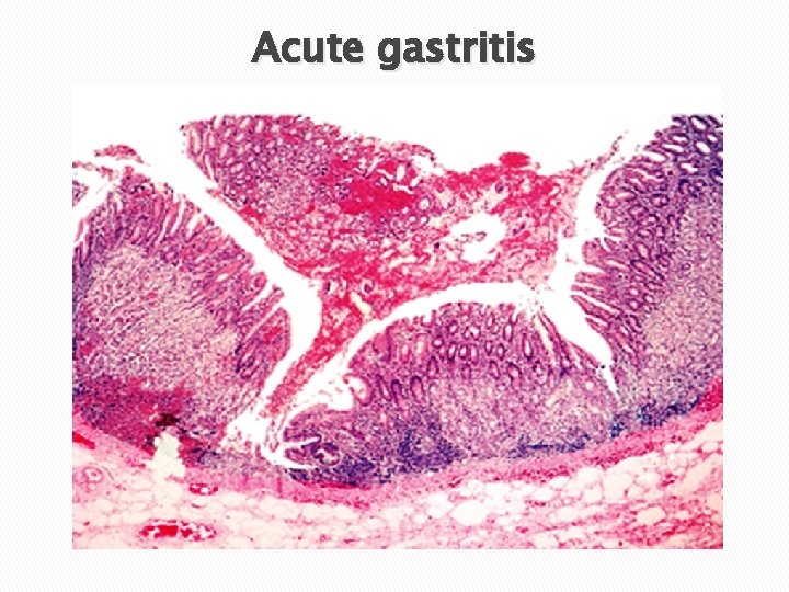
Acute gastritis
Clinical Features � Depends on the severity. q May be asymptomatic q Epigastric pain, nausea and vomiting q Hematemesis, melena or potentially fatal blood loss. � It is one of the major causes of hematemesis.
Chronic Gastritis �Definition: “The presence of chronic inflammatory changes in the gastric mucosa leading to mucosal atrophy + epithelial metaplasia”.
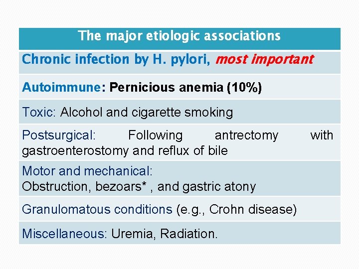
The major etiologic associations Chronic infection by H. pylori, most important Autoimmune: Pernicious anemia (10%) Toxic: Alcohol and cigarette smoking Postsurgical: Following antrectomy gastroenterostomy and reflux of bile Motor and mechanical: Obstruction, bezoars* , and gastric atony Granulomatous conditions (e. g. , Crohn disease) Miscellaneous: Uremia, Radiation. with
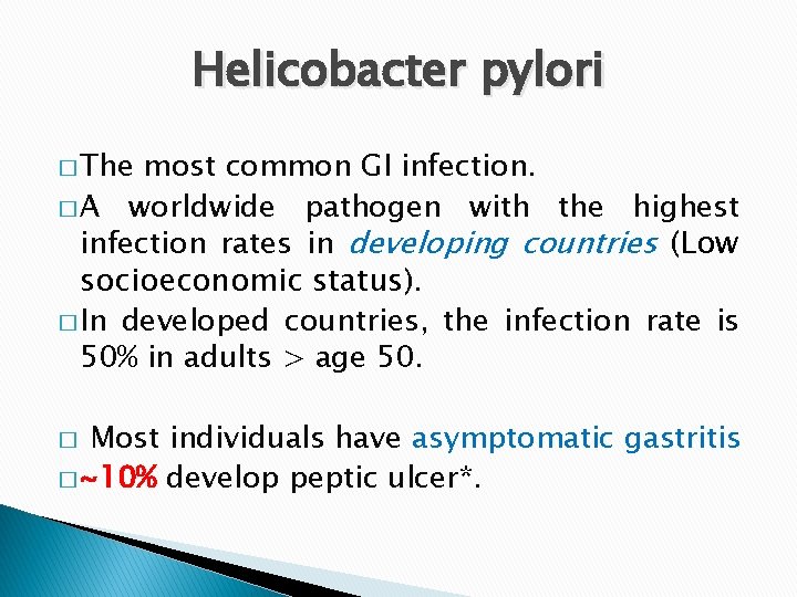
Helicobacter pylori � The most common GI infection. � A worldwide pathogen with the highest infection rates in developing countries (Low socioeconomic status). � In developed countries, the infection rate is 50% in adults > age 50. Most individuals have asymptomatic gastritis � ~10% develop peptic ulcer*. �
Pathophysiology � � q • • q H. pylori is a noninvasive curvilinear gram-negative rod. Mechanism of tissue injury Direct (Bacterial virulence): Flagella: allow the bacteria to be motile in viscous mucus. Urease: generates ammonia from urea elevating gastric p. H around the organisms and protecting it from the acidic secretions. Adhesins: enhance bacterial adherence to surface foveolar cells. Toxins, such as that encoded by cytotoxin-associated gene A (Cag. A), proteases and phospholipase. Indirect: Bacterial products lead to: • ↑ production of cytokines as IL-1, IL-6 … that recruits inflammatory cells with subsequent inflammation and tissue damage.
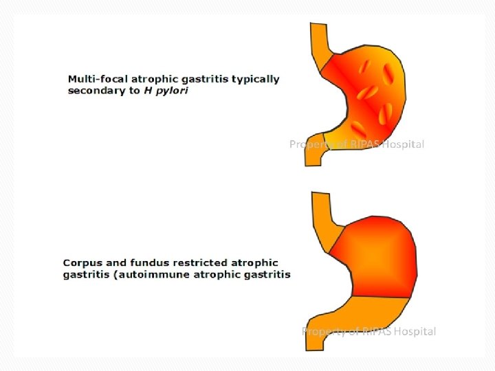
H. Pylori associated gastritis � Antral-type gastritis (commoner): q High HCL production (hyperchlohydria)*. q Higher risk for the development of DU. � Pangastritis: q Multifocal mucosal atrophy q low HCL production (hypochlorhydria) q Increased risk for intestinal metaplasia & adenocarcinoma.
Peptic duodenitis � H. pylori ↑ gastric acid secretion and impairs duodenal bicarbonate production. �↓ luminal p. H in the duodenum. � This favor gastric metaplasia in the first part of the duodenum D 1. � Such metaplastic foci provide areas for H. pylori colonization.
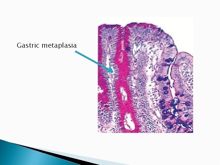
Gastric metaplasia
Morphology of chronic gastritis � Chronic inflammatory infiltrate in the lamina propria. � Predominance of lymphocytes & plasma cell � Neutrophilic infiltration indicates activity. � Gland loss and mucosal atrophy.
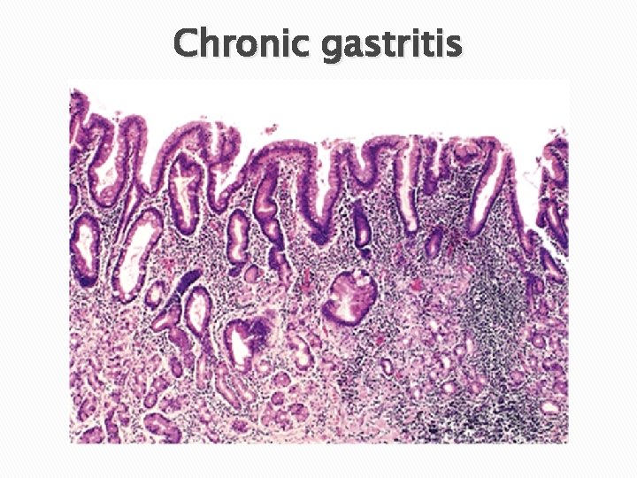
Chronic gastritis
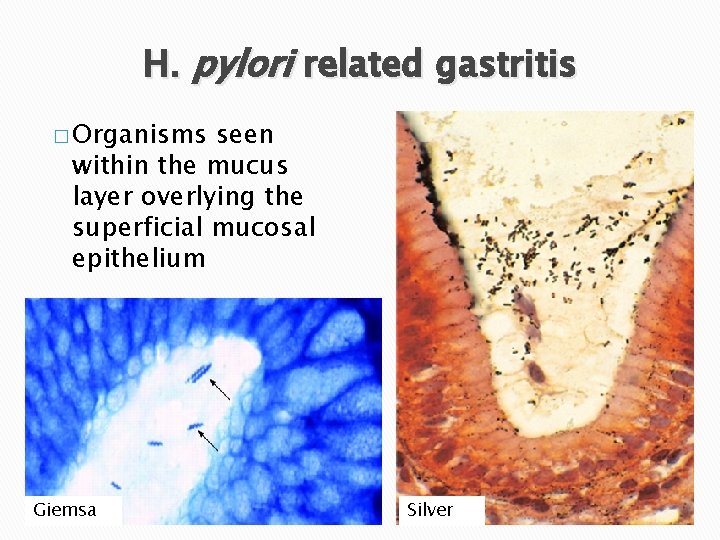
H. pylori related gastritis � Organisms seen within the mucus layer overlying the superficial mucosal epithelium Giemsa Silver
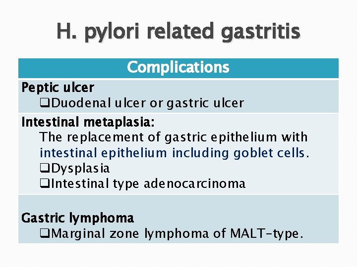
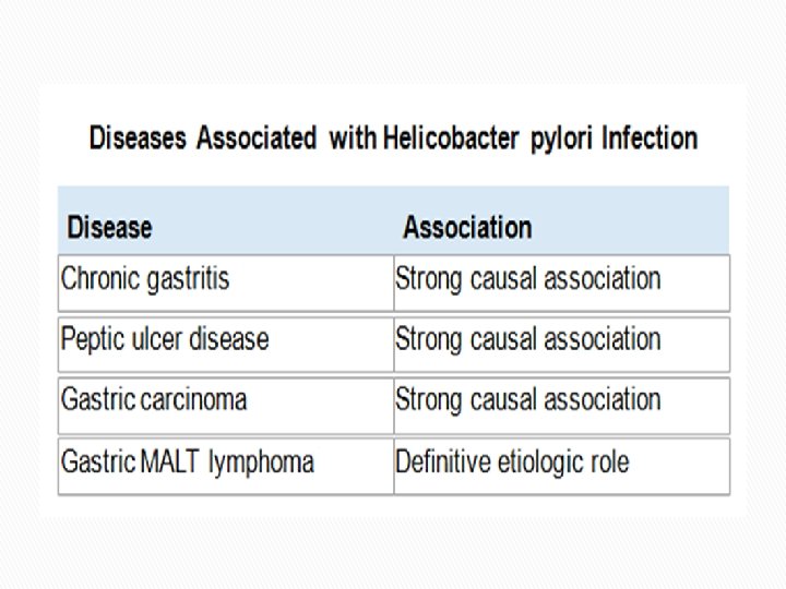
H. pylori related gastritis Complications Peptic ulcer q. Duodenal ulcer or gastric ulcer Intestinal metaplasia: The replacement of gastric epithelium with intestinal epithelium including goblet cells. q. Dysplasia q. Intestinal type adenocarcinoma Gastric lymphoma q. Marginal zone lymphoma of MALT-type.
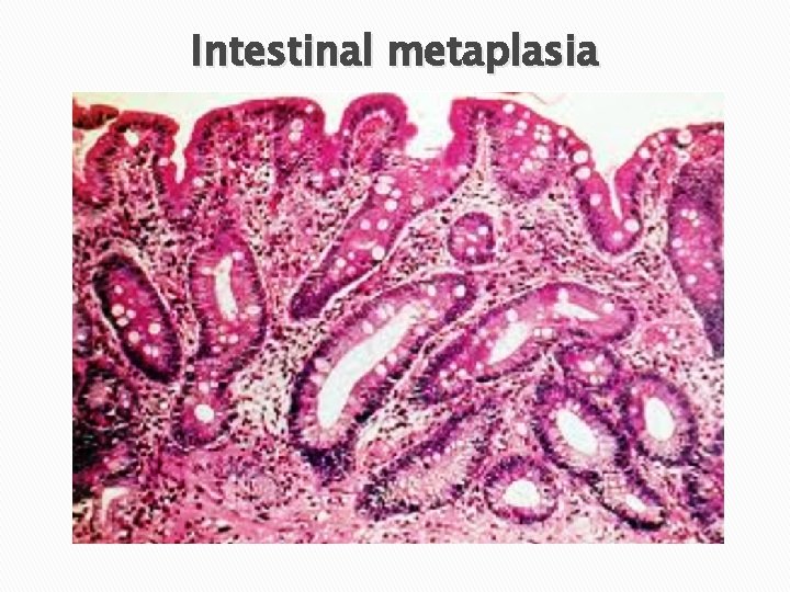
Intestinal metaplasia
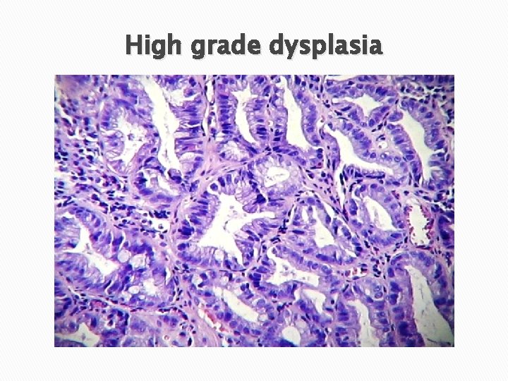
High grade dysplasia
H. pylori associated gastritis Clinical Features � Asymptomatic � Upper abdominal discomfort, N & V � May be hypochlorhydric* (pangastritis), but these persons do not develop achlorhydria or pernicious anemia. � Serum gastrin levels are either normal or only modestly elevated.
H. Pylori associated gastritis � Diagnosis: q Noninvasive • • • tests: Serologic test for anti–H. pylori antibodies Fecal bacterial detection The urea breath test based on the generation of ammonia by bacterial urease. q Invasive • • tests: (Antral biopsy specimen*): The rapid urease test, bacterial culture, or PCR Histologic identification of the organism � Treatment: § Triple therapy (two antibiotics and PPIs)
Autoimmune gastritis � 10% of cases of chronic gastritis � Common in Scandinavians. F>M. � In association with pernicious anemia*. � May be seen with other autoimmune diseases as Hashimoto thyroiditis and Addison disease � Production of autoantibodies to parietal cells and intrinsic factor that can be detected in serum and gastric secretions.
Autoimmune gastritis � Spares the antrum and cardia. � The autoimmune injury leads to gland destruction and diffuse mucosal atrophy � Loss of parietal cells & intrinsic factor � Loss of gastric acid (hypochlorhydria or achlorhydria) � Reduced serum pepsinogen I levels. � Hypergastrinemia, antral endocrine cell hyperplasia & ↑ risk of gastric carcinoid � Increased risk of gastric carcinoma.
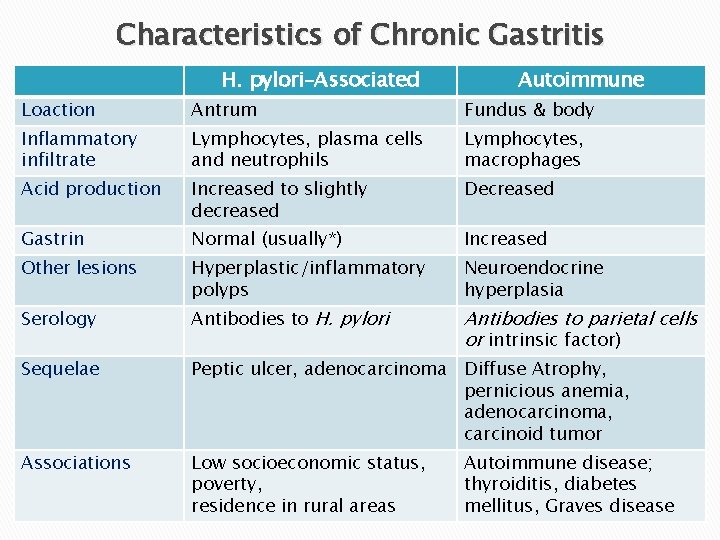
Characteristics of Chronic Gastritis H. pylori–Associated Autoimmune Loaction Antrum Fundus & body Inflammatory infiltrate Lymphocytes, plasma cells and neutrophils Lymphocytes, macrophages Acid production Increased to slightly decreased Decreased Gastrin Normal (usually*) Increased Other lesions Hyperplastic/inflammatory polyps Neuroendocrine hyperplasia Serology Antibodies to H. pylori Antibodies to parietal cells or intrinsic factor) Sequelae Peptic ulcer, adenocarcinoma Diffuse Atrophy, pernicious anemia, adenocarcinoma, carcinoid tumor Associations Low socioeconomic status, poverty, residence in rural areas Autoimmune disease; thyroiditis, diabetes mellitus, Graves disease
Clinical Features � Periumbilical pain that ultimately localizes to the right lower quadrant. � Nausea, vomiting & low-grade fever. � Mc. Burney’s sign. � Mildly elevated peripheral white cell count. Complications: � Appendiceal perforation. � Peritonitis. � Portal venous thrombosis. � Liver abscess. � Bacteremia.

- Slides: 34