DISEASES OF THE CORNEA Dr Neeti Gupta Associate

 Asso.")


 ULCER n n n n Form of peripheral ulcerative keratitis Immune response to")


")


 n n Common in people exposed to hot &")


- Slides: 56
DISEASES OF THE CORNEA Dr. Neeti Gupta Associate Professor Department of Ophthalmology
IMMUNE MEDIATED KERATITIS n n n n Phlyctenular keratitis Marginal ulcer( catarrhal ulcer) Asso. with acne rosacea Asso with systemic collagen vascular disease Chronic serpiginous(Mooren) ulcer Interstitial keratitis Disciform keratitis
INTERSTITIAL KERATITIS n n n Non ulcerative deep stromal keratitis Infective or allergic in origin Causes: Syphilis congenital 90%, acquired 10% Tuberculosis Cogan’s syndrome Leprosy Sarcoidosis § Local Ag –Ab reaction
n n n Progressive stage Florid stage Stage of regression Bilateral Keratitis is secondary to uveitis
Treatmemt n n Systemic : penicillin Local: lubricants steroids cycloplegics
MARGINAL(CATARRHAL) ULCER n n n n Form of peripheral ulcerative keratitis Immune response to staphylococcal toxins, Moraxella, Haemophilus Old age Shallow, slightly infiltrated Typically located at the points of contact of lids with cornea i. e 4, 7, 10 and 2 o’ clock Clear zone b/w lesion & limbus Vascularization + in recurrent cases Treatment Antibiotic+ steroids
ROSACEA KERATITIS n n Ds. of sebaceous glands of skin Women > men Facial eruptions in butterfly configuration Keratits in 5 -10% cases
ROSACEA KERATITIS n n n Infiltrates + small corneal ulcers near limbus Prominent vascularization Blepharoconjunctivitis Frequent recurrences Local treatment - low dose steroids Systemic therapy- Tetracycline 3 -6 months Doxycycline
KERATITIS IN RHEUMATOID ARTHRITIS n n Sclerosing keratitis Perpheral corneal thinning( contact lens cornea) Acute stromal keratitis Acute corneal melting
SHIELD ULCER IN VKC
MOOREN’S ULCER n n § Form of inflammatory peripheral ulcerative keratitis Two forms Limited form/ Benign Progressive/Virulent Etiology Autoimmune Ischaemic necrosis Enzymes produced by conj. Degenerative
MOOREN’S ULCER
n n Severe pain Signs Superficial infiltration Shallow ulceration Overhanging edge Vascularization at base Perforation rare Sclera uninvolved NO CLEAR ZONE b/w ulcer & limbus DIAGNOSIS OF EXCLUSION
TREATMENT OF MOOREN’S ULCER n n n Topical antibiotics for infiltration Frequent topical steroids Systemic steroids/Cyclosporin Conjunctival peritectomy Bandage contact lens Lamellar corneal grafts
CORNEAL ECTASIAS n Secondary to inflammations anterior staphyloma § Non inflammatory keratoconus keratoglobus pellucid marginal degeneration
KERATOCONUS n Non inflammatory, self limiting, ectasia of central cornea n Cornea becomes conical n Congenital weakness of the cornea n Manifest after puberty
Ø Central/paracentral stromal thinning Ø Apical protrusion Ø Irregular astigmatism
Keratoconus n
Bilateral in 90% cases, onset asymmetrical n Etiology Destruction of stromal tissue by collagenaze n Ø Ø Heredity Asso. with atopic diseases Hormonal influences Systemic ds i. e Marfans, Downs, Ehlers Danlos syndrome
KERATOCONUS SEVERITY n Mild K reading < 48 D § Moderate K reading 48 -54 D § Severe >54 D K reading MORPHOLOGY n Nipple cones <5 mm § Oval cones 5 -6 mm § Globus cones >6 mm
SYMPTOMS n Frequent change of glasses n Decreased tolerance to CL wear n Monocular diplopia n Distortion for distant and near objects
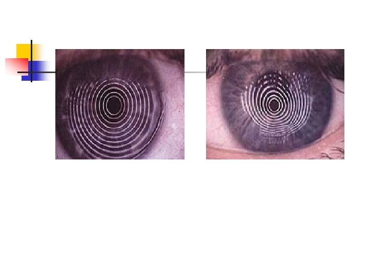
SIGNS n Streak retinoscopy scisssoring reflex § DDO oil droplet reflex § Keratometry malalignment of mires n Slit lamp examination corneal thinning stress lines of vogt Fleischers ring corneal scarring hydrops Munsons sign Rizzuti’s sign § Placido disc unevenly placed circles § Corneal topography
MUNSONS SIGN
FLEISCHER’S RING
VOGT’S STRIAE
HYDROPS
TOPOGRAPHY n
TREATMENT OF KERATOCONUS n Spectacles early cases n Contact lenses n Epikeratoplasty n Penetrating keratoplasty n U V cross linking
DEGENERATIONS AND DESTROPHY
Degenerations Dystrophies 1. Unilateral and asymmetric 1. Bilateral and symmetric 2. Not inherited 2. Inherited (AD) 3. Located eccentrically 3. Centrally located 4. Usually accompanied by vascularization 4. Avascular 5. Middle life or later 5. Early onset 6. Progressive lesions 6. Slower in progression 7. ass. With systemic conditions Ageing, infllamation or trauma 7. Not related to any systemic or local conditions
CORNEAL DEGENERATIONS n Age related n Pathological Band shaped keratopathy Arcus senilis Climatic droplet keratopathy Salzmann’s nodular degeneration Terrien marginal degeneration
Arcus senilis n n n M/c peripheral corneal opacity Lipoid infiltration of corner seen in elderly Present almost universally in people above 60 yrs of age It is a yellowish white deposit that occurs first in the inferior then in the superior aspect finally joining to form an arc Lucid interval of Vogt is characteristic, being sharply defined on the periphery, fading in the center
• U/L arcus-associated with carotid ds or ocular hypotony Histology –lipid first deposited in the ant. 1/2 of descemet’s membrane and then in ant. stroma
ARCUS SENILIS
Band keratopathy n n Hyaline infiltration of superficial parts of cornea followed by deposition of calcium salts in the ant part of Bowmans menbrane Causes - ocular – Chr. Ant. Uveitis - Pth. Bulbi - Silicone oil in AC - Chr. keratitis
n n n - Age related - Metabolic – increase ca, CRF - Hereditary Peripheral inter-palpeberal calcification with clear cornea separating sharp peripheral margins of the band from the limbus Spread centrally to form band like chalky plaque Advance lesion – nodular & elevated with discomfort d/t epi. breakdown
Treated by chelation- mild cases n Sodium EDTA – applied with cotton bud n Exc. Laser keratectomy
BAND SHAPED KERATOPATHY
Salzmann nod. degn n n Bluish white avasc. nodule- Sup. Layer of stroma & Bowmann memb. Seen with previous corneal Ds Slow progression Treat- lam. KP
SALZMANN’S NODULAR DEGENERATION
Spheroidal degn( climatic droplet KP) n n Common in people exposed to hot & dry weather Exposed inter- palp. cornea sparing the limbus Sign- small amber colored granules in sup. Stroma, Treat- lamellar KP, PRK
Terrien marginal degeneration n n n Usually bilateral Slow progressive thinning of peripheral cornea sparing the limbus More frequently seen in males Eye is quite with no inflammation Vascularised pannus is seen with yellow deposit of lipid May cause myopic or irregular astigmatism Perforation can occur with mild trauma
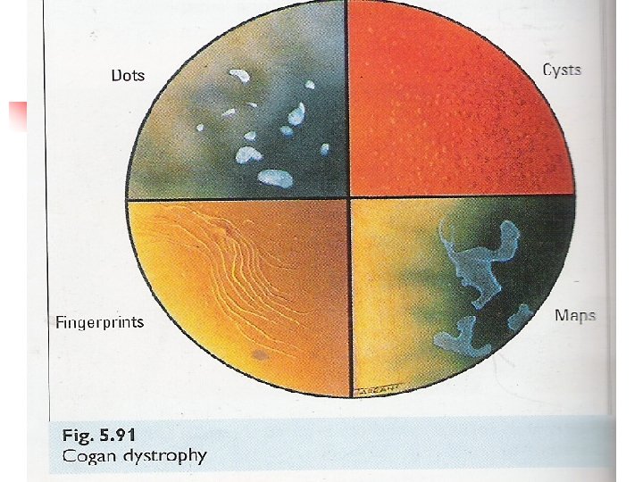
CORNEAL DYSTROPHIES n Anterior Cogans microcystic dystrophy Reis – Buckler § Stromal Granular Macular Lattice § Endothelial Fuchs endothelial dystrophy Posterior polymorphous dystrophy
Epi. BM dys n n n Also k/a - Map dot fingerprint dys. m/c dys. Onset - 2 nd decade Recurrent corneal erosions – 10% Signs- dot like opacity , epi. microcysts, sub-epi map like pattern Treat- same as rec. corneal erosions( saline , bandage 48 hrs, ointment)
Reis – Buckler dys n n n n AD Onset- early childhood Arise in region of bow. Memb Recurrent erosions Cornea- irregular dense grey sub-epi. Opacity arranged in fish net pattern Treat- PRK, lam KP HIGH RECURRENCE AFTER CORNEAL TRANSPALANT
REIS-BUCKLER DYSTROPHY
Hereditary stromal corneal dys n n n n Bilateral Around puberty Central area of cornea, chr. By discrete areas of opacity in sup. Areas of stroma Hyaline deposits b/w the cornel lamellae Symptom less without inflammation Progress slowly until visual impairment Treat- KP
GRANULAR CORNEAL DYSTROPHY
MACULAR CORNEAL DYSTROPHY
GRANULAR MACULAR
LATTICE CORNEAL DYSTROPHY
ENDOTHELIAL CORNEAL DYS n M/C Fuch’s dys- n AD, seen in elderly n m/c in females n n d/t changes in endothelium with formation of hyaline excrescences on DM ( corn. Guttata) TREAT- Nacl 5% drop or ointment, bandage contact lens. -Pen KP
FUCH’S ENDOTHELIAL DYSTROPHY n n Stage of cornea guttata Stage of endothelial decompensation Stage of bullous keratopathy Stage of scarring