DISEASES OF THE CORNEA Dr Neeti Gupta Associate


 Atmospheric oxygen")


 CORNEAL ULCERS Ø Etiology Ø Causative agent Ø Indolent course Ø Trauma")


- Slides: 39
DISEASES OF THE CORNEA Dr. Neeti Gupta Associate Professor Department of Ophthalmology
DIMENSIONS n Ant surface elliptical n Posterior surface circular 11. 5 mm 11 -12 mm 10 - 11 mm • Shape - Prolate 11. 5 mm
n n Thickness centre periphery Radius of curvature ant. surface post surface Refractive index 0. 5 -0. 6 (thinner) 0. 7 -1. 0 mm 7. 8 mm 6. 5 mm (steeper) 1. 376 Refractive power - 40 -44 D (70% Of total refractive power of the eye
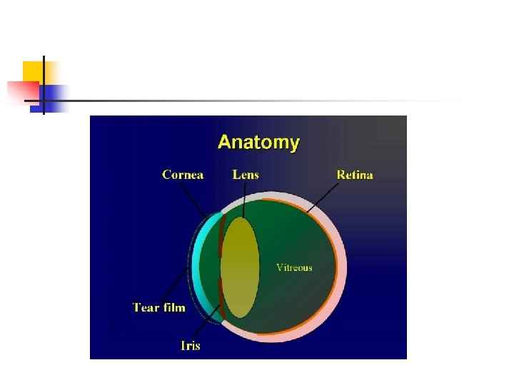
LAYERS OF CORNEA
n n n Epithelial Layer –Regenerates Bowman’s Layer- Resistant to trauma and infection Stroma –Collagen bundles with keratocytes Descemets layer- very tough Endothelium – hexagonal cells – 3000 cells/mm 2
Limbus n n 1 -1. 5 mm anatomy Cells at limbus are unique – Limbal stem cells Responsible for growth and regeneration of epithelial cells
NERVE SUPPLY OF CORNEA Cornea has body’s highest no. of nerve endings 5 th cranial nerve (Trigeminal) Ophthalmic division Nasociliary branch Long ciliary nerves Annular plexus around limbus Subepithelialplexus Intraepithelial plexus
NUTRITION n n n Perilimbal capillaries METABOLISM n Aqueous humour (glucose diffussion) Atmospheric oxygen (tear film) n Epithelium & endothelium metabolically very active Both aerobic & anaerobic metabolism
CORNEAL TRANSPARENCY n n Avascularity Uniform refractive Index of the cornea Arrangement of corneal lamellae State of relative dehydration(78%) Barrier effect Endothelial of epithelium pump & endothelium Osmotic gradient
FUNCTIONS OF CORNEA n Transmission of light/Refractive medium n Structural integrity of globe/Protects the eye
PATHOLOGICAL CHANGES IN THE CORNEA n n n Keratitis Superficial Deep Stromal Endothelial Corneal abrasion/erosion Corneal ulcer Corneal opacity Nebular, Macular, Leucomatous Corneal oedema Vascularisation
KERATITIS MORPHOLOGICAL CLASSIFICATION ULCERATIVE n n Suppurative/non suppurative Superficial/deep/ perforated NON ULCERATIVE Superficial Diffuse sup. Keratitis SPK Deep Non suppurating Interstitial/disciform Suppurating Central/posterior corneal abscess
KERATITIS ETIOLOGICAL CLASSIFICATION § Infective § Allergic § § § Asso. with systemic collagen vascular ds § Traumatic § Idiopathic Trophic Asso. with skin & mucous memb. ds Moorens ulcer
INFECTIVE KERATITIS PATHOGENESIS Epithelial damage Infection Corneal abrasion Exogenous Epith. Drying Spread from ocular tissue Epith. Necrosis Epith. desquamation Endogenous
PREDISPOSING FACTORS n Ocular - Trauma - Contact lens - lids and adenexal infections - Topical medications n Ocular surface diseases - Dry eyes – Sjogrens syndrome, SJ synd. , Vit A def - Prolonged Corneal Exposure - Proptosis, Lagophthalmos , ectropion - Epi. Defect – Entropion , Trichiasis
PREDISPOSING FACTORS n Systemic - Diabetes mellitus - Sjögren’s syndrome - Steven johnsons syndrome - Connective tissue disorders - AIDS - Measles malnutrition n Occupational - Farmers - Animal handlers - Gardeners
HISTORY n n n Pain Redness Photophobia Discharge Lacrimation Decrease visual aquity
EXAMINATION n n Eyelids Lacrimal Sac Conjuntiva Corneal ulcer - size - shape - location - margins - infiltration - corneal sensation
EXAMINATION n n n Anterior chamber Iris Pupil and Lens Scleral involvment Posterior segment/ USG
Bacterial corneal ulcers n Agents : Staphlococcus aureus/ albus Streptococcus Pseudomonas Pneumococcus N. gonorrhoeae C. diphtheriae E. coli
PATHOLOGY OF CORNEAL ULCER n Stage of ulceration- desquamation of the epithelium and tissue necrosis resulting in saucer shaped ulceration n Progressive infiltration- progression of ulceration with leucocytes infiltration and purulent suppuration n Regression –characterized by relatively smooth and transparent ulcer area n Cicatrization –Scar formation
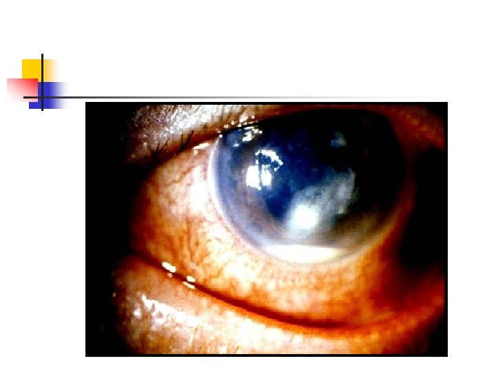
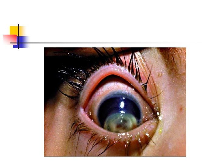
BACTERIAL CORNEAL ULCER SYMPTOMS Pain/ FB sensation Redness Watering Photophobia Blurred vision SIGNS Lid oedema Blepharospasm Conj. chemosis Infiltration Corneal oedema Hypopyon +/ -
n n n Symptoms are acute Severe clinical signs Rapid progression Wet looking ulcer area Purulent discharge
TREATMENT OF BACTERIAL KERATITIS UNCOMPLICATED ULCER Ø Ø Identify & treat the cause Corneal scraping staining/culture Antibiotics Rest to eye Cycloplegics Antiglaucoma medications Systemic antibiotics
PERFORATED ULCER Small < 3 mm Ø Ø Ø IOP lowering drugs Pressure bandage Bandage contact lens Tissue adhesives Conj. flap Large Ø >3 mm Therapeutic PK
Fungal keratitis n Incidence is low n Most common organism is Aspergillus n Infections are more common when there is high humidity
Classification n Filamentous 1. Septate - Nonpigmented – Fusarium Aspergillus Penicillium - Pigmented - Curvularia Alternaria 2. Nonseptate Rhizopus n Yeast Candida
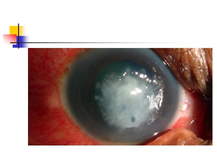
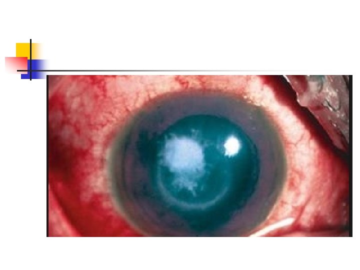
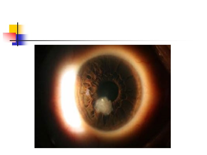
FUNGAL (MYCOTIC) CORNEAL ULCERS Ø Etiology Ø Causative agent Ø Indolent course Ø Trauma with organic matter Injury with animal tail Systemic/ local immune suppresion Aspergillus , Fusarium Candida , Cryptococcus Curvularia, Alternaria Symptom – foreign body sensation , photophobia , blurred vision and discharge
SIGNS MORE THAN SYMPTOMS SIGNS Soft creamy raised exudates Dry looking Feathery margins Satellite lesions Immune ring of Wesseley Hypopyon +/Endothelial plaque Posterior abscess Fungal ulcer
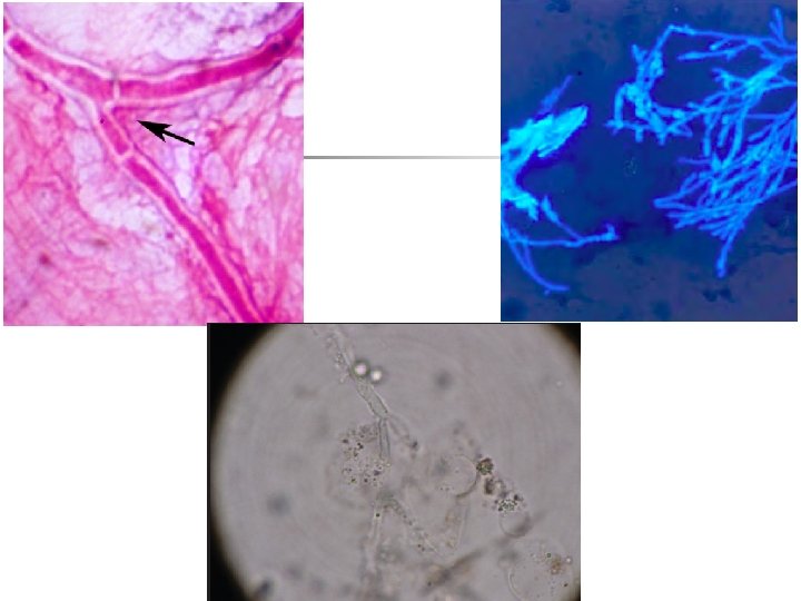
DIAGNOSIS n History matter Organic n Typical clinical picture n Corneal scrapings KOH wet mount Gram, Giemsa staining Calcoflour white Culture on SDA TREATMENT § § § Topical antifungals Natamycin 5% Itraconazole 1% Fluconazole 0. 2% Amphotericin B 0. 10. 2% Systemic antifungals Cycloplegics Anti inflammatory drugs Therapeutic PK in unresponsive cases
COURSE OF CORNEAL ULCER Healing Deep penetration Descemetocele Perforation Adherent leucoma Sloughing Pseudocornea Ant. Staphyloma
COMPLICATIONS n n § Toxic iridocyclitis Secondary glaucoma Descemetocele Perforation Iris prolapse Ant. Capsular cataract Corneal fistula Spontaneous expulsion of lens & vitreous Intraocular haemorrhage Expulsive hmg. Purulent uveitis Endophthalmitis/ Panophthalmitis Corneal scarring/ opacification