Discuss the markers for bone formation and Resorption

Discuss the markers for bone formation and Resorption and their clinical use in diagnosis Describe the molecular basis of: 1. 2. 3. 4. Duchene Muscular Dystrophy. Glycogen storage diseases of muscle. Muscle Mitochondrial diseases. Describe the molecular basis of: Osteogenesis imperfecta and Ehlar Danlos syndromes

Clin Biochem Rev Vol 26 November 2005

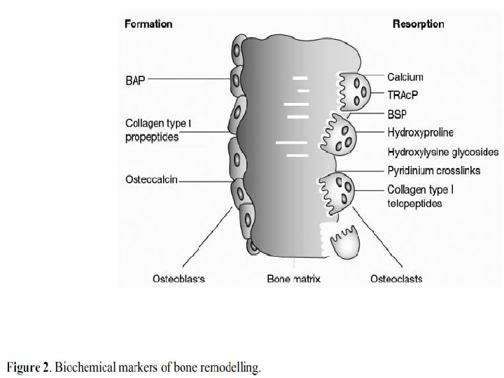
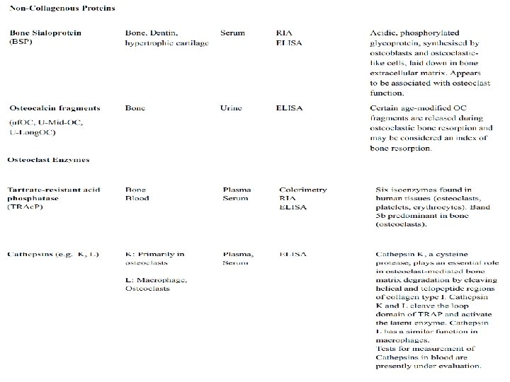
Biochemical Markers of Bone Turnover The currently available markers of bone turnover include both enzymes and non-enzymatic peptides derived from cellular and non-cellular compartments of bone, they are usually classified according to the metabolic process they are considered to reflect.

Markers of bone resorption are related to: • Collagen breakdown products such as hydroxyproline or the various collagen cross-links and telopeptides. • Other markers of bone resorption include noncollagenous matrix proteins such as bone sialoprotein (BSP), or osteoclast-specific enzymes like tartrate-resistant acid phosphatase or cathepsin K.

Markers of bone formation are either by-products of collagen neosynthesis: (e. g. propeptides of type I collagen), or osteoblastrelated proteins such as osteocalcin (OC) and alkaline phosphatase (ALP).
.")
Bone formation and bone resorption markers: (e. g. hydroxyproline, certain OC fragments).
: membrane-bound tetrameric enzyme attached to")
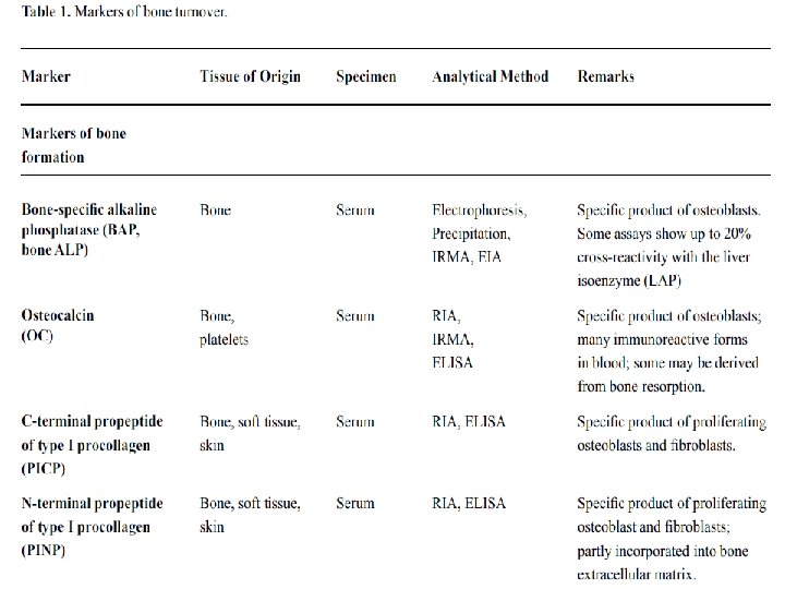
Markers of Bone Formation Serum Total Alkaline Phosphatase (AP): membrane-bound tetrameric enzyme attached to glycosyl-phosphatidylinositol moieties located on the outer cell surface Osteocalcin (OC): OC is a 5. 8 k. Da, hydroxyapatitebinding, protein exclusively synthesised by osteoblast, odontoblasts and hypertrophic chondrocytes Procollagen Type I Propeptides: The procollagen type I propeptides are derived from collagen type I, the most abundant form of collagen found in bone.
: Is formed intracellularly from the post-translational hydroxylation of")
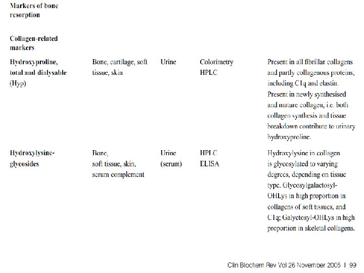
Markers of Bone Resorption Hydroxyproline (OHP): Is formed intracellularly from the post-translational hydroxylation of proline and constitutes 12 -14% of the total amino acid content of mature collagen. Ninety percent of the OHP liberated during the degradation of bone collagen is primarily metabolised in the liver. Hydroxylysine-Glycosides: arise during the posttranslational phase of collagen synthesis and occur in two forms, glycosyl-galactosyl-hydroxylysine (GGHL) and galactosyl-hydroxylysine (GHL). Both components are released into the circulation during collagen degradation
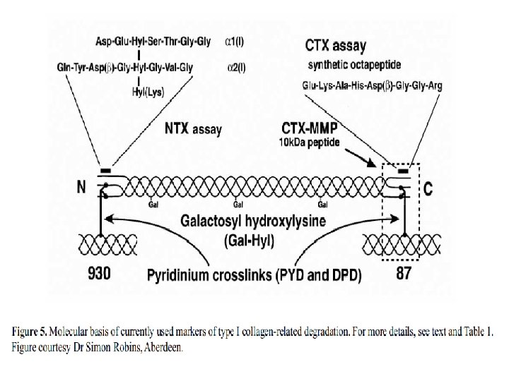
 and Deoxypyridinoline (DPD): PYD and DPD are")
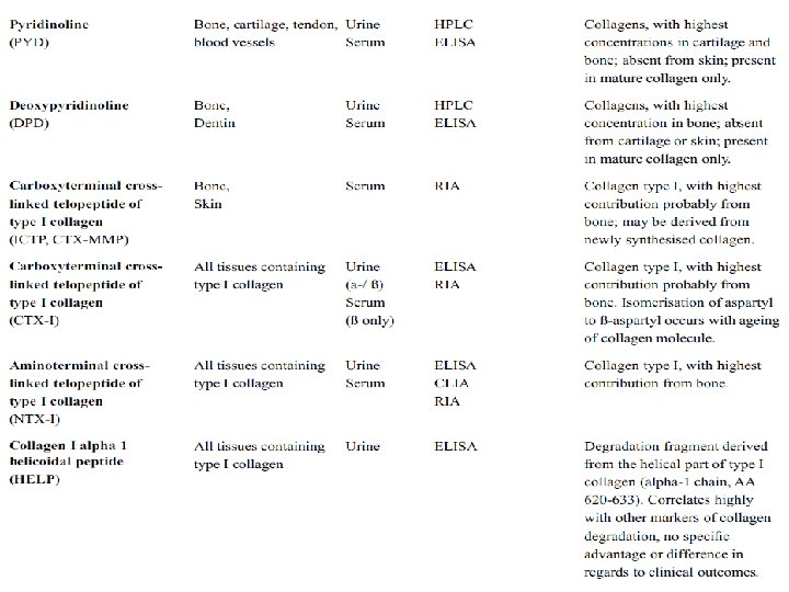
3 -Hydroxypyridinium Crosslinks of Collagen Pyridinoline (PYD) and Deoxypyridinoline (DPD): PYD and DPD are formed during the extracellular maturation of fibrillar collagens Crosslinked Telopeptides of Type I Collagen The: Crosslinked telopeptides of type I collagen are derived from specific regions of the collagen type I molecule, namely the amino terminal (NTP) and the carboxyterminal (CTP) telopeptide (Figure 5).

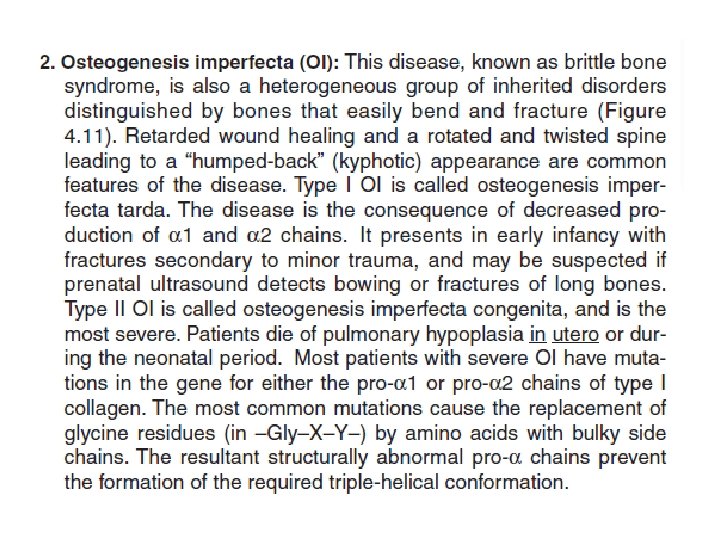
: Genetic disorder characterized by progressive muscle degeneration and weakness. There")
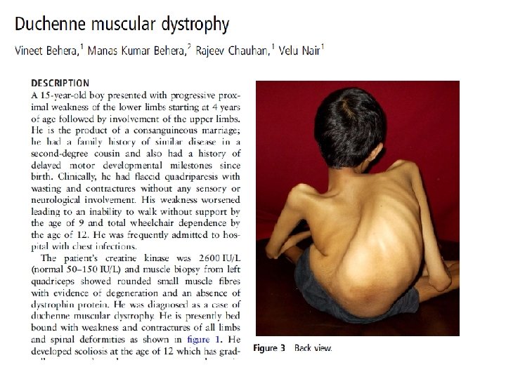
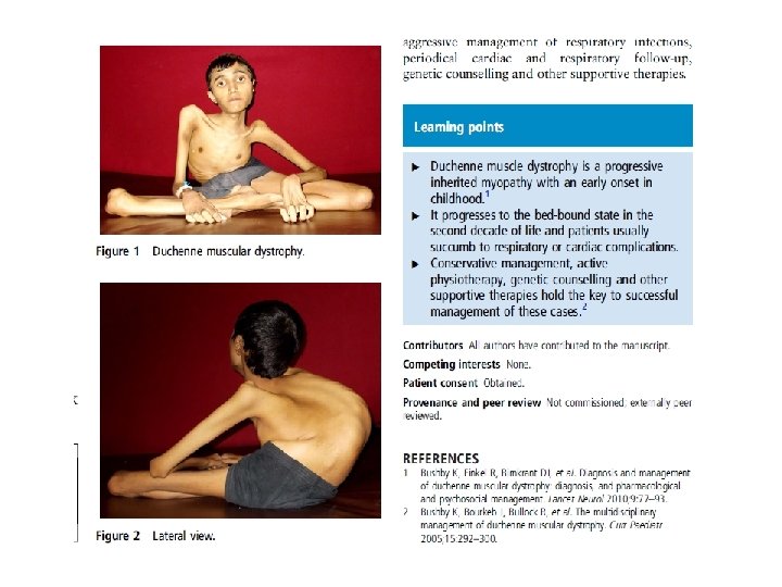
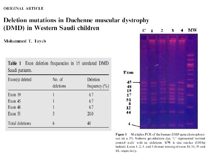
Duchenne muscular dystrophy (DMD): Genetic disorder characterized by progressive muscle degeneration and weakness. There are nine types of muscular dystrophy. Caused by an absence of dystrophin. Early childhood onset X-linked Prevalence 1: 3, 600 male infants.

Sarcolemmal proteins and sarcomere structure. Fedik Rahimov, and Louis M. Kunkel J Cell Biol 2013; 201: 499 -510 © 2013 Rahimov and Kunkel


Mitochondrial Myopathy Group of neuromuscular diseases caused by damage to the mitochondria. Most mitochondrial myopathies occur before the age of 20, and often begin with exercise intolerance or muscle weakness.
 q. Leigh syndrome and maternally-inherited Leigh")
Types of Mitochondrial Myopathies q. Kearns-Sayre syndrome (KSS) q. Leigh syndrome and maternally-inherited Leigh syndrome (MILS) q. Mitochondrial DNA depletion syndrome (MDS) q. Mitochondrial encephalomyopathy, lactic acidosis and strokelike episodes (MELAS) q. Mitochondrial neurogastrointestinal encephalomyopathy (MNGIE) q. Myoclonus epilepsy with ragged red fibers (MERRF) q. Neuropathy, ataxia and retinitis pigmentosa (NARP) q. Pearson syndrome q. Progressive external ophthalmoplegia (PEO)
- Slides: 30