
DIRECT OPHTHALMOSCOPE BY MOHAMMED SUHAIL ALSALAM LEARNING OBJECTIVES
DIRECT OPHTHALMOSCOPE BY MOHAMMED SUHAIL ALSALAM
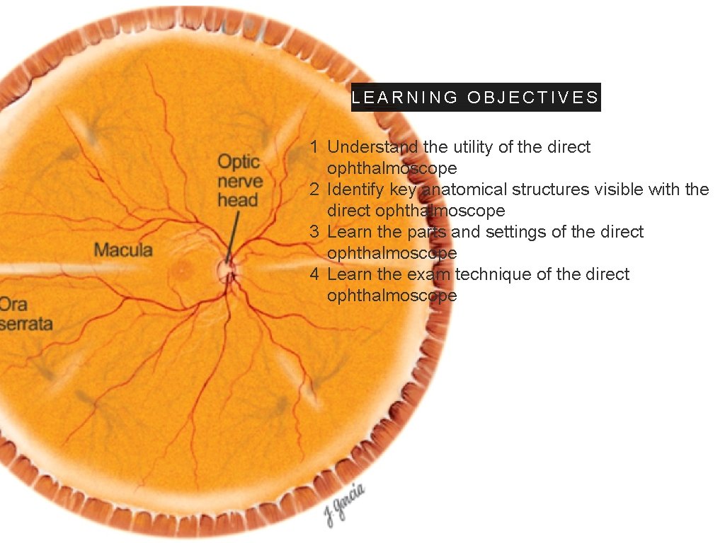
LEARNING OBJECTIVES 1 Understand the utility of the direct ophthalmoscope 2 Identify key anatomical structures visible with the direct ophthalmoscope 3 Learn the parts and settings of the direct ophthalmoscope 4 Learn the exam technique of the direct ophthalmoscope
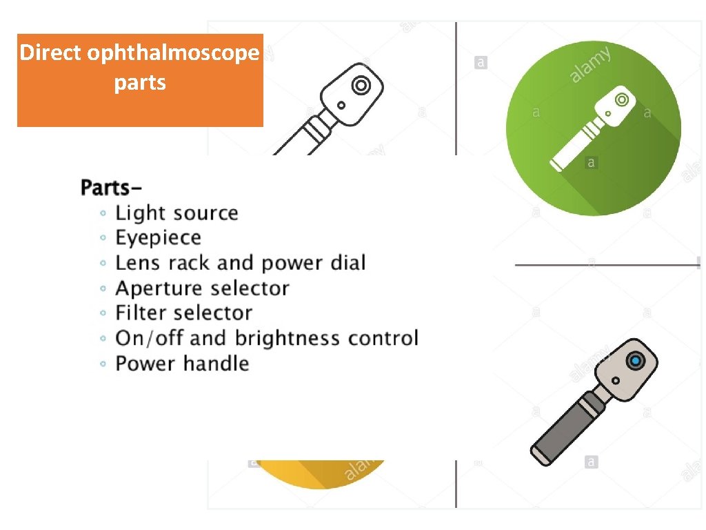
Direct ophthalmoscope parts “Type a quote here. ” –JOHNNY APPLESEED
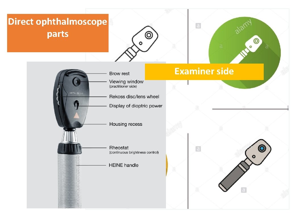
Direct ophthalmoscope parts “Type a quote here. ” – J O H N N Y A P P L E S Examiner EED side
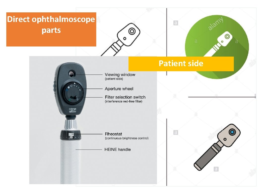
Direct ophthalmoscope parts “Type a quote here. ” – J O H N N Y A P P L E S EPatient ED side
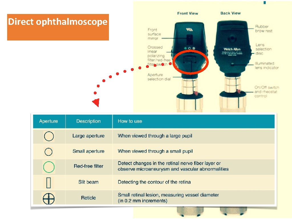
Direct ophthalmoscope “Type a quote here. ”
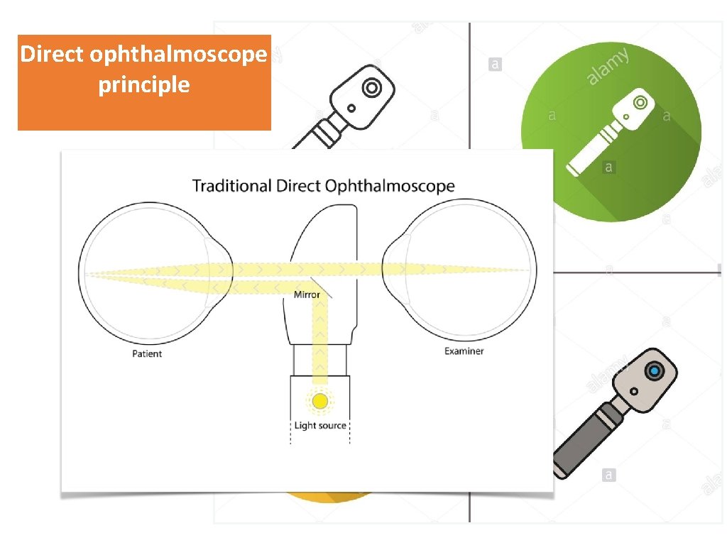
Direct ophthalmoscope principle “Type a quote here. ” –JOHNNY APPLESEED
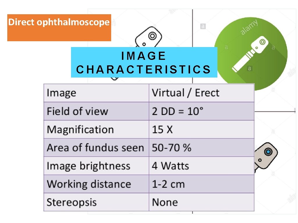
Direct ophthalmoscope “Type a quote here. ” IMAGE C H A– R T AEPR TEIDC S J OA H NC NY P LIES SE
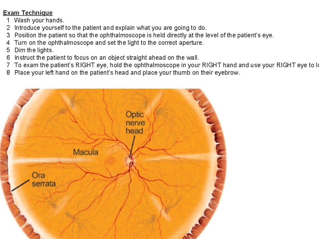
Exam Technique 1 Wash your hands. 2 Introduce yourself to the patient and explain what you are going to do. 3 Position the patient so that the ophthalmoscope is held directly at the level of the patient’s eye. 4 Turn on the ophthalmoscope and set the light to the correct aperture. 5 Dim the lights. 6 Instruct the patient to focus on an object straight ahead on the wall. 7 To exam the patient’s RIGHT eye, hold the ophthalmoscope in your RIGHT hand use your RIGHT eye to lo 8 Place your left hand on the patient’s head and place your thumb on their eyebrow.
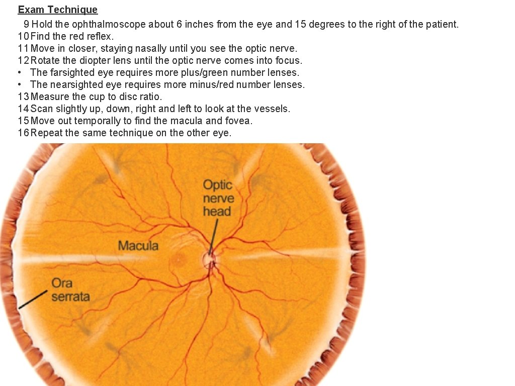
Exam Technique 9 Hold the ophthalmoscope about 6 inches from the eye and 15 degrees to the right of the patient. 10 Find the red reflex. 11 Move in closer, staying nasally until you see the optic nerve. 12 Rotate the diopter lens until the optic nerve comes into focus. • The farsighted eye requires more plus/green number lenses. • The nearsighted eye requires more minus/red number lenses. 13 Measure the cup to disc ratio. 14 Scan slightly up, down, right and left to look at the vessels. 15 Move out temporally to find the macula and fovea. 16 Repeat the same technique on the other eye.
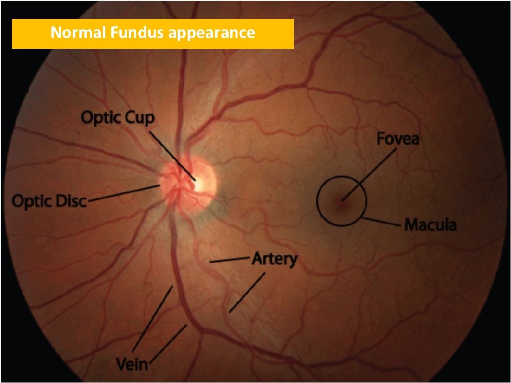
Normal Fundus appearance
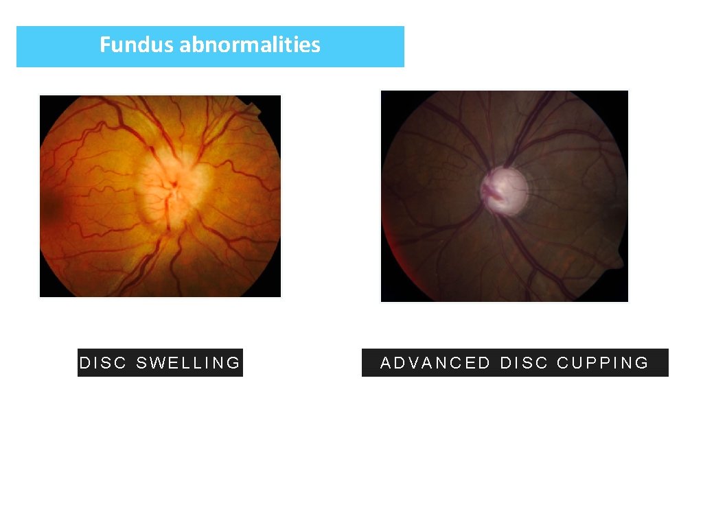
Fundus abnormalities DISC SWELLING ADVANCED DISC CUPPING

Fundus abnormalities
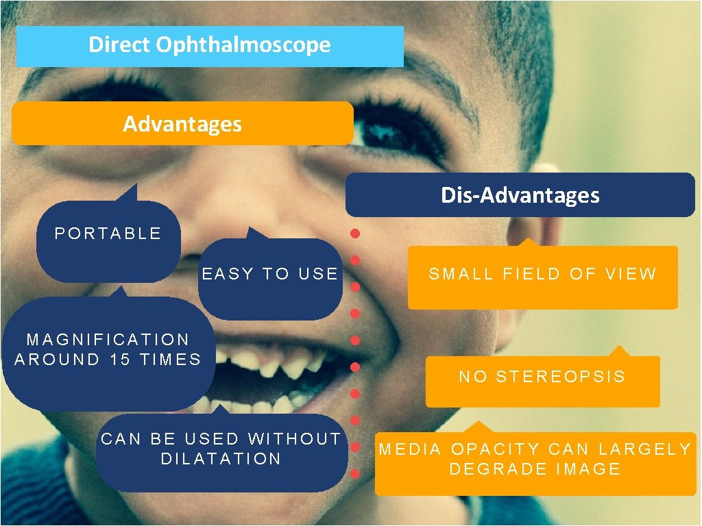
Direct Ophthalmoscope Advantages Dis-Advantages PORTABLE EASY TO USE MAGNIFICATION AROUND 15 TIMES CAN BE USED WITHOUT DILATATION SMALL FIELD OF VIEW NO STEREOPSIS MEDIA OPACITY CAN LARGELY DEGRADE IMAGE
T H AN K Y O U
- Slides: 15