DIGGIN UP BONES CHAPTER 5 The Skeletal System

DIGGIN’ UP BONES CHAPTER 5

The Skeletal System n Divided into two divisions n Axial skeleton n Appendicular skeleton

Functions of Bones n n n Support of the body Protection of soft organs Movement due to attached skeletal muscles Storage of minerals and fats Blood cell formation

Classification of Bones Long bones; Examples: Femur, humerus n Short bones; Examples: Carpals, tarsals n Flat bones; Examples: Skull, ribs, sternum n Irregular bones; Exam. : Vertebrae and hip n

Classification of Bones on the Basis of Shape Figure 5. 1 Copyright © 2003 Pearson Education, Inc. publishing as Benjamin Cummings Slide 5. 5 c

Gross Anatomy of a Long Bone · Diaphysis · Shaft · Composed of compact bone · Epiphysis · Ends of the bone · Composed mostly of spongy bone Copyright © 2003 Pearson Education, Inc. publishing as Benjamin Cummings Figure 5. 2 a Slide 5. 6

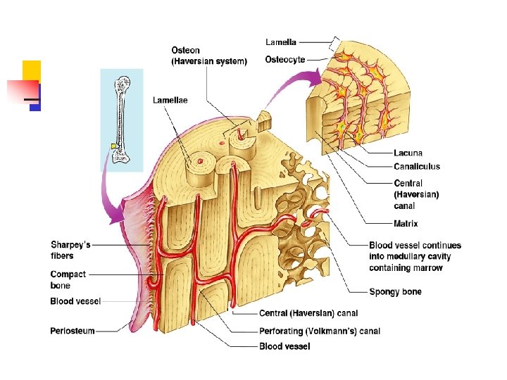
Structures of a Long Bone n Periosteum n n Sharpey’s fibers n n n Outside covering of the diaphysis Secure periosteum to underlying bone Arteries n Supply bone cells with nutrients Articular cartilage n Covers the external surface of the epiphyses

Structures of a Long Bone · Periosteum · Outside covering of the diaphysis · Fibrous connective tissue membrane · Sharpey’s fibers · Secure periosteum to underlying bone · Arteries · Supply bone cells with nutrients Copyright © 2003 Pearson Education, Inc. publishing as Benjamin Cummings Figure 5. 2 c Slide 5. 7
 in adults")
Medullary cavity Cavity of the shaft n Contains yellow marrow (mostly fat) in adults n Contains red marrow (for blood cell formation) in infants n

Bone Markings Surface features of bones n Sites of attachments for muscles, tendons, and ligaments n Passages for nerves and blood vessels n
 n n Central (Haversian) canal n")
Microscopic Anatomy of Bone n Osteon (Haversian System) n n Central (Haversian) canal n n n A unit of bone Opening in the center of an osteon Carries blood vessels and nerves Perforating (Volkman’s) canal n n Canal perpendicular to the central canal Carries blood vessels and nerves
")
Microscopic Anatomy of Bone n Lacunae n n n Cavities containing bone cells (osteocytes) Arranged in concentric rings Lamellae n n Rings around the central canal Sites of lacunae

Microscopic Anatomy of Bone n Canaliculi n n n Tiny canals Radiate from the central canal to lacunae Form a transport system

Types of Bone Cells Osteocytes n Mature bone cells n Osteoblasts n Bone-forming cells n

Types of Bone Cells Osteoclasts n Bone-destroying cells n Break down bone matrix for remodeling and release of calcium n Bone remodeling is a process by both osteoblasts and osteoclasts n
 is formed Break is splinted")
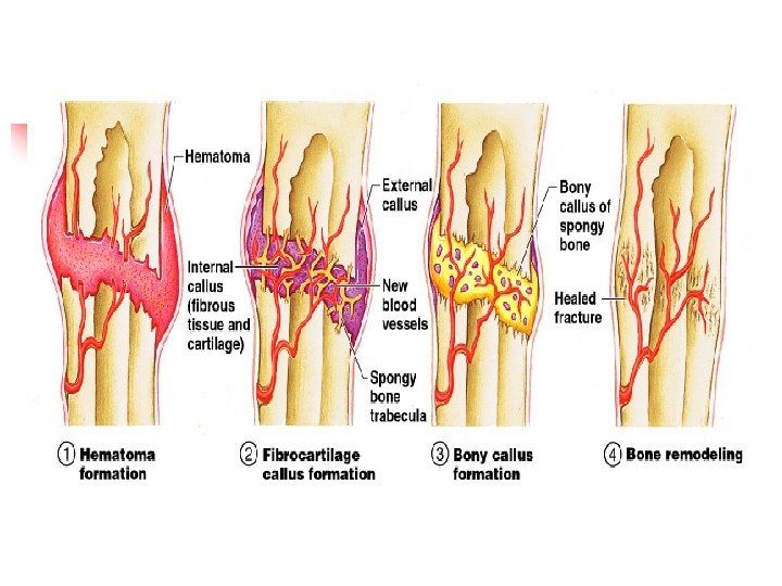
Repair of Bone Fractures n n Hematoma (blood-filled swelling) is formed Break is splinted by fibrocartilage to form a callus Fibrocartilage callus is replaced by a bony callus Bony callus is remodeled to form a permanent patch

Common Types of Fractures Table 5. 2 Copyright © 2003 Pearson Education, Inc. publishing as Benjamin Cummings Slide 5. 17
- Slides: 19