Digestive Tract Development 3 rd week 4 th


 small intestine")
 labium superius labium inferius rima oris anguli oris sulcus nasolabialis sulcus")
 m. buccinator covered by fascia buccopharyngea corpus adiposum buccae (buccal fat pad)")
 rima oris (oral fissure) isthmus faucium (isthmus of fauces) borders:")
 mucosa covering the alveolar processes of the jaws, firmly grows together with")
 I arcus dentalis superior (elipsoid) arcus dentalis inferior (parabolic) dentes permanentes (32)")

 II parts of the tooth corona dentis (crown) – cuspides cervix dentis (neck)")

 occlusalis x vestibularis lingualis directions x mesialis distalis")
 Lips –")


 muscle organ covered by mucosa radix, corpus, apex dorsum linguae sulcus medianus")


 NO submucosa")
vallate papillae The largest,")

 Pharynx at the")
 hard palate (palatum durum) bony base plicae palatinae transversae, raphe palati (seam)")


 valleculae epiglotticae plica glossoepiglottica mediana + laterales 3.")


 „ring“ of the lymphatic tissue first protective barrier")
")
- Slides: 56
Digestive Tract
Development 3 rd week 4 th week The primordial gut forms during the 4 th week as the folds incorporate the dorsal part of the yolk sac into the embryo
Development Foregut Hindgut Oropharyngeal membrane Foregut Celiac trunk Cloacal membrane From oral cavity to duodenum (opening of the bile duct) Midgut Superior mesenteric artery From duodenum to transverse colon Hindgut Inferior mesenteric artery The rest of colon
Development Foregut Stomodeum Hindgut Proctodeum The endoderm of the primordial gut The epithelium and glands of the digestive tract The ectoderm of the stomodeum The epithelium in the cranial part The ectoderm of the proctodeum The epithelium in the caudal part
Wall 4. Serosa / adventitia 3. Muscularis 2. Submucosa 1. Mucosa The wall is made up of four layers
Mucosa 1. Epithelial lining 2. Lamina propria mucosae Connective tissue Blood vessels, lymphatics, macrophages and lymphocytes, sometimes glands 1. Mucosa 3. Lamina muscularis mucosae Smooth muscles Movements of the mucosa – better contact with food
Submucosa Connective tissue Blood and lymph vessels, glands, lymphoid tissue 2. Submucosal plexus (Meissner´s plexus) of autonomic nerves Function: secretion
Muscularis Longitudinal Circular 3. Muscularis Cajal´s cells - pacemaker Smooth muscle cells 2 sublayers 1) Internal - circular 2) External - longitudinal Myenteric plexus – (Auerbach´s plexus) contraction of the muscularis Enteric nervous system: submucosal and myenteric plexus, Plexus – aggregates of nerve cells that form parasympathetic ganglia (contains autonomic neurons) Origin: neural crest
Enteric nervous system Chagas disease Parasite injures the plexuses – dilatations: Megaesophagus, Megacolon Romaňa´s sign Kissing bug Hirschprung disease Cells from neural crest don´t migrate well: Congenital megacolon
Serosa / Adventitia 4. Serosa / adventitia Serosa Simple squamous covering epithelium + Connective tissue rich in vessels and adipose tissue Is continuous with the mesenterium and the peritoneum Organs which are inside the abdominal cavity Adventitia – connective tissue Organs which are outside the abdominal cavity
Parts of the digestive tract oral cavity pharynx esophagus stomach (ventriculus, gaster) small intestine (intestinum tenue) large intestine (intestinum crassum) rectum liver (hepar) pancreas gallbladder (vesica fellea)
Lips (labia oris) labium superius labium inferius rima oris anguli oris sulcus nasolabialis sulcus mentolabialis philtrum tuberculum labii superioris transtion to the keratizing epithelium pars cutanea, intermedia (sebaceous glands), mucosa (salivary glands - glandulae labiales) m. orbicularis oris http: //www. botulinumtoxinambulanz. de/hemispasmus. htm
Cheek (bucca) m. buccinator covered by fascia buccopharyngea corpus adiposum buccae (buccal fat pad) – reaches under ramus mandibulae into fossa infratemporalis there are glandulae buccales in the mucosa – small salivary glands papilla parotidea – at the level of the 2 nd upper molar
Cavitas oris (oral cavity) rima oris (oral fissure) isthmus faucium (isthmus of fauces) borders: ventrally and externally: lips (labia oris) and cheeks (buccae) roof: palate floor: m. mylohyoideus and m. geniohyoideus vestibulum oris (oral vestibule) fornix vestibuli sup. + inf. frenulum labii sup. + inf. cavitas oris propria (oral cavity proper)
Gum (gingiva) mucosa covering the alveolar processes of the jaws, firmly grows together with periosteum margo gingivalis sulcus gingivalis papillae gingivales http: //medicaldictionary. thefreedictionary. com/Gum+%28 anatomy%29
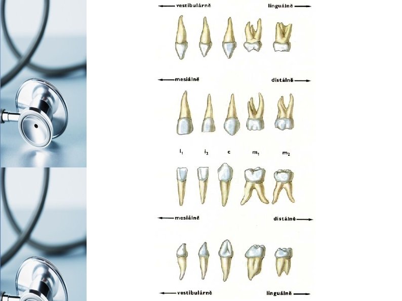
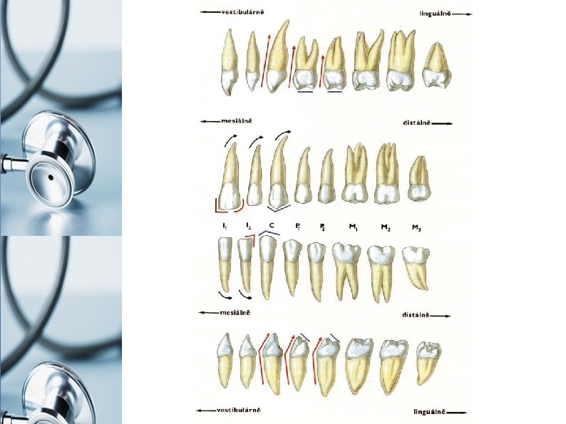
Teeth (dentes) I arcus dentalis superior (elipsoid) arcus dentalis inferior (parabolic) dentes permanentes (32) + dentes decidui (20) dens incisivus (cutter) 8/8 dens caninus (cuspid) 4/4 dens premolaris (bicuspid) 8/0 dens molaris (molar) 12/8 teething (eruptio) dentes decidui 6 th-30 th month dentes permanentes 6 th-30 th year dental formula i 1, i 2, c, m 1, m 2 I 1, I 2, C, P 1, P 2, M 1, M 2, M 3 occlusion: psalidodontia – „scissors occlusion“ (labidodontia – „pincer occlusion“, …)
Dental formula for deciduous teeth Dental formula for permanent teeth
Teeth(dentes) II parts of the tooth corona dentis (crown) – cuspides cervix dentis (neck) radix dentis (root) – apex, canalis cavitas dentis – pulp (vessels, nerves) gomphosis = dentoalveolar juncture periodontium - ligaments between the alveolus and the tooth, run in many directions, hold the tooth in place parodontium - all structures around the tooth (bone, connective tissue, gum)
Periodontium
facies (surfaces) occlusalis x vestibularis lingualis directions x mesialis distalis
Oral cavity Stratified squamous epithelium Non keratinized (Keratinized - gingiva, hard palate) Lips – transition from the oral non-keratinized epithelium to the keratinized skin
Teeth Enamel Dentin Pulp cavity Cement Apical foramen Crown Neck Root
Dentin Calcified tissue harder than bone 70% calcium hydroxyapatite Organic matrix: collagen I and glycosaminoglycans Who makes the organic matrix? Odontoblasts – tall cells that line the pulp cavity Their long processes (Tomes processes) lie within dentinal tubules
Enamel 98% hydroxyapatite (fluorid incorporated by the crystals – fluorapatite is more resistent to acidic disolution caused by microorganism) Organic matrix: no collagen, proteins: amelogenin, enamelin Who makes the enamel? Ameloblasts – one ameloblast produces one prism After finishing the synthesis of enamel, ameloblasts cover the crown until the eruption of the tooth
Tooth formation Dental lamina Tooth bud 6 th week: thickening of the oral ectoderm = dental lamina 10 centers of proliferation In one jaw Tooth buds grow into the underlying mesenchyme These tooth buds develop into the deciduous teeth
Tooth formation 2. Cap stage 1. Bud stage Ectoderm Enamel organ Enamel Mesenchyme Dental papilla Enamel organ Stellate reticulum Outer enamel epithelium Inner enamel epithelium 3. Bell stage
One layer of the mesenchymal cells in the dental papilla dfferentiates into odontoblasts They produce dentin Cells of the inner enamel epithelium differentiate into ameloblasts They produce enamel – the basal part of the cells produces it
Dental sac Cementum Dental sac Periodontal ligament Cementum covers the dentin of the root. It is similar to bone, but has no osteons. Periodontal ligament connects the cementum and the alveolar bone. Collagen has an unusually high turnover rate Alveolar bone – primary (immature) bone Gum – mucous membrane bound to the periosteum
Tongue (lingua) muscle organ covered by mucosa radix, corpus, apex dorsum linguae sulcus medianus sulcus terminalis foramen caecum papillae linguales (spits of the mucosa) pp. filiformes pp. fungiformes pp. foliatae pp. vallatae tonsilla lingualis margo linguae facies inferior frenulum linguae (caruncula, plica sublingualis), plica fimbriata
Muscles of the tongue I intraglossal muscles: m. longitudinalis sup. + inf. m. transversus linguae m. verticalis linguae aponeurosis linguae – on the dorsal surface septum linguae – incomplete!
Muscles of the tongue II extraglossal muscles: m. genioglossus m. hyoglossus m. styloglossus m. palatoglossus
Supply of the tongue arteries: a. car. ext-> a. lingualis veins: v. lingualis -> v. jugularis int. lymph drainage: n. l. submentales, submandibulares, cervicales profundi contralateral connections!!! innervation: motor: n. hypoglossus (XII), apart from m. palatoglossus (X) sensitive: n. lingualis (V 3), IX, X senzory: chorda tympani (VII), IX, X
Tongue Muscle covered by a mucous membrane (lamina propria penetrates the muscles) NO submucosa Muscle fibres cross one another in three planes
Tongue The dorsal surface is covered by eminences called papillae (Circum)vallate papillae The largest, 7 -12, V-shaped line before the terminal sulcus Foliate papillae Poorly developed, parallel ridges on the sides Filiform papillae Numerous, rough surface Fungiform papillae Mushroom shaped
Taste buds In all papillae except for the filiform Most of them in the vallate papillae Serous salivary glands – von Ebner, empty into the groove around the papilla They wash food particles
Development of the tongue 1. arch: Median tongue bud (tuberculum impar) Pharynx at the end of the 4 th week Distal tongue buds (lateral swellings) Lateral swellings Tuberculum impar Cupola Hypopharyngeal eminence The copula is overgrown by the hypopharyngeal eminence and disappears 2. arch: Cupola 3. , 4. arch: hypopharyngeal eminence
Palate (palatum) hard palate (palatum durum) bony base plicae palatinae transversae, raphe palati (seam) soft palate (palatum mole) aponeurosis palatina uvula palatina (uvula) muscles: m. tensor veli palatini (n. V 3) m. levator veli palatini m. uvulae m. palatoglossus m. palatopharyngeus all innervated by (n. X – plexus pharyngeus)
Isthmus faucium arcus palatoglossus arcus palatopharyngeus sinus tonsillaris tonsilla palatina – capsula – fossulae, cryptae
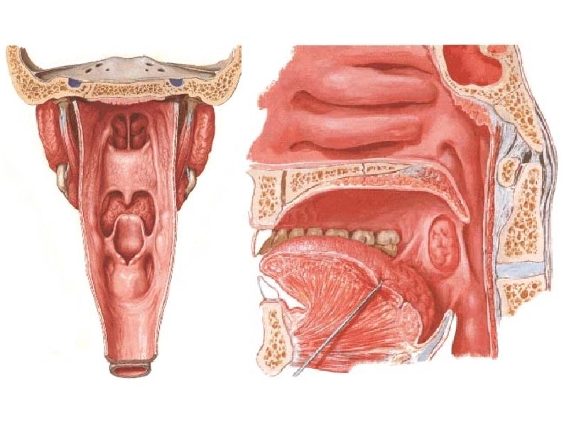
Pharynx I 1. pars nasalis = nasopharynx fornix fascia pharyngobasilaris sinus Morgagni recessus pharyngeus Luschkae (remnant of notochord) tonsilla pharyngea Luschkae tuba auditiva Eustachii torus tubarius tonsilla tubaria Gerlachi recessus pharyngeus Rosenmülleri pseudostratified columnar with the cilia
Pharynx II 2. pars oralis („oropharynx“) valleculae epiglotticae plica glossoepiglottica mediana + laterales 3. pars laryngea („hypopharynx, laryngopharynx“) recessus piriformis aditus laryngis both stratified sqamous non-keratinizing epithelium
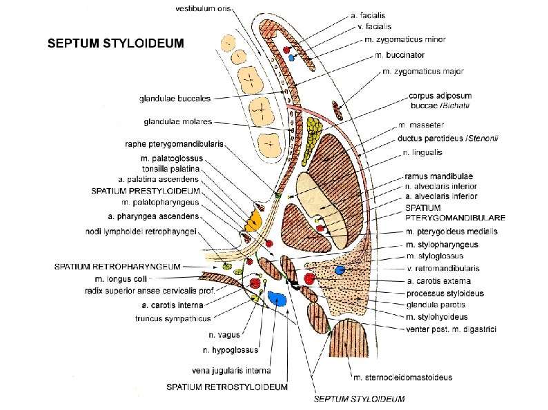
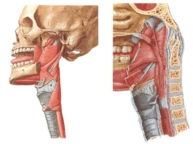
Pharynx surrounding spaces spatium parapharyngeum spatium prestyloideum styloid septum 5 muscles + ligament + proc. styloideus spatium retrostyloideum spatium retropharyngeum
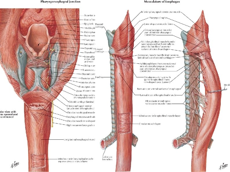
Pharynx muscles raphe pharyngis, fascia pharyngobasilaris, Luschka´s space mm. constrictores /3/ m. c. superior – 4 parts – origin at skull /3/ and tongue /1/ m. c. medius – 2 parts – origin at hyoid bone m. c. inferior – 2 parts – origin at laryngeal cartilages mm. levatores /3/ m. palatopharygeus (part of soft palate muscles, mounting of the palatopharyngeal arch) m. salpingopharyngeus m. stylopharyngeus (!exception! – innervated by n. IX !) innervation: plexus pharyngeus – n. X - except m. stylopharyngeus /n. IX /
Pharynx blood supply arteries: a. carotis ext. a. pharyngea ascendens a. facialis a. palatina ascendens a. lingualis rr. dorsales linguae a. maxillaris a. palatina major, a. canalis pterygoidei, r. pharyngeus veins: plexus (venosus) pharyngeus v. facialis v. jugularis int.
Pharynx Lymph and Nerves lymph n. l. retropharyngei n. l. paratracheales n. l. cervicales profundi nerves form plexus pharyngeus motor n. X (plexus pharyngeus), n. IX (m. stylopharyngeus) sensory n. X + n. IX (plexus pharyngeus), n. V 2 (n. pharyngeus for nasopharynx) autonomic n. X (plexus pharyngeus) = parasympathetic, rr. laryngopharyngei = sympathetic
Anulus lymphoideus pharyngis (Waldeyer lymphatic ring) „ring“ of the lymphatic tissue first protective barrier of an organism tonsilla pharyngea (Luschkae) tonsillae tubariae (Gerlachi) tonsillae palatinae tonsilla lingualis
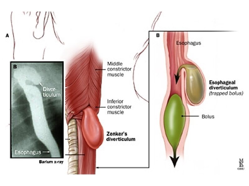
Sites with weakened wall trigonum Killiani cranially: m. thyropharyngeus (m. constrictor ph. inf. ) caudally: m. cricopharyngeus (m. constrictor ph. inf. ) diverticulum of Zenker (= pharyngo-oesophageal diverticle; dehiscence of Killian) trigonum Laimeri cranially: m. cricopharyngeus caudally: upper oblique fibres of longitudinal muscle layer of oesophagues area Killian-Jamieson at lateral side of oesophagus diverticulum of Killian-Jamieson