
Digestive system Presented by Mirza Salman Baig Faculty
Digestive system Presented by— Mirza Salman Baig Faculty of Pharmacy School of Pharmacy AIKTC, New Panvel
THE DIGESTIVE SYSTEM • The digestive system is the collective name used to describe the alimentary canal, some accessory organs and a variety of digestive processes which take place at different levels in the canal to prepare food eaten in the diet for absorption. • Alimentary canal begins at the mouth, passes through the thorax, abdomen and pelvis and ends at the anus.
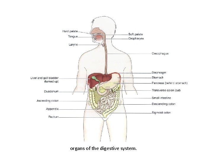
organs of the digestive system.
FUNCTION OF DIGESTIVE SYSTEM Ingestion. This is the process of taking food into the alimentary tract. Propulsion. This moves the contents along the alimentary tract. Digestion. • mechanical breakdown of food by, e. g. mastication (chewing) • chemical digestion of food by enzymes present in secretions produced by glands and accessory organs of digestive system Absorption. This is the process by which digested food substances pass through the walls of some organs of the alimentary canal into the blood and lymph capillaries for circulation round the body. Elimination. Food substances which have been eaten but cannot be digested and absorbed are excreted by the bowel as faeces.
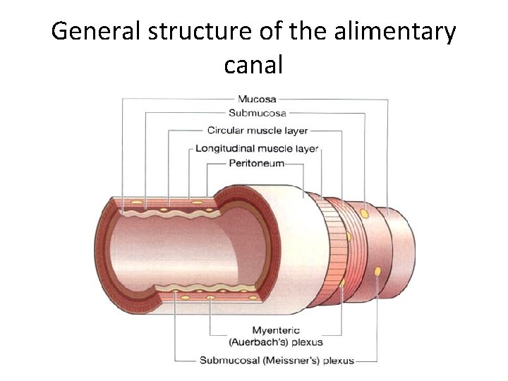
General structure of the alimentary canal
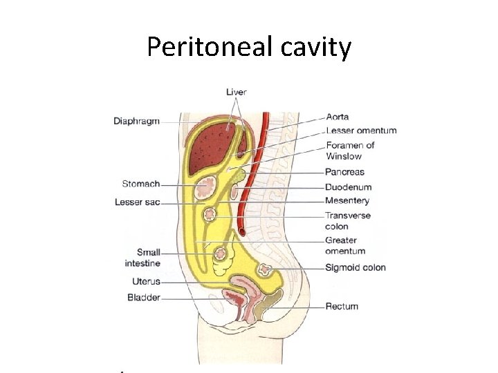
Peritoneal cavity

Peritoneum(1) Outer • The peritoneum is the largest serous membrane of the body • It consists of a closed sac, containing a small amount of serous fluid, within the abdominal cavity. • It is richly supplied with blood and lymph vessels & lymph nodes. • It provides a physical barrier to local spread of infection.

Other layers(3) • Muscle layer two layers of smooth (involuntary) muscle • Submucosa loose areolar connective tissue containing collagen. • Mucosa • Mucous membrane formed by columnar epithelium (some part also by stratified columnar epithelium) Functions: protection, secretion and absorption • Lamina propria consisting of loose connective tissue, which supports the blood vessels that nourish the inner epithelial layer, and varying amounts of Lymphoid tissue that has a protective function • Muscularis mucosa, a thin outer layer of smooth muscle that provides involutions of the mucosa layer, e. g. gastric glands, villi.

Mucous membrane • Mucus lubricates the walls of the tract and protects them from digestive enzymes • The surface in the regions lined with columnar epithelium are collections of specialised cells, or glands, which pour their secretions into the lumen of the tract-– saliva from the salivary glands – gastric juice from the gastric glands – intestinal juice from the intestinal glands – pancreatic juice from the pancreas – bile from the liver.
NERVE SUPPLY TO GIT The parasympathetic supply. • This supply to most of the alimentary tract is provided by one pair of cranial nerves, the vagus nerves. • Stimulation causes smooth muscle contraction and the secretion of digestive juices. • The most distal part of the tract is supplied by sacral nerves The sympathetic supply. • This is provided by numerous nerves which emerge from the spinal cord in the thoracic and lumbar regions
BLOOD SUPPLY TO GIT • Arterial Blood supply • Venous drainage
Arterial Blood supply • In the thorax. The oesophagus is supplied by paired oesophageal arteries, branches from the thoracic aorta. • In the abdomen and pelvis. The alimentary tract, pancreas, liver and biliary tract are supplied by the unpaired branches from the aorta: the coeliac artery and the superior and inferior mesenteric arteries
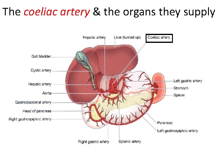
The coeliac artery & the organs they supply
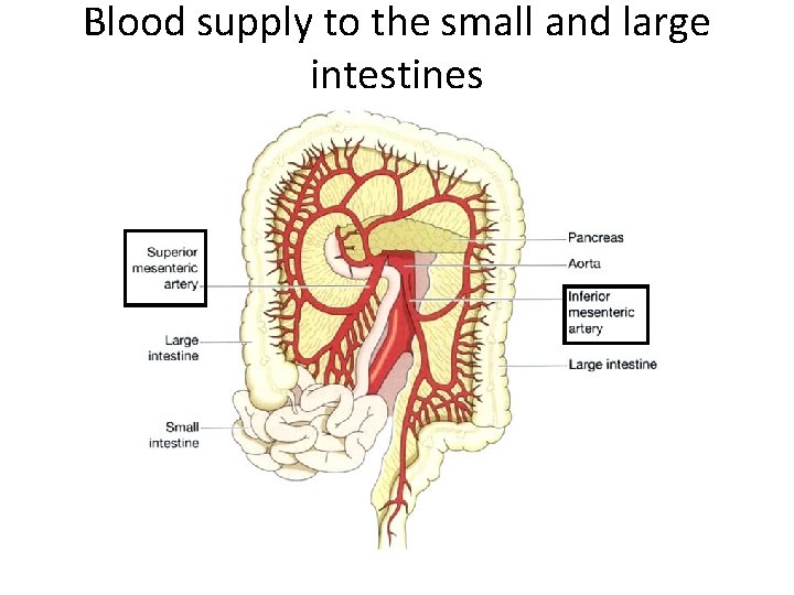
Blood supply to the small and large intestines
Venous drainage • In the thorax. Venous blood from the oesophagus passes in the oesophageal veins to the azygos and hemiazygos veins, hemiazygos joins the left brachiocephalic vein • In the abdomen and pelvis. The veins that drain blood from the lower part of the oesophagus, the stomach, pancreas, small intestine, large intestine and most of the rectum join to form the portal vein
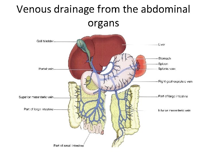
Venous drainage from the abdominal organs
TONGUE
• The tongue is a voluntary muscular structure which occupies the floor of the mouth. • It is attached by its base to the hyoid bone and by a fold of its mucous membrane covering, called the frenulum, to the floor of the mouth.
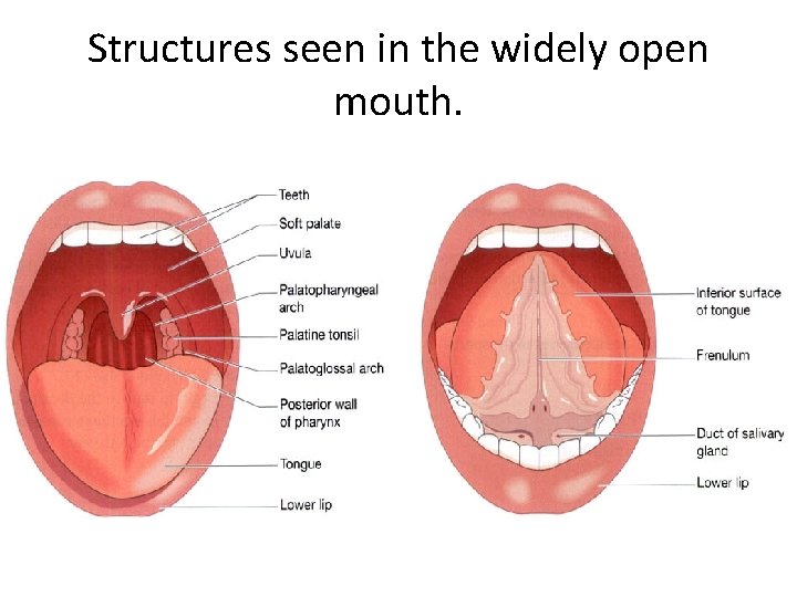
Structures seen in the widely open mouth.
Blood supply • The main arterial blood supply to the tongue is by the lingual branch of the external carotid artery. • Venous drainage is by the lingual vein which joins the internal jugular vein
Nerve supply • The hypoglossal nerves (12 th cranial nerves) which supply the voluntary muscle tissue • The lingual branch of the mandibular nerves which are the nerves of somatic (ordinary) sensation, i. e. pain, temperature and touch • The facial and glossophanryngeal nerves (7 th and 9 th cranial nerves) which are the nerves of the special sensation of taste.
Functions of the tongue The tongue plays an important part in- • Mastication (chewing) • Deglutition (swallowing) • Speech • Taste

TEETH
• The teeth are embedded in the alveoli (sockets) of the mandible and the maxilla. • Functions of the teeth – The incisor and canine teeth are the cutting teeth and are used for biting off pieces of food, – The premolar and molar teeth, with broad, flat surfaces, are used for grinding • Two dentitions, – the temporary or deciduous teeth (20) – the permanent teeth (32)
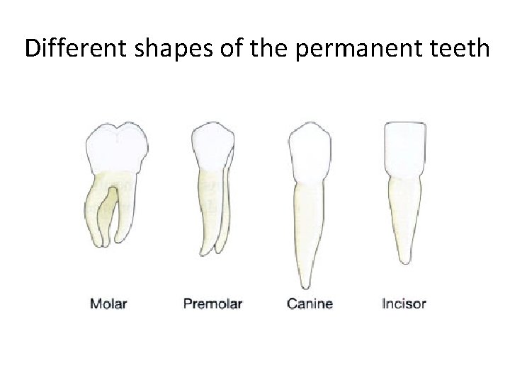
Different shapes of the permanent teeth
Structure of a tooth The crown — the part which protrudes from the gum The root — the part embedded in the bone The neck — the slightly narrowed region where the crown merges with the root.
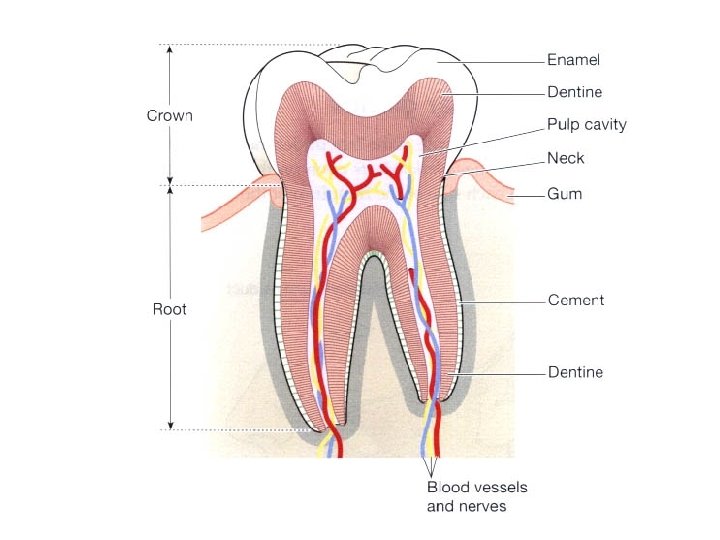

Blood supply to Teeth • The arterial blood supply to the teeth is by branches of the maxillary arteries • The venous drainage is by a number of veins which empty into the internal jugular veins.
SALIVARY GLANDS
• Parotid glands • Submandibular glands • Sublingual glands
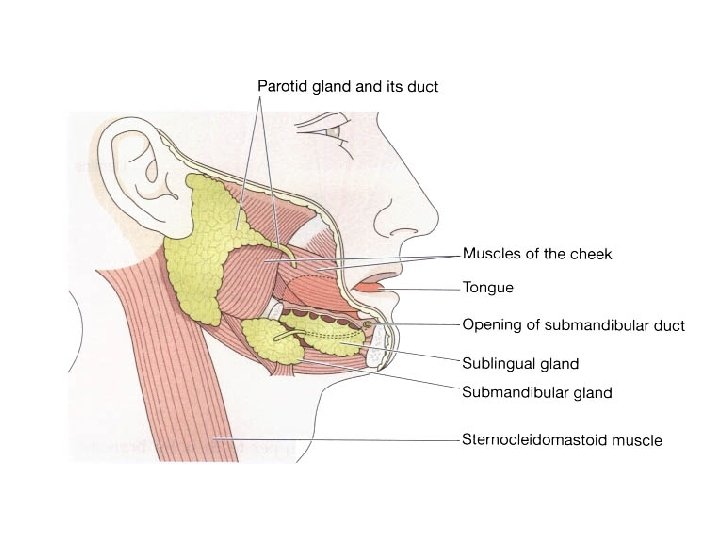
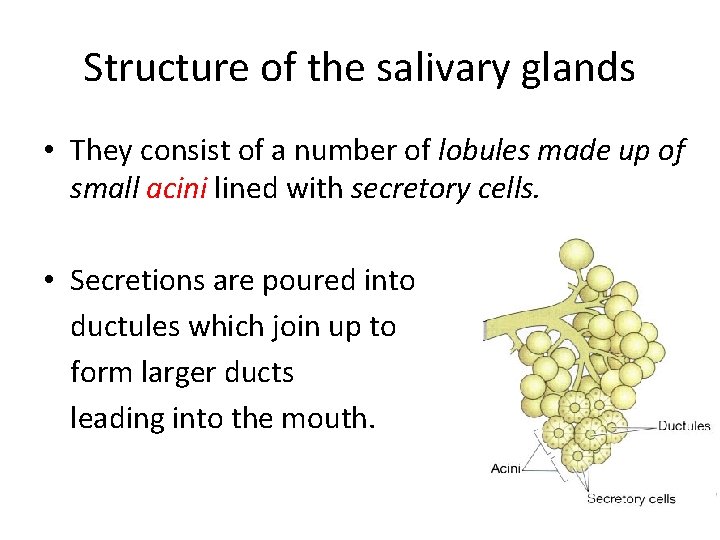
Structure of the salivary glands • They consist of a number of lobules made up of small acini lined with secretory cells. • Secretions are poured into ductules which join up to form larger ducts leading into the mouth.
Composition of saliva About 1. 5 litres of saliva is produced daily and it consists of • water • mineral salts • enzyme: salivary amylase • mucus • lysozyme • immunoglobulins • blood-clotting factors.
Secretion of saliva • Parasympathetic stimulation causes vasodilatation and profuse secretion of watery saliva with a relatively low content of enzymes. • Sympathetic stimulation causes vasoconstriction and secretion of small amounts of saliva rich in organic material.
Functions of saliva • Chemical digestion of polysaccharides • Cleansing and lubricating-to prevent damage to the mucous membrane by rough or abrasive foodstuffs • Non-specific defence- Lysozyme, immunoglobulins and clotting factors combat invading microbes. • Taste- The taste buds are stimulated only by chemical substances in solution.
PHARYNX
• nasopharynx, • oropharynx • laryngopharynx
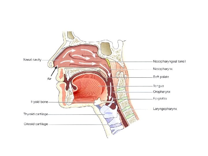
• lining membrane (mucosa) is stratified squamous • middle layer consists of fibrous tissue • outer layer consists of a number of involuntary constrictor muscles
OESOPHAGUS
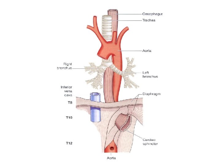
Oesophgus • The oesophagus is about 25 cm long and about 2 cm in diameter and lies in the median plane in the thorax in front of the vertebral column behind the trachea and the heart. • It is continuous with the pharynx above and just below the diaphragm it joins the stomach • The upper cricopharyngeal sphincter prevents air passing into the oesophagus during inspiration and the aspiration of oesophageal contents. • The cardiac or lower oesophageal sphincter prevents the reflux of acid gastric contents into the oesophagus.
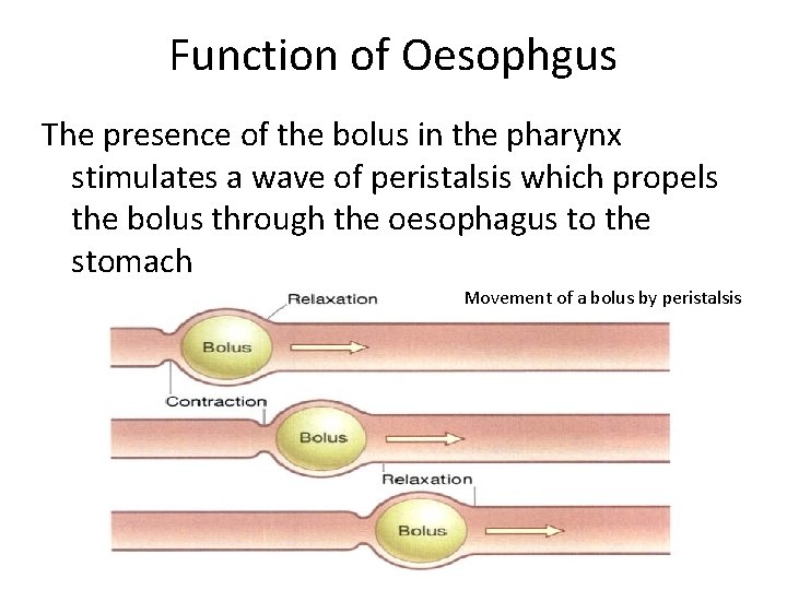
Function of Oesophgus The presence of the bolus in the pharynx stimulates a wave of peristalsis which propels the bolus through the oesophagus to the stomach Movement of a bolus by peristalsis

Thank You
- Slides: 44