Digestive System Overview mouth pharynx esophagus stomach small


- Slides: 45
Digestive System: Overview • – mouth, pharynx, esophagus, stomach, small intestine, and large intestine • – teeth, tongue, gallbladder, salivary glands, liver, and pancreas
Digestive Process • The GI tract is a ___________________ line – Nutrients become more available to the body in each step • There are six essential activities: – Ingestion – – mechanical digestion – – – defecation
G. I. Tract Activities • Ingestion – • Propulsion – swallowing and peristalsis – Peristalsis – ___________ of muscles in the organ walls • Mechanical digestion –
Gastrointestinal Tract Activities • Chemical digestion – catabolic _ • – movement of nutrients _ • Defecation – elimination of _
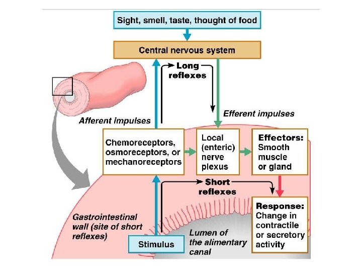
GI Tract • __________________ for the digestive process • Regulation of digestion involves: – Mechanical and chemical stimuli – _________________, osmolarity, and presence of substrate in the lumen – Extrinsic control by _ – Intrinsic control by _
Receptors of the GI Tract • Mechano- and chemoreceptors respond to: – Stretch, osmolarity, and p. H – Presence of substrate, and end products of digestion • They initiate reflexes that: – –
Nervous Control of the GI Tract • Intrinsic controls – ___________________ initiate short reflexes – Short reflexes are mediated by local enteric plexuses (gut brain) • Extrinsic controls – Long reflexes arising within or outside the GI tract – ______________ and extrinsic _
Peritoneum and Peritoneal Cavity • Peritoneum – ___________________ of the abdominal cavity – • covers external surface of most _ – • lines the _ • Peritoneal cavity – ________________ digestive organs – Allows them to slide across one another
Peritoneum and Peritoneal Cavity • Mesentery : – supplies _______________ to the viscera – Holds digestive organs in place and _
Histology of the Alimentary Canal • From esophagus to the anal canal the walls of the GI tract have the _ – From the lumen outward they are the _________________________, muscularis externa, and ______________ • Each tunic has a predominant tissue type and a specific digestive function
Figure 23. 6
Mucosa • Moist epithelial layer that _______________ of the alimentary canal • Three major functions: – – – ________________ against infectious disease • Consists of three layers: a lining epithelium, lamina propria, and muscularis mucosae
Mucosa: Epithelial Lining • ________________ and mucus -secreting goblet cells • Mucus secretions: – ____________________ from digesting themselves – Ease food along the tract • Stomach and small intestine mucosa contain: – – _________________ -secreting cells (making them endocrine and digestive organs)
Mucosa: Lamina Propria and Muscularis Mucosae • – Nourishes the epithelium and absorbs nutrients – Contains lymph nodes _______________ important in defense against bacteria • Muscularis mucosae – _________________ that produce local movements of mucosa
Mucosa: Other Sublayers • – dense connective tissue containing elastic fibers, blood and lymphatic vessels, lymph nodes, and nerves • Muscularis externa – responsible for _ • Serosa – the _ – Replaced by the fibrous adventitia in the esophagus – Retroperitoneal organs have both an adventitia and serosa
Enteric Nervous System • two major intrinsic nerve plexuses: • – regulates glands and smooth muscle in the mucosa • _______________ – Major nerve supply that controls GI tract mobility
Enteric Nervous System • Segmentation and peristalsis are largely _______________ involving local reflex arcs • Linked to the CNS via long _______________ reflex arc
Mouth • Oral or ___________ cavity: – Is bounded by lips, cheeks, palate, and tongue – oral orifice • – continuous with the oropharynx posteriorly
Mouth • To withstand _ – The mouth is lined with _ – The gums, hard palate, and dorsum of the tongue are _
Lips and Cheeks • Have a core of skeletal muscles – Lips: – Cheeks: • – bounded by the lips and cheeks externally, and teeth and gums internally
Lips and Cheeks • Oral cavity proper – area that lies _ • – median fold that joins the internal aspect of each lip to the gum
Palate • Hard palate – palatine bones and palatine processes of the maxillae – – Slightly _____________ on either side of the raphe (midline ridge)
Palate • Soft palate – mobile fold _ – Closes off the nasopharynx during swallowing –
Tongue • Occupies the _ • fills the oral cavity when mouth is closed • Functions include: – __________________ food during chewing – ___________________ and forming the bolus – Initiation of _
Tongue • _______________ muscles change the _ • ________________ muscles alter the tongue’s _ • __________________ secures the tongue to the floor of the mouth
Tongue • three types of papillae – • give the tongue roughness and provide friction – • scattered widely over the tongue and give it a reddish hue – • V-shaped row in back of tongue
Tongue • – groove that separates the tongue into two areas: – Anterior 2/3 residing in the _ – Posterior third residing in the _
Tongue Figure 23. 8
Salivary Glands • Produce and secrete saliva that: – – Moistens and dissolves food chemicals – Aids in bolus formation – Contains _
Salivary Glands • Three pairs of __________ glands – – – • Intrinsic salivary glands (____________ glands) – scattered throughout the oral mucosa
• Parotid Salivary Glands – lies ________________ between the masseter muscle and skin – _________________ opens into the vestibule next to second upper molar • Submandibular – lies along the medial aspect of the mandibular body – ducts open at the _
Salivary Glands • Sublingual – lies anterior to the submandibular gland _ – It opens via 10 -12 ducts into the _
Salivary Glands Figure 23. 9 a
Saliva: Source and Composition • Secreted from ____________ cells of salivary glands • contains – ________________ – Na+, K+, Cl–, PO 42–, HCO 3– – Digestive enzyme – – Proteins – mucin, lysozyme, defensins, and Ig. A – __________________ – urea and uric acid
Control of Salivation • Intrinsic glands keep the mouth _ • Extrinsic salivary glands secrete serous, enzyme-rich saliva in response to: – Ingested food which stimulates chemoreceptors and pressoreceptors – The thought of food • Strong ________________ inhibits salivation and results in dry mouth
Teeth • Primary – _________________ that erupt at intervals between 6 and 24 months • Permanent – enlarge and develop causing the root of deciduous teeth to be resorbed – fall out between the ages of _ – All but the third molars have erupted by the end of adolescence – Usually _
Classification of Teeth • Based on shape and function • – chisel-shaped teeth for cutting or nipping • Canines – fanglike teeth that _ • Premolars (bicuspids) and molars – have ________________; best suited for grinding or crushing
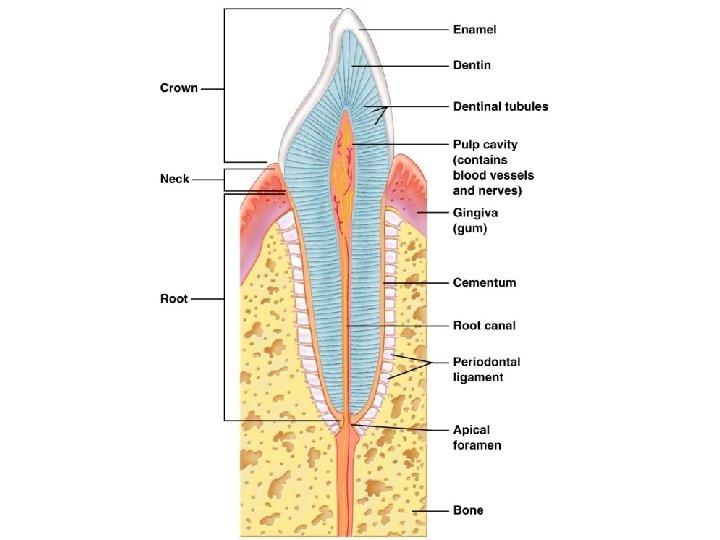
Tooth Structure • Two main regions – • Crown – _______________ above the gingiva • Enamel – acellular, brittle material composed of calcium salts and hydroxyapatite crystals; – – • Root – portion of the tooth _
Tooth Structure • Neck – constriction _ • Cementum – – – Attaches it to the periodontal ligament
Tooth Structure • Periodontal ligament – ________________ in the alveolus of the jaw – Forms the _ • Gingival sulcus – depression where the gingiva borders the tooth
Tooth Structure • Dentin – bonelike material ________________ that forms the bulk of the tooth • – cavity surrounded by dentin that contains pulp • Pulp – connective tissue, _
Tooth Structure • Root canal – portion of the pulp cavity that extends into the root • Odontoblasts – secrete and maintain dentin throughout life
Tooth and Gum Disease • Dental _ – gradual ______________ of enamel and dentin by bacterial action – Dental plaque adheres to teeth • a film of _ – Acid from the bacteria dissolves calcium salts – Without calcium salts, organic matter is digested by _ – Daily flossing and brushing help prevent caries by removing forming plaque