Digestive System J Hinson Human Anatomy and Physiology


- Slides: 36

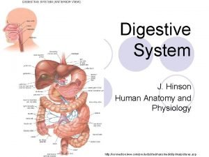
Digestive System J. Hinson Human Anatomy and Physiology http: //connection. lww. com/products/stedmansmedict/primalpictures. asp

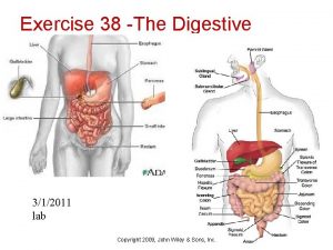
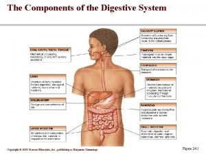
I. Introduction A. Digestion B. Includes 1. Alimentary Canal (aliment/o = nourish) : mouth, pharynx, esophagus, stomach, small intestine, large intestine 2. Accessory Organs: salivary glands, liver, gallbladder, pancreas http: //wps. prenhall. com/wps/media/objects/488/500694/CDA 40_1. jpg

II. The Alimentary Canal A. Muscular tube ~ 9 m B. Tissues 1. Mucosa a. Glands secrete b. Functions: protection, absorption and secretion 2. Submucosa a. Contains blood vessels, lymphatic vessels, and nerves b. Nourishes

II. The Alimentary Canal B. Tissues 3. Muscular a. circular smooth muscle controls diameter; longitudinal controls length 4. Serosa a. visceral peritoneum b. contains serous fluid C. Movements: mixing or propelling - peristalsis http: //medsci. indiana. edu/c 602 web/602/c 602 web/scans/19 f. jpg

III. Oral Cavity A. Mouth (or/o): mechanical digestion B. Cheeks (bucc/o): muscular for expression and chewing C. Lips (cheil/o): movement and sensation; color from blood vessels

III. Oral Cavity D. Tongue (lingu/o; gloss/o): mixes food w/ saliva 1. Connects via frenulum 2. Papillae contain taste buds (bitter, sweet, sour, salty) http: //content. answers. com/main/content/img/gale. Neurology/gend_01 _img 0040. jpg http: //health. yahoo. com/media/healthwise/n 5551320. jpg

III. Oral Cavity E. Palate (palat/o) 1. anterior – hard posterior – soft projection is the uvula (uvul/o) 2. palatine tonsils – lateral 3. pharyngeal (pharyg/o) tonsils - posterior http: //biology. clc. uc. edu/fankhauser/Labs/ Microbiology/Strep_Detection/oropharynx _P 2253089_lbd. JPG

III. Oral Cavity F. Teeth (dent/i; dont/o; odont/o) 1. 20 primary (deciduous); 32 secondary (permanent) 2. increase surface area 3. Types: a. incisor (front) b. cuspids (canines) c. bicuspids d. molars 4. Consists of: crown, root, enamel, dentin, pulp 5. peridontal ligament attaches http: //www. dentalgentlecare. com/_derived/tooth_anatomy. htm_txt_Tooth 2. gif

Teeth

IV. Pharynx (pharyng/o) A. Cavity behind mouth (aka – throat) 1. Nasophar ynx 2. Oropharyn x 3. Laryngopharynx http: //www. levelfive. com/ZINA/IMAGES/Anatomical/pharynx. jpg

V. Swallowing A. Three Stages http: //greenfield. fortunecity. com/rattler/46/upali 4. htm 1. Food is mixed with saliva; forms a bolus; forced into the phayrnx – voluntarily initiated! 2. Involuntary reflex moves food to esophagus; epiglottis (skin flap) closes 3. Food to stomach via peristalsis.

VI. Esophagus (esophag/o) A. Collapsible tube ~ 25 cm 1. Penetrates diaphragm B. Circular smooth muscle http: //home. hawaii. rr. com/dochazenfield/images/esophagus%20 B. jpg

VII. Stomach (gastr/o) A. General Info 1. J-shaped, pouch-like, organ in upper L abdomen 2. Holds ~ 1 L 3. Folded rugae 4. Mixes bolus w/ HCl and gastric juice to become chyme http: //www. sciencebob. com/lab/bodyzone/stomach. gif

VII. Stomach B. Regions 1. Cardiac 2. Fundus 3. Body 4. Antrum http: //training. seer. cancer. gov/module_anatomy/images/illu_stomach. jpg 5. Pyloric a. Pyloric sphincter prevents regurgitation

VII. Stomach C. Gastric Secretions 1. Glands secrete juice a. Goblet – mucus b. Chief – enzymes c. Parietal – HCl Acid 2. Gastric Juice 3. Regulated by gastrin 4. food inhibits secretion D. Begin protein digestion; Some absorption: H 20, glucose, salts, alcohol, lipid-soluble drugs http: //intmed. muhealth. org/gast/images/stomach. gif

Component Source Function Chief cells Inactive form of pepsin Formed from pepsinogen when mixed with HCl Protein-splitting enzymes Parietal cells Changes pepsinogen to pepsin; gives acidic environment for pepsin Goblet cells; mucous glands Provides viscous, alkaline protective layer on stomach wall Parietal cells Aids in B 12 absorption Pepsinogen Pepsin Hydrochloric Acid Mucus Intrinsic Factor

VIII. Small Intestine (enter/o) A. Extends from pyloric sphincter to LI B. Functions: 1. Receive secretions 2. Completes digestion in chyme 3. Absorbs nutrients 4. Transports wastes to LI http: //www. exn. ca/news/images/1999/02/01/19990201 -smallintestine. jpg

VIII. Small Intestine C. 3 portions 1. Duodenu m 2. Jejunum 3. Ileum a. Mesentery suspends

VII. Small Intestine D. Secretions 1. Include mucus and enzymes 2. Split macromolecules a. Peptidases: proteins to amino acids a. Sucrase/maltase/lactase: disaccharides to monosaccharides b. Lipase: lipids to fatty acids and glycerol 3. Enhanced by gastric juices, chyme, and distension http: //bio. bd. psu. edu/cat/Circula tory_System/vesels%20 of%20 s mall%20 intestine. jpg

VII. Small Intestine E. Absorption 1. Villi increase SA 2. Monomers absorbed via diffusion or active transport 3. Also water and electrolytes 4. Malabsorption a. Bile problems; Celiac disease – gluten reaction b. Sxs: weakness, etc. http: //publications. royalcanin. com/images/2/2343/p 7_image. gif

IX. Large Intestine (col/o) A. ~ 5 feet a. Cecum a. Appendix b. Colon (i) Ascending (ii)Transverse (iii)Descending (iv)Sigmoid

IX. Large Intestine A. 1. c. Rectum: ~ 5 cm d. Anal canal: ~ 2 -3 cm (i) anus (a) internal (b) external (ii) hemorrhoids http: //www. nutrigenesis. com/page_images/hemorrhoids. jpg

IX. Large Intestine C. Functions 1. no digestive function 2. mucus secretions 3. absorbs water and electrolytes 4. forms, stores, and expels feces

http: //mi 2 mm 00. eng. shizuoka. ac. jp/ matsumaru 2/research/21 cvalsalva. jpg IX. Large Intestine D. Movement 1. Mixing and peristalsis 2. 2 -3 movements daily 3. Defecation (initiated by distension) a. valsalva maneuver E. Feces 1. Composition: 75% water, undigested material, mucus, & bacteria 2. bile salts

Ostomy http: //www. youtube. com/w atch? v=k. Uv. HDdj. ZJkc Colonoscopy http: //www. youtube. com/w atch? v=rl 4 s 1 D 4 MGH 8 http: //www. dhmc. org/dhmc-internet-upload/file_collection/colonscopy. jpg

Digestive System Accessory Organs

X. Salivary (sial/o) Glands A. Function of fluids: 3 1. 2. 3. 4. Moisten food particles Bind Dissolve carbs Dissolve chemicals for taste 5. Cleansing B. Cells: 3 1 2 1. Serous – secrete salivary amylase to break down carbs 2. Mucous – mucus to bind and lubricate http: //www. salivary-glands-disease. com/assets/images/img. SGDcauses. jpg

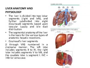
XI. Liver (Hepat/o) http: //www. livercancer. com/images/anterior. liver. gif A. Below diaphragm B. Functions: 1. Metabolism of macromolecules 2. Storage – vitamins A, D, B 12 3. Filtering blood 4. Destruction of toxins (alcohol) 5. Secretion of bile

IX. Liver C. Structure 1. Reddish-brown color 2. Lobes a. Separated into hepatic lobules 3. Hepatic ducts carry bile a. Common bile duct (cholagi/o) formed by merging http: //www. liverdoctor. com/images/detox_pathways. jpg

IX. Liver http: //www. pathguy. com/l ectures/fatty_liver. jpg D. Bile (chol/e, o) 1. Secreted by hepatic cells 2. Contains bile salts, pigments, etc. a. bile salts digest http: //www. pathguy. com/lectures/liver. htm

XII. Gallbladder (cholecyst/o) http: //www. lbah. com/images/liver/gallbladder. jpg A. Attached to liver by cystic duct B. Functions: 1. Stores bile 2. Concentrates bile 3. Release bile 4. Lipid

X. Gallbladder C. Common Bile Duct = cystic + hepatic ducts D. Gallstones = choleliths! http: //www. emedicine. com/med/images/1226 gs 1. jpg http: //images. medicinenet. com/images/illustrations/stomach. jpg

XIII. Pancreas (pancreat/o) A. Both endocrine and exocrine function B. Structure 1. Extends horizontally 2. Pancreatic duct and Bile duct from liver and gallbladder empties into duodenum http: //training. seer. cancer. gov/module_anatomy/images/illu_pancrease. jpg

XIII. Pancreas (pancreat/o) C. Pancreatic juice 1. Contains enzymes that can split carbs, proteins, fats, and nucleic acids Regulation 2. 1. 2. Secretin stimulates release of pancreatic juice high in bicarb. Cholecystokinin stimulates release of pancreatic juice high in digestive enzyme. http: //training. seer. cancer. gov/module_anatomy/images/illu_pancrease. jpg

VIII. Pancreas (pancreat/o) D. Islets of Langerhans (endocrine cells) secrete: 1. Insulin: stimulates body cells to take in glucose from the blood stream to decrease blood sugar levels a. Damage or underproduction: hyperglycemia and/or Diabetes Mellitus (DM) b. Overproduction: hypoglycemia 2. Glucagon: stimulates liver to release glucose to increase blood sugar levels

XIV. Clinical Terms A. B. C. D. E. F. G. H. Aphagia Cholecystitis Cirrhosis Diverticulitis Dyspepsia Gingivitis Hepatitis Diabetes http: //www. dental-health-index. com/images/Ging 1. jpg
 Holly hinson md
 Endomysium
 Waistline
 Holes essential of human anatomy and physiology
 Distal and proximal
 Chapter 2 human reproductive anatomy and physiology
 Human anatomy and physiology 10th edition
 Anatomy and physiology edition 9
 Anatomy and physiology unit 7 cardiovascular system
 Types of respiration in human
 Appendicular skeleton pectoral girdle
 Digestive system circulatory system and respiratory system
 Serves as a mixing chamber and holding reservoir
 Exercise 38
 Functional anatomy of the digestive system
 Ileocecal junction cat
 Mucus c
 Intestine
 Emulsification
 Digestive physiology ppt
 Upper and lower airway
 Tattoo anatomy and physiology
 International anatomy olympiad
 Structure anatomy and physiology in agriculture
 Anatomy and physiology of bone
 Pud triple therapy
 Segmental anatomy of the liver
 Difference between anatomy and physiology
 Epigastric vs hypogastric
 Rbc anatomy and physiology
 The central sulcus divides which two lobes? (figure 14-13)
 Http://anatomy and physiology
 Physiology of appendix
 Aohs foundations of anatomy and physiology 1
 Aohs foundations of anatomy and physiology 2
 Anatomy and physiology of swine
 Anatomy and physiology chapter 8 special senses

