Digestive System Gastrointestinal tract or Alimentary canal ingest


– lower jaw (submandibular), under tongue (sublingual) and near")


, muscular tube wrapped in smooth muscle- first part")


 • Jejunum and ileum: Contain many tiny, fingerlike projections")

 and exocrine (ducts) functions.")
- made-up of hepatic cells and has 2")


- Slides: 29
Digestive System Gastrointestinal tract or Alimentary canal ingest, breakdown mechanically and chemically, absorb, and eliminate Structures, functions, and nutrition
Gastrointestinal Tract Objectives: students will be able to. . • Trace food from the mouth to the anus • Determine the make-up and function of each structure. • Determine what happens to the food in each structure. • Explain what kind of organic compound is broken down by the enzymes/secretions produced by the structure. • Discuss diseases http: //highered. mheducation. com/sites/0072495855/student_view 0/chapter 26/animation__org ans_of_digestion. html
Mouth • Oral cavity consisting of teeth, tongue, hard and soft palate and salivary glands • FX: – Ingestion – Both chemical and mechanical breakdown of food • Mechanical action allows molecules to become exposed to digestive enzymes • Chemicals/enzymes increase the rate of breakdown into organic molecules – Prepares food for the GI tract • Forms ball of food: bolus • Rough projections on tongue papillae – Aids in handling and contains taste buds for sensory perception • Teeth: mechanical breakdown (grinding)
Mouth
Digestive enzymes/secretions • Salivary glands (3)– lower jaw (submandibular), under tongue (sublingual) and near (parotid) • FX: produces saliva containing…. . • Amylase: breaks down starches • saliva: made of water and mucus to moisten and dissolve – Contains antibacterial lysozyme » Bursitis_: cancer of salivary glands
Pharynx passageway between the mouth and trachea/esophagus and is made up of 3 parts: • Nasopharynx: air • Oropharynx: air and food • laryngopharynx: food – FX: food and air passageway/prevents food from entering the trachea and air from entering GI tract • Respiratory and digestive tracts meet • Tongue will push down on the epiglottis so that the opening into respiratory tract is closed when swallowing
Pharynx
Esophagus • Long (25 cm- 11”), muscular tube wrapped in smooth muscle- first part of the “gut” • FX: FX transport bolus from pharynx to stomach • Peristalsis: wave like motion to move bolus— (5 -10 seconds) • Sphincter: ring of muscle that allows bolus to pass into stomach • Disorders: – Esophageal etresia: missing part of the esophagus – Esophagitis: regurgitates stomach contents – Heartburn: reflux of stomach contents
Peristalsis
Gut Lining • 4 layers of the gut – Mucosa • Mucus membrane and secretes digesting enzymes – Submucosa • Loose connective tissue with blood and lymph vessels – Muscularis • Smooth musclecontractions – Serosa • moisture
Stomach • Found on left side between the esophagus and small intestines – Sac-like organ that’s 10 inches long – J shaped, thick walled – Folds: rugae: increase surface area • Capacity is around 1 gallon • FX: mechanical (peristaltic waves) and chemical breakdown – Alcohol and water absorption • 2 -6 hrs. bolus becomes chyme or thick, soupy liquid • Chyme leaves the stomach through pyloric valve/sphincter
Enzymes and Secretions Gastric juices • Mucus – lubrication/protect stomach lining • HCL (parietal cells): increase acidity of stomach to help pepsin work better (p. H 2) and breaks protein bonds (peptide bonds)/kills bacteria • Pepsinogen to pepsin (chief cells): protein • Gastrin: hormone that regulates muscular contractions and secretions • Intrinsic factor: binds to B 12 to prevent being broken down
Stomach • Parts of the stomach – cardiac – Fundic – body – Pylorus Disorders: • Indigestion and heartburn • Ulcers • Gastritis: inflammation of stomach
Small Intestines
Small Intestines • 18 -20 feet long • Found in between the stomach and large intestines and made-up of finger-like projections/folds called villi and microvilli • FX. : MOST chemical breakdown and absorption of nutrients • Made up of 3 parts: • Duodenum: Duodenum ( 10 “) chemical breakdownpancreatic and liver enzymes/secretions • Jejunum: Jejunum (3 ‘) villi and microvilli- MOST absorption of nutrients into bloodstream • Ileum: ( 6 -7’) some absorption – indigestible material: water, cellulose, and other Segmentation and peristalsis • localized movement increases absorption and mixing • moves indigestible material
http: //time. com/4621074/mesentery-organ-human-body/ New body organ found: mesentery
Enzymes and secretions • Peptidase: Peptidase proteins • Maltase, sucrase, lactase: lactase carbohydrates • Secretin: Secretin stimulates pancreas Small intestines receives digestive enzymes from both the liver, via the gall bladder, and the pancreas
Peristalsis occurs here- (1 -6) • Jejunum and ileum: Contain many tiny, fingerlike projections called villi or folds in the intestinal lining to help with absorption- these folds are then divided into microvillus -Contains many blood vessels for absorption Small Intestines
Accessory organs
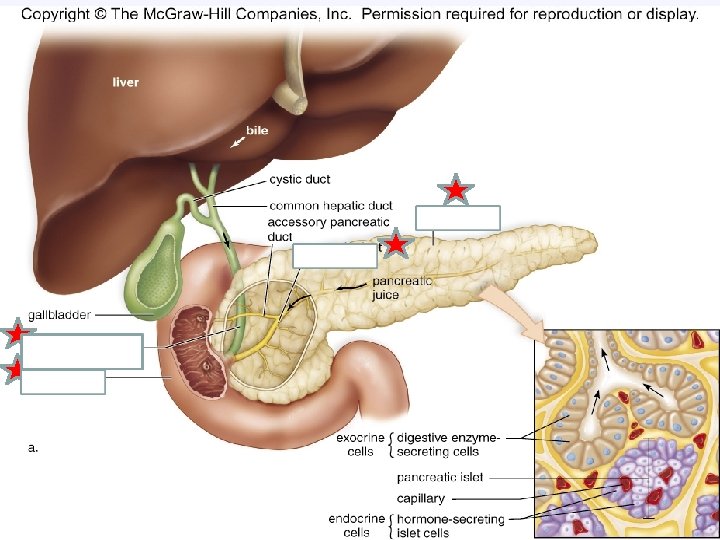
Pancreas • Structure made-up of pancreatic islets. Has endocrine (ductless) and exocrine (ducts) functions. • FX: produces digestive enzymes called pancreatic juices that go into the duodenum • Pancreatic amylase: carbohydrates • Pancreatic lipase: fats • Peptidases: trypsin and chymotrypsin and carboxypeptidase's: proteins • Sodium bicarbonate: neutralizes the intestines (p. H of 7) http: //q 13 fox. com/2016/02/03/pos » Pancreatic cancer » diabetes sible-diabetes-cure-being-testedon-small-number-of-patients/ http: //www. mayoclinic. org/diseases-conditions/diabetes/multimedia/blood-sugar/vid-20084642
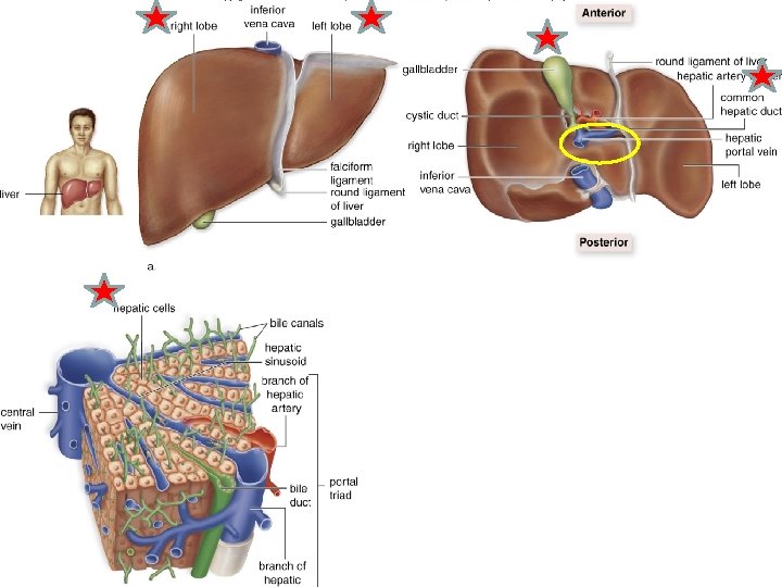
Liver • Large brown organ(size of football)- made-up of hepatic cells and has 2 main lobes. • FX: produces yellow, bile salts that aids lipase in emulsifying fats. – Bile also promotes absorption of fatty acids and fat soluble vitamins – also responsible for converting glycogen to glucose – detoxification of substances – Produces urea
Disorders of the liver • Hepatitis A, B, and C: caused by a bacterial pathogen (A in contaminated food/water) (B and C- blood)– effect: inflammation of liver/causes jaundice • Cirrhosis of the liver: scarring due to alcohol, drugs, or infections • Jaundice: Jaundice yellowing of skin, eyes, and urine due to increase bile pigments in the blood
Gallbladder • Pear shaped, muscular sac • Located in the depression on the inferior surface of the liver • FX: stores bile from the liver – Bile emulsifies fats Disorders: Gall stones (crystalized cholesterol) can form and block the bile duct
Large Intestine Ascending colon cecum
Large Intestine • Also known as the colon • NO CHECMIAL DIGESTION!!!!!!!!! • 3 -6 feet in length: larger diameter than SI/ 12 -24 hours • Fx: remove water (75%), minerals, salts from the indigestible material and turn it into solid fecal matter-feces – Contains helpful bacteria: breakdown cellulose for energy and also make vitamin K and B – Vermiform appendix is a small projection from cecum: fight infection – Rectum: stores feces
Large Intestines • Peristalsis occurs • Disorders: • Diarrhea • Constipation • Ulcerative colitis: ulcers in lining • Colon cancer: polyps • Crohn’s Disease • Diverticulosis
Anus • The end of the GI tract – Sphincter muscle • FX: expels waste • Disorders: • Hemorrhoids