Digestive System By Prof Nabil A Hasan Organs

Digestive System By Prof. Nabil A Hasan

Organs of Digestive system

Organs of Digestive system The digestive system includes the alimentary canal and some accessory organs Alimentary canal is a long tube starting from the mouth down to the anus. Its parts are: Mouth ( Vestibule & Oral cavity) Boundaries: Anteriorly: Lips Posteriorly: contineous with the pharynx Laterally: muscles of the check Inferiorly: Mylohoid muscle (floor of mouth) Superiorly: hard and soft palate

Palate The palate is formed of two parts: 1. Hard palate 2. Soft palate the posterior end of the soft palate is called Uvula From the soft palate, 4 arches arise (2 on each side); palatoglossal and palatopharyngeal Between these arches, a collection of lymphoid tissue exists (palatine tonsils)

Tongue It is a muscular organ attached to the floor of the mouth by the frenulum Its superior surface carries numerous papillae called taste buds Blood supply Arterial: lingual artery Venous: lingual vein
teeth: 20 in number, 10 in each jaw, begins")
Teeth Types of teeth Temporary (deciduous)teeth: 20 in number, 10 in each jaw, begins to erupt at 6 th month and completely present by the end of 24 th month. Permanent teeth: 32 in number, 16 in each jaw, begins to replace the decidua’s teeth in the 6 th year and usually completed by the 24 th year old Structure of the teeth: 1. Crown: the part that protrudes from the gum 2. Root: the part embedded in the bone 3. Neck: between the crown and the root In the centre of the teeth there is a pulp cavity containing blood vessels, nerves and lymph vessels The pulp cavity is surrounded by dentine which is surrounded by enamel from outside

Teeth

Salivary glands Three pairs of glands: 1. Two parotid: lie below the external acoustic meatus and open in the vestibule of the mouth opposite the upper 2 nd molar tooth 2. Two submandibular: lie on the inner surface of the lower jaw, open in the floor of the mouth on both sides of the frenulum 3. Two sublingual: lie in front of submandibular salivary glands and open by numerous glands into the floor of the mouth PAROTID GLAND SUBLINGUAL GLAND SUBMANDIBULAR GLAND

Pharynx * It is a common fibromuscular tube in respiratory and digestive systems *it lies just below the base of the head, on front of the vertebral column down to 6 th cervical vertebra & behind nose, mouth & larynx *It is divided into 3 parts: Nasopharynx, Oropharynx & Laryngopharynx * It continues with esophagus below 6 th cervical vertebra

Oesophagus • It is 25 cm long and 2 cm in diameter • It is continuous above with the pharynx and below with the stomach It descends in front of the vertebral column at the level of 6 th cervical vertebra • and behind the trachea and heart • It enters the abdomen by passing through the diaphragm at the level of the 10 th thoracic vertebra • Its lower sphincter (cardiac sphincter) prevents reflux of gastric acid into the oesophagus

Stomach Greater curvature Cardiac sphincter Pyloric antrum Fundus Body • It is the most dilated portion of the alimentary tract • It is situated in the upper part of the abdominal cavity (mainly in the left) under the diaphragm • Continuous with the oesophagus at the cardiac sphincter and with the duodenum at the pyloric sphincter • Has anterior and posterior surfaces, lesser and greater curvatures, • It is composed of fundus, body and pyloric parts Lesser curvature

Small & large intestine Small intestine Large intestine It is continuous with the stomach at the pyloric orifice and with the large intestine at the ileocecal valve It begins at the caecum and terminates at the rectum and anal canal It has the following parts: It is 6 meters long, described in three parts: 1. Duodenum 2. Jejunum 3. Ilium 1. 2. 3. 4. 5. 6. 7. 8. 9. Caecum & appendix Ascending colon Right colic flexure Transverse colon Left colic flexure Descending colon Sigmoid colon Rectum Anal canal

Pancreas Spleen It weighs 60 gm, and 12 -15 cm long It lies in the curve of the duodenum, behind the body of the stomach, reaches the spleen Pancreas Duodenum It is a mixed gland (exocrine) secretes digestive enzymes and (endocrine) called islets of Langerhan secretes Insulin & glucagon hormones Nerve supply: Parasympathetic: increases secretions Sympathetic: decreases secretions
 necessary for")
Liver Function • Detoxification • Protein synthesis • Production of biochemicals (Bile) necessary for digestion via emulsification of lipids • Glycogen storage Liver from the front Right lobe Left lobe the Greek word for liver, hēpar Anatomy It is a wedge shaped reddish brown organ. A human liver normally weighs 1. 44 -1. 66 kg. It is situated in the upper part of the abdominal cavity (mainly in the right) under the diaphragm. The apex of the wedge lies close to the apex of the heart. The base forms the right lateral surface

Liver • The liver has 4 lobes: large right lobe and smaller left lobe and on the posterior surface of the right lobe there are two, caudate and quadrate lobes Caudate lobe • porta hepatis (hilum of the liver) Lt. Lobe Rt. Lobe it is the part on the posterior surface where structures enter and leave. These structures Quadrate lobe are: Gall bladder 1 - Portal vein (enters) carrying venous blood from stomach, spleen, pancreas, and intestine Liver from the back 2 - Hepatic artery (enters) carrying arterial blood 3 - Rt. and lt. Hepatic ducts (leave) carrying bile from the liver to the gall bladder 4 - Lymph vessels (leave) 5 - sympathetic and para-sympathetic nerves (enter)

Biliary system

Composition of the Biliary System 1 - Gall Bladder: It lies on the inferior surface of liver & is composed of fundus, body & neck 2 - Biliary ducts: which are cystic duct, hepatic ducts( right, left & common) and common bile duct 11) carrying arterial blood

Renal & Reproductive systems By Prof. Nabil A Hasan

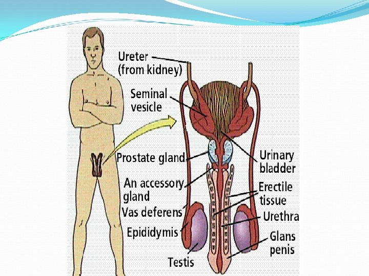
Renal system_Kidneys • It is the main excretory system in the body • It consists of the following structures: 2 kidneys, 2 ureters, 1 urinary bladder, 1 urethra Kidneys • They are bean shaped, lie on the posterior abdominal wall one on each side of the vertebral column just below the diaphragm • They are surrounded by a fibrous capsule

Structure of the Kidney The kidney substance is composed of about 1 million of functional units (Nephrons) A longitudinal section of the kidney shows: A- Fibrous capsule surrounding the kidney B- Cortex, under the capsule C- Medulla, innermost layer

Ureters & Urinary Bladder Ureters • It is about 25 -30 cm long and about 3 mm diameter • It is continuous with the renal pelvis in the abdomen. • It descends behind the peritoneum down to the pelvis to open into the urinary bladder Urinary bladder • It is a reservoir of urine, pear shaped but becomes oval when it fills with urine • It lies in the pelvic cavity behind the symphysis pubis • It opens into the urethra at its lowest point

Renal system_ Urethra urethra It is a canal that commences at the neck of the bladder down to the exterior Female urethra: • It is approximately 4 cm long • There are two urethral sphincters 1 - Internal urethral sphincter (involuntary) 2 - External urethral sphincter (under voluntary control) Male urethra: • It is approximately 19 -20 cm long • It consists of three parts; 1 - Prostatic urethra 2 - Membranous urethra 3 - Penile urethra • Like female urethra, the internal sphincter of male urethra is not under voluntary control but the external sphincter is under voluntary control

Male Renal & Reproductive System

Female Renal & Reproductive systems

Female Reproductive System The female reproductive organs are divided into external, internal and accessory organs External genitalia: 1 - Labia majora 2 - Labia minora 3 - Clitoris 4 - Vestibule 5 - Hymen 6 - Greater vestibular glands (Bartholin’s glands) Internal genitalia: 1 - Vagina 2 - Uterus 3 - Uterine (Fallopian tubes) 4 - Ovaries Accessory glands: (Breasts or Mammary glands)
 Vagina • It is a fibro-mascular tube that connects external")
Vagina & Uterus (1) Vagina • It is a fibro-mascular tube that connects external with internal genital organs • It lies between the urinary bladder anteriorly and rectum posteriorly • Its anterior wall is about 7. 5 cm long and its posterior wall is about 9 cm long • It is lined with stratified squamous epithelium (2) Uterus • Hollow mascular pear shaped organ, lies in the pelvic cavity behind the urinary bladder and in front of the rectum • It is continuous inferiorly with the vagina • It consists of three parts; fundus, body and cervix • Its wall is formed of three layers (Perimetrium, Myometrium, and Endometrium)

Reproductive system_Uterus FEMALE REPRODUCTIVE SYSTEM
 Uterine tubes • It is about 10 cm long")
Fallopian tubes & Ovaries (3) Uterine tubes • It is about 10 cm long • It extends from the sides of the uterus between the fundus and the body (4) Ovaries • It is the female sex glands, almond shaped, one on each side of the pelvic cavity • It is the site of production of ova and secretion of estrogen and progesterone hormones which are responsible for female sexual characters and functions and control pregnancy and regulate menses • It consists of an outer cortex (contain the ovarian follicles which produce ova) and an inner medulla (secretes sex hormones)

Female External Genital Organs UTERUS CERVIX VAGINA URINARY BLADDER SYMPHYSIS PUBIS RECTUM ANAL CANAL URETHRA

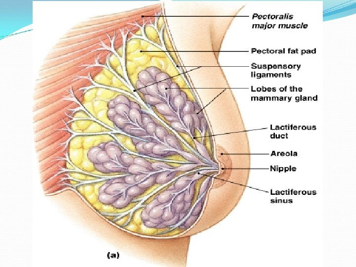
Breast or mammary gland • accessory glands of the female reproductive system • It lies in front of the chest at the level of 2 nd – 6 th costal cartilages • The Nipple is a small conical eminence at the center surrounded by a pigmented area called Areola

Male Reproductive system Male reproductive system • The male reproductive system consists of the following organs: 1. 2. 3. 4. 5. 6. 7. 8. Two testes (lie in a sac called scrotum) Two epididymis Two vas deference Two spermatic cords Two seminal vesicles Two ejaculatory ducts One prostate One penis Scrotum • It is a deeply pigmented skin pouch • It is divided into two compartments each of which contains one testis

Testis Testes • They are the reproductive glands of the male • They are suspended to the scrotum by the spermatic cord Vas deference • They are the site of production of sperms and secretion of male sex hormone (Aldosterone) responsible for male sexual characters and functions Spermatic cords One from each testis, suspends it to the scrotum Seminal vesicles - Fibro- mascular puches lying on the posterior aspect of the urinary bladder - They secrete viscous nutrient fluid to sperms Testis

Ejaculatory ducts & Prostate Ejaculatory ducts - Each is formed by the union of a duct from the seminal vesicle and vas deference - It passes through the prostate gland joins prostatic urethra carrying seminal fluid and sperms to the urethra Prostate gland - It lies around the neck of the urinary bladder and beginning of the urethra - It secretes lubricating fluid that passes into the urethra through neumerous ducts

Male Renal & Reproductive Systems

- Slides: 37