Digestion Gastrointestinal System The Digestive Tract The digestive


- Slides: 54
Digestion Gastrointestinal System
The Digestive Tract The digestive system contributes to homeostasis using four steps. n n Ingesting food Digesting food into nutrients that cells can use Absorbing nutrients Eliminating indigestible remains
Fig. 14. 1 3
The Digestive Tract Digestion involves two processes. n Mechanical digestion Begins with the chewing of food in the mouth Continues with the churning and mixing of food in the stomach n Chemical digestion Enzymes break down macromolecules into smaller molecules that can be absorbed
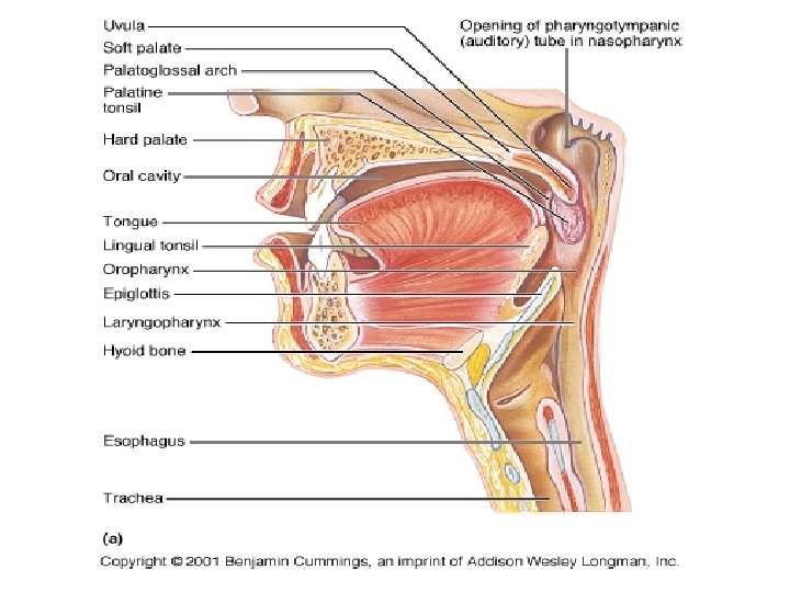
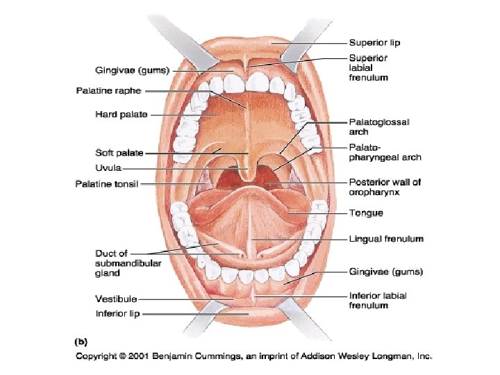
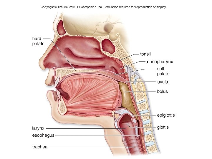
The Mouth The mouth, which ingests food, is bounded externally by the lips and cheeks. n The tongue Comprised of skeletal muscles to change shape of the tongue Taste buds are sensory receptors that allow people to enjoy eating by taste and texture
The Mouth The roof of the mouth separates the nasal cavity from the mouth. n n Prevents ingested food from entering the nasal cavity Divided into two parts Hard palate (anterior) n Contains several bones Soft palate (posterior) n n n Made of muscle Uvula: finger-shaped projection at back of the mouth Tonsils: Help protect the body from infections
Mastication • Mechanical digestion • Teeth, tongue Deglutition • Bolus formation for swallowing Peristalsis in esophagus
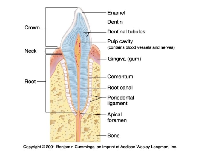
The Teeth are used to chew food into pieces suitable for swallowing. Adults have 32 teeth. Each tooth has two main divisions. n n A crown A root
The Teeth 20 baby teeth 32 adult teeth Tooth divided into n Crown Enamel Dentin Pulp n Root n Dentin Pulp Gingiva (gums) Copyright © The Mc. Graw-Hill Companies, Inc. Permission required for reproduction or display. enamel crown dentin pulp gum jawbone root canal root periodontal membrane cementum
The Teeth Bacteria that adhere to teeth metabolize sugar and release acids, which erode enamel. Dental caries (cavities) result from eroded enamel, a slow process. n Once the dentin layer is reached, the damage spreads more rapidly Gingivitis is inflammation of the gums. Periodontitis is inflammation of the periodontal membrane, characterized by bone loss.
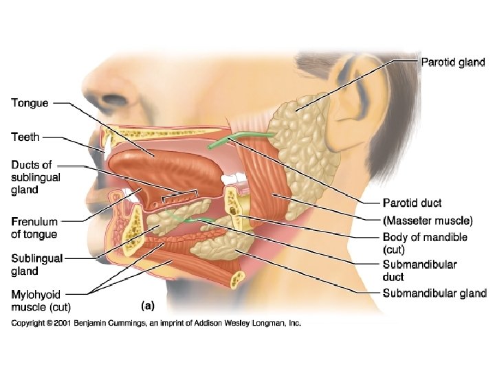
Salivary Glands Sublingual and Submandibular glands secrete Amylase n Breaks down complex carbohydrates into simple carbohydrates
The Pharynx n n Passageway that receives air from the nasal cavities and food from the mouth Swallowing Reflex action – performed automatically Soft palate closes off the nasopharynx Trachea moves up causing the epiglottis to cover the glottis As a result, food can enter esophagus only
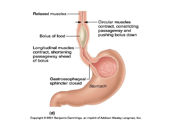
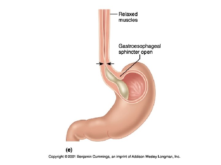
The Esophagus n The esophagus is a muscular tube that extends from the pharynx to stomach. Usually collapsed except during swallowing n n Peristalsis, rhythmic muscular contractions, pushes food along the digestive tract. The sphincter muscle closes the esophagus from the stomach Relaxation of the sphincter allows food to enter the stomach If contents of the stomach escape into the esophagus, this causes heartburn
Deglutition
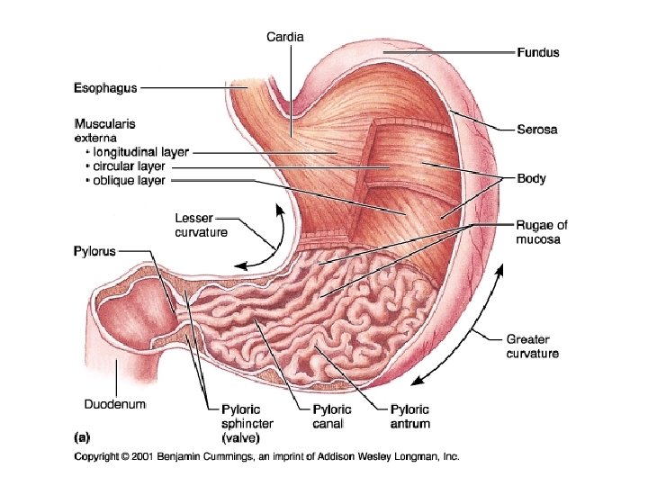
The Stomach n n n Thick-walled, J-shaped organ Continuous with esophagus and duodenum of small intestine Receives food from the esophagus Starts digestion of proteins Moves food into the small intestine Expands to hold about four liters when full. Rugae or folds allows for expansion
Reflux Esophagitis = Heartburn = GERD Gastroesphogeal Reflux Disease Lower esophageal sphincter dysfunction Why reflux against gravity?
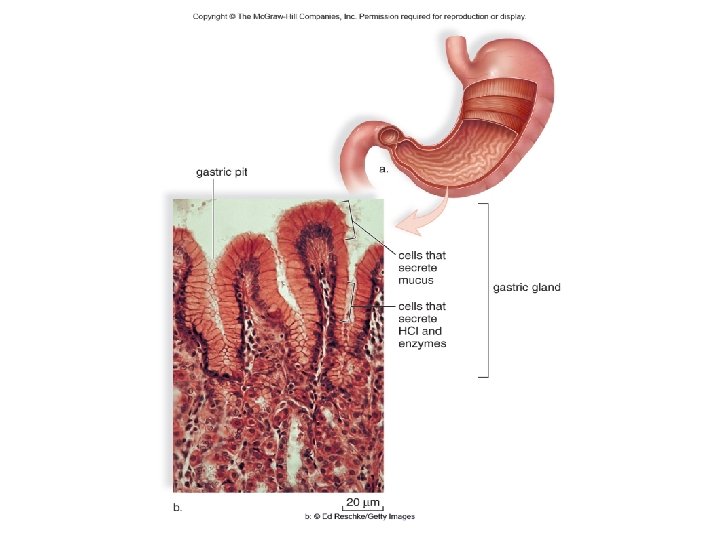
The Stomach Copyright © The Mc. Graw-Hill Companies, Inc. Permission required for reproduction or display. The columnar epithelium lines the stomach. n n a. gastric pit Contains gastric pits which lead into gastric glands Gastric glands produce gastric juice Pepsinogen (becomes pepsin) cells that secrete mucus cells that secrete HCl and enzymes Hydrochloric acid (HCl) Mucus 20 µm b. b: © Ed Reschke/Peter Arnold Figure 14. 5 gastric gland
Chemical Digestion Gastric Pits n n Invaginations of the stomach wall Lined with several different types of cells Chief Cells n n At the bottom of the pits Secretes pepsinogen Inactive form of the enzyme pepsin Pepsinogen is converted into pepsin upon mixing with HCl n Pepsin breaks down proteins into amino acids
Chemical Digestion Cont. Parietal Cells n n n Above Chief Cells Secret H+ and Cl- into the Gastric Pits Used for Digestion of foodstuff Gastric Glands or G cells n n Secretes the hormone Gastrin secretion increases when the contents of the stomach is basic Gastrin secretion decreases when the contents of the stomach is acidic Gastrin’s functions are To increase HCl secretion from the Parietal Cells To increase Gastric Motility
Mucous Cells Secrete Mucous to coat the lumen of the stomach n Prevents ulceration H. Pylori also responsible for ulcers
31
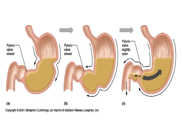
The Stomach The stomach acts both mechanically and chemically on food. The wall has three layers of muscles. n n Circular, longitudinal, and oblique Churns the food, mixing it with gastric juices Most food is not absorbed by the stomach. n Alcohol and other liquids are absorbed in the stomach The stomach normally empties in two to six hours. n Chyme – food leaving stomach n Enters small intestine through pyloric sphincter
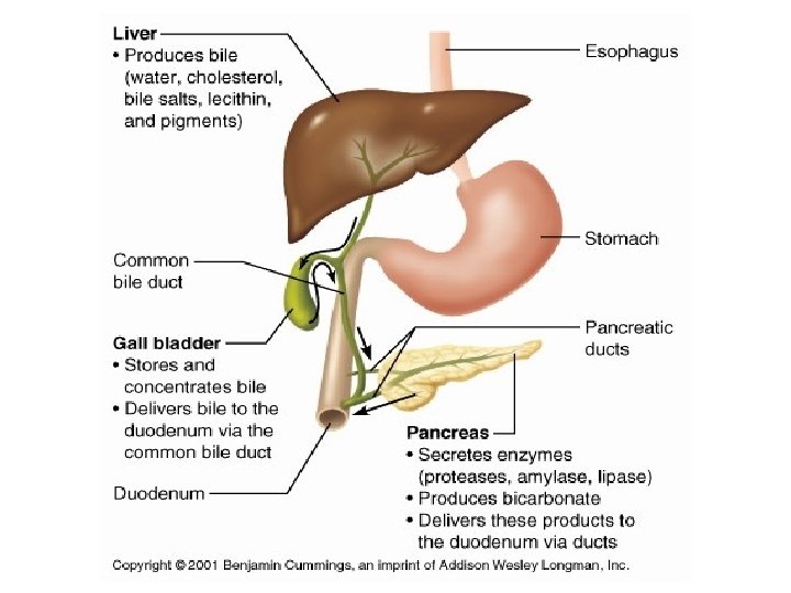
The Small Intestine n Approximately six meters long Smaller in diameter compared to the large intestine n Duodenum The first 25 centimeters of the small intestine Receives bile from the liver n Bile emulsifies fat Receives pancreatic juice from the pancreas n n Many enzymes for digestion of nutrients Bicarbonate to neutralize acidic p. H of chyme
The Small Intestine Jejunum n Middle section Ileum n n Remainder leading to large intestine Contains Peyer’s patches – immune response to intestinal pathogens
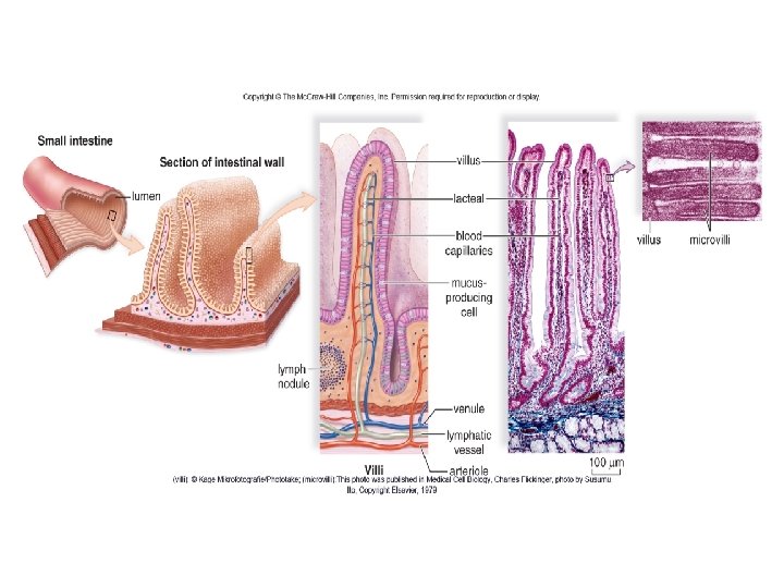
The Small Intestine The small intestine has large surface area. n Villi – fingerlike projections Blood capillaries for nutrient absorption n Carries sugars and amino acids Lacteals (lymph/blood capillaries) n Part of lymphatic system n Carries digested fats n Microvilli – microscopic extensions on surface of epithelial cells of villi
Copyright © The Mc. Graw-Hill Companies, Inc. Permission required for reproduction or display. Small intestine Section of intestinal wall lumen lymph nodule (villi): © Manfred Kage/Peter Arnold; (microvilli): Photo published in Medical Cell Biology, Charles Flickinger, photo by Susumu Ito, Copyright Elsevier, 1979. Figure 14. 6
Copyright © The Mc. Graw-Hill Companies, Inc. Permission required for reproduction or display. villus lacteal blood capillaries villus microvilli mucusproducing cell lymph nodule venule lymphatic vessel Villi arteriole 100 µm (villi): © Manfred Kage/Peter Arnold; (microvilli): Photo published in Medical Cell Biology, Charles Flickinger, photo by Susumu Ito, Copyright Elsevier, 1979. Figure 14. 6
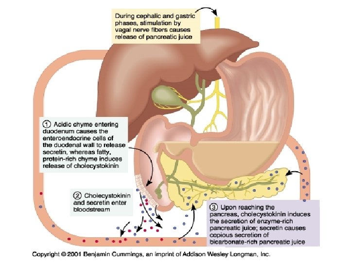
Regulation of Digestive Secretions Digestive secretions are regulated by the nervous system and by hormones. n Hormone – substance produced by a group of cells that affects a different group of cells (target cells) Hormone released into the blood for transport Ex: Gastrin released by stomach after protein rich meal n Stomach churns, gastric gland activity increased Ex: GIP (gastric inhibitory peptide) produced by duodenal wall n Inhibits gastric gland secretion
Regulation of Digestive Secretions Two other hormones that regulate digestion n Secretin - produced by duodenal wall Release stimulated by entrance of HCl in chyme n CCK (cholecystokinin) - produced by duodenal wall Release stimulated by proteins and fat in chyme n Together, secretin and CCK act on three organs. n n n Pancreas to increase pancreatic juice output Liver to increase bile output Gallbladder to contract to release bile
Copyright © The Mc. Graw-Hill Companies, Inc. Permission required for reproduction or display. Secretin and CCK together stimulate actions of three organs. gallbladder liver stomach Pancreas Liver Gallbladder pancreas duodenum blood vessel Figure 14. 7 secretin CCK gastrin
The Liver Acts as gatekeeper for the blood n n n Detoxifies and removes poisonous substances Removes and stores iron and vitamins A, D, E, K, and B 12 Makes plasma proteins Regulates cholesterol Regulates blood glucose – stores as glycogen Produces bile Bilirubin – hemoglobin breakdown product Bile salts – emulsify fat
The Gallbladder n The liver produces 400 -800 ml of bile each day. n The gallbladder stores excess bile. n Water is reabsorbed so that bile is thickened. n Bile is secreted through the common bile duct into the duodenum.
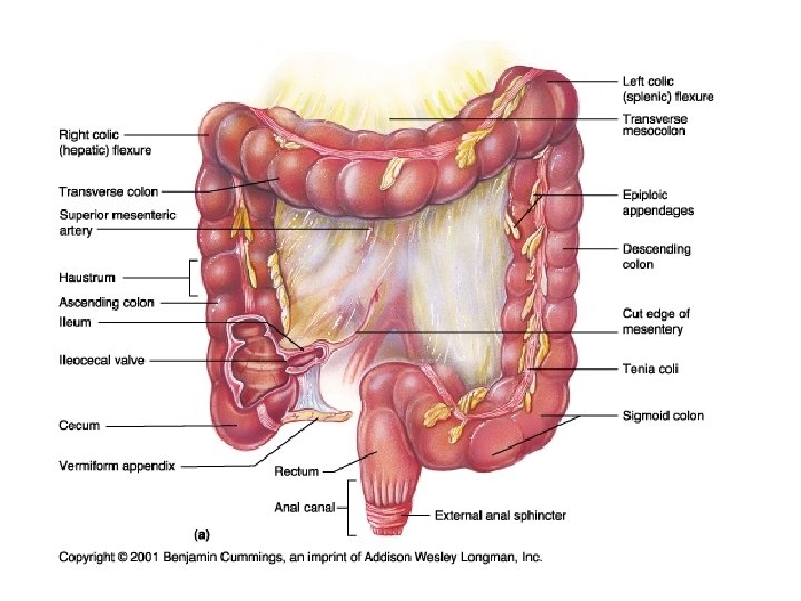
The Large Intestine n n Includes cecum, colon, rectum and anal canal Larger in diameter, but shorter length compared to the small intestine Functions: n n Absorbs water, salts, and some vitamins Stores indigestible materials until eliminated as feces
The Large Intestine Cecum n n n Lies below junction with small intestine Small pouch (6 cm long) that forms first part of the large intestine Human cecum has projection called appendix or veriform appendix Like tonsils, may play a role in fighting infection Subject to inflammation, appendicitis If inflamed it should be removed before rupturing
Copyright © The Mc. Graw-Hill Companies, Inc. Permission required for reproduction or display. Cecum n n Lies below junction with small intestine Human cecum has vermiform appendix large intestine Role in fighting infection small intestine cecum Figure 14. 8 vermiform appendix
The Large Intestine Colon n Ascending, transverse, descending, and sigmoid Rectum n n Last 20 cm of large intestine Opens to anus Anus n n Where defecation (expulsion of feces) occurs Reflex triggered as feces are forced into rectum
Colon Three Major Functions of the Large Intestine n n Absorption of H 2 O Storage of Feces Usually 24 hours n Production of Vitamin K From E. Coli Used in the Blood Clotting Process
The Large Intestine Feces are typically ¾ water and ¼ solids n n n Bacteria, plant fiber and indigestible materials are in the solid portion. Bacterial breakdown of indigestible materials account for the odor also for the presence of gas. Metabolism of bilirubin and oxidized iron account for the brown color.