Digestion Absorption Sphincter A sphincter is a ring

Digestion / Absorption

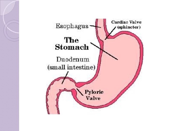
Sphincter �A sphincter is a ring of muscle that controls the passage of material. Relaxed = open, Contracted = closed �At the junction of esophagus and stomach is the cardiac sphincter (AKA Lower Esophageal Sphincter). Stops stomach contents from going into esophagus

�At the junction of stomach and duodenum is the pyloric sphincter. Regulates movement of stomach contents from stomach small intestine.

Draw This!

Heart Burn �Caused by a cardiac sphincter that doesn’t tighten as it should stomach acid into esophagus �Often happens when too much food in the stomach (overeating) or to much pressure on the stomach (obesity, pregnancy) �Certain foods act as a trigger (relaxes the sphincter or causes more acid production smoking causes both!)

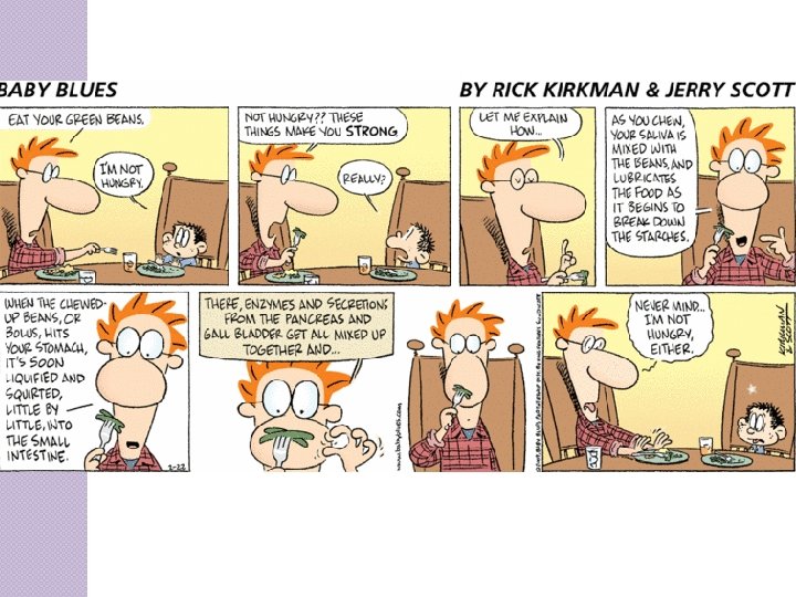
The Stomach �Site of initial protein digestion and food storage. �Smooth muscle contractions cause ingested food to be crushed, ground, and mixed, liquefying it to form Chyme

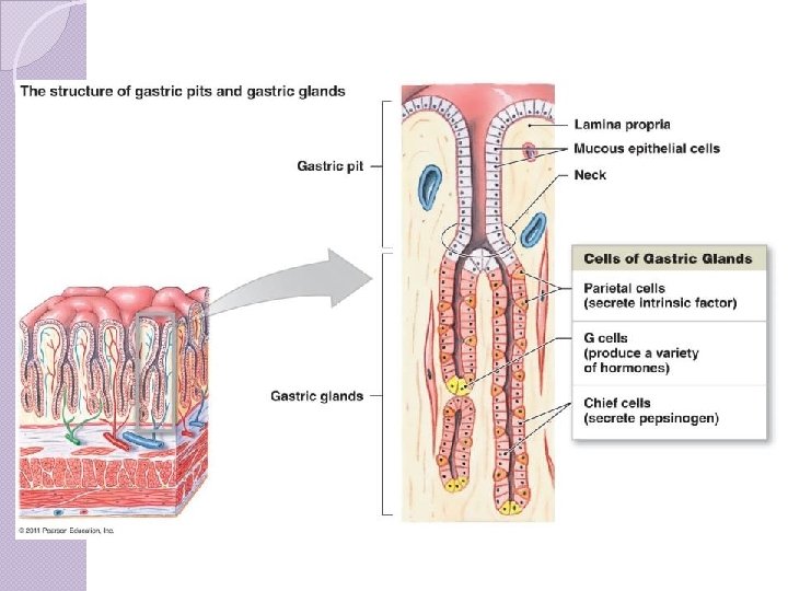
Gastric Pits

Gastric Pits �Stomach contains GASTRIC PITS that have GLANDS and MUCUS cells. Glands contain: �Parietal cells – secrete HCl (hydrochloric acid) �Chief cells – secrete pepsinogen, the zymogen (inactive) form of the digestive enzyme pepsin. (Pepsinogen Pepsin under low p. H)

�Pepsin breaks down proteins into short amino acid chains

�G cells – secretes gastrin, a hormone. Gastrin stimulates the secretion of HCl and aids in stomach motility. �It’s released in response to stomach stretching or the presence of proteins in the stomach. It is inhibited by HCl in the stomach. What kind of feedback is this? ?
, initiates the digestion")
The J-shaped stomach stores food (the semidigested mass is called chyme), initiates the digestion of proteins, has only minimal absorption, and moves materials on to the small intestine.

Absorption �Some water, specific vitamins and alcohol

Stomach Composition �The stomach has four layers that surrounds the space called the lumen. 1) 2) 3) 4) Mucosa Submucosa Muscle layer Serosa �The stomach has folded membranes on the inside called Rugae allows stomach to expand

Why doesn’t the stomach digest itself? �Mucus cells in the gastric pits secrete a thick layer of mucus which protects the walls of the stomach �also secrete bicarbonate solution which neutralizes stomach acid (acid base reaction)

Peptic Ulcers
")
Peptic Ulcer �Most commonly caused by bacterial infection Helicobacter pylori �NSAIDS (non-steroidal antiinflammatory drugs) aspirin and IBProfen �Smoking �Alcohol �Genetics

A Very Famous Stomach! �Alexis St. Martin, 1822 �https: //www. youtube. com/watch? v=pq gc. EIa. XGME

Mechanical vs Chemical recap Mechanical – churning of stomach Chemical : �HCl denatured proteins and kills ingested bacteria �Pepsin begins protein digestion
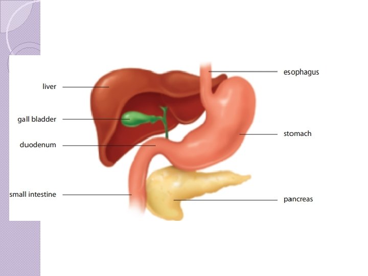

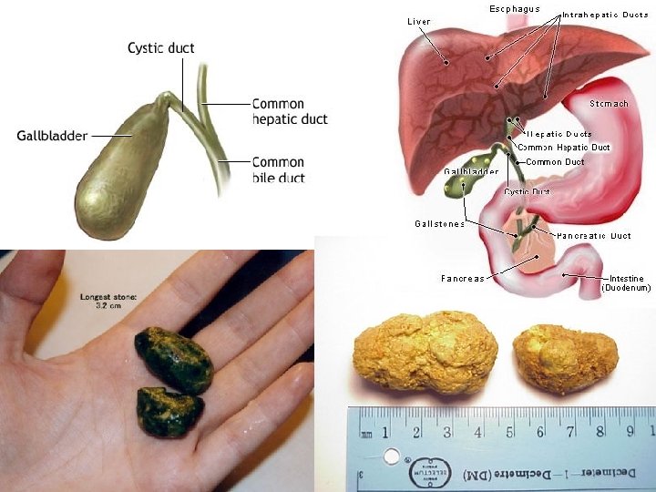
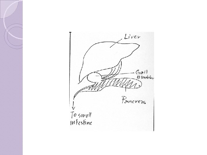
Liver & Gall Bladder �Liver produces Bile mainly water and some bile salts �Bile is stored in the Gall Bladder the gall bladder absorbs water making the bile more concentrated �Bile releases into duodenum via bile duct

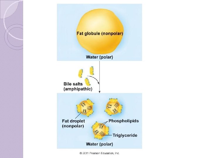
Bile Salts �Emulsify Fats (Emulsify - To make a suspension of small globules of one liquid in a second liquid in which the first will not mix)

Bile continued Mechanical digestion: �Emulsification – bile salts break down fat globules into smaller droplets so that they can be digested by enzymes. �Purpose – To increase surface area!

Check your understanding �What is the function of the 2 sphincters in the stomach �What role does each cell in the gastric pit play? �Describe the chemical digestion that occurs in the stomach �What is the main purpose of bile salts. Explain.
 �Breakdown and synthesis of lipids")
Other Liver Functions �Regulate blood glucose levels (stores glycogen) �Breakdown and synthesis of lipids and fats �Protein breakdown and amino acid synthesis �Stores vitamins (A, B 12, D) and iron �Breaks down hormones, antibiotics, and other toxic substances (detoxifier) �Breaks down the by-products of RBC recycling Note** these are main functions. Liver is

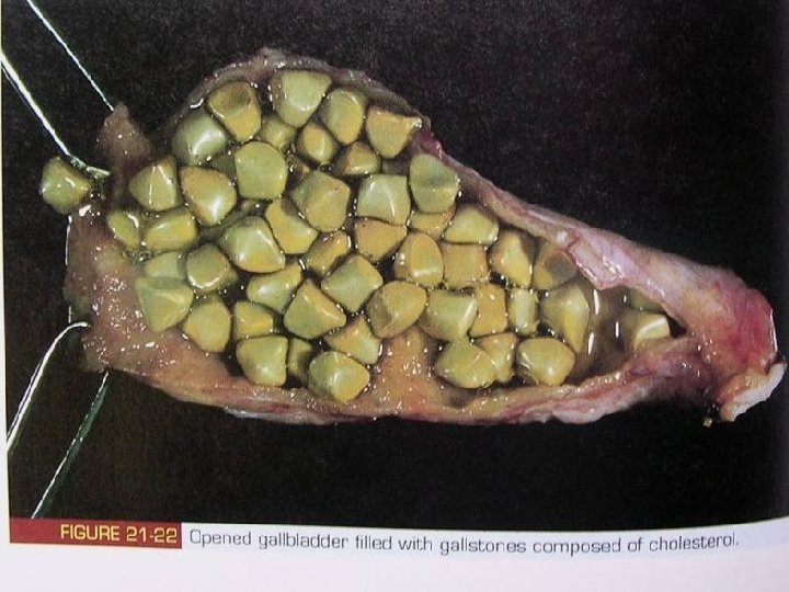
Gall Stones �Crystalline mass formed from bile pigments, cholesterol, and calcium salts. �Cause severe pain and blockage of bile duct

Liver Cirrhosis �Most commonly caused by alcohol, hepatits B, hepatits C, and fatty liver disease. �Gradual build up of scar tissue that replaces normal tissue �Leads to liver failure

Ethyl alcohol is toxic to the liver

Healthy Liver

Liver cirrhosis Note extensive scarring

Cirrhosis of the liver Chronic alcoholism will typically lead to damage of the liver characterized by scarring. The damaged liver often turns an orange color. This damage of the liver is called “cirrhosis”.
 �The liver")
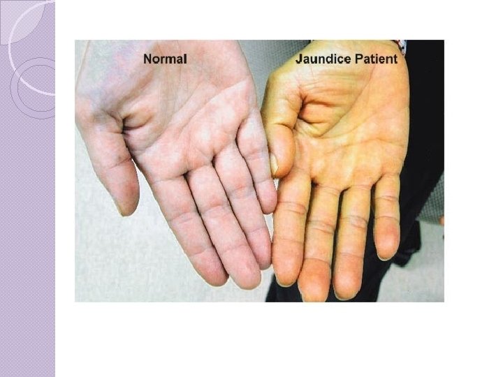
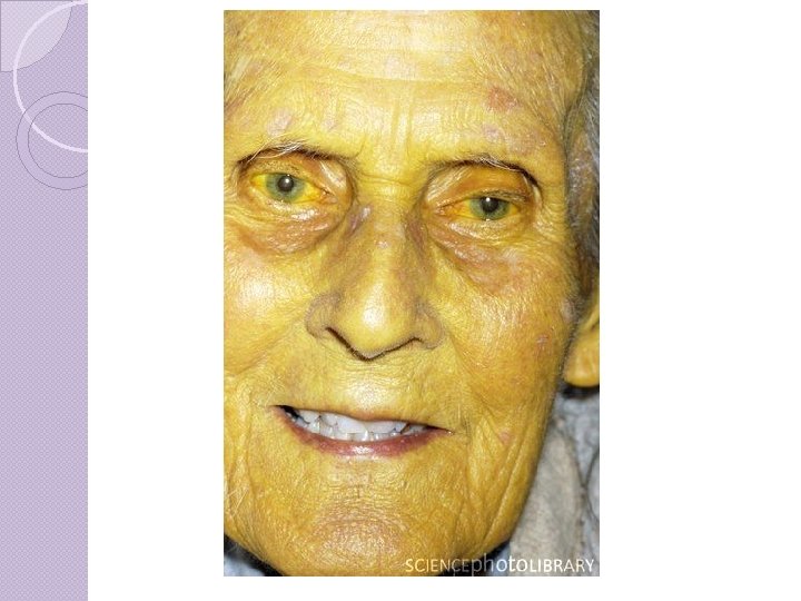
Jaundice �One of the by-products of RBC destruction is bilirubin (yellow color) �The liver filters out bilirubin from blood, and excretes in bile. �If liver is failing, or bile duct is blocked, bilirubin builds up in body, causing jaundice a yellowing of skin and whites of eyes.
 a lot about the normal")
Gastrointestinal Disorder Project �So now that you know (almost) a lot about the normal functioning of the digestive system, it’s time to investigate some disorders!

Plagiarism �Source all the materials you use! �In University if caught plagiarizing you will automatically receive a zero on the assignment with the potential of receiving a failing grade in the course or a suspension from the University!

�http: //www. youtube. com/watch? v=y. Q Hx 9 WEf. T-Y&safe=active

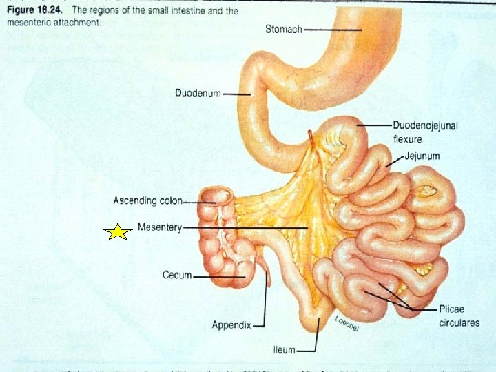
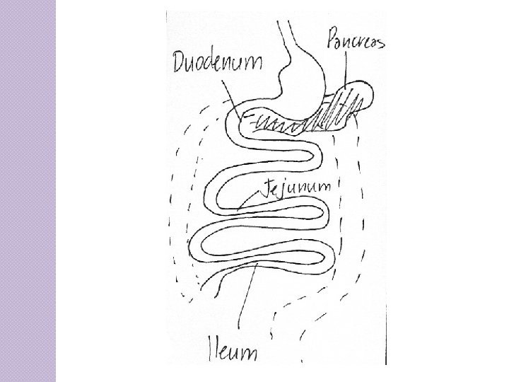
Small Intestine is 7 meters in length • Three parts to small intestine: 1. Duodenum – first 25 -30 cm. - Area of most digestion. 2. Jejunum 3. Ileum •

�When chyme enters the small intestine it stimulates the release of secretin and cholecystokinin by the duodenum walls. �Secretin regulates p. H inhibits gastric HCl production and stimulates bicarbonate ion secretion (pancreas) Cholecystokinin causes release of bile (gall bladder) and digestive enzymes (pancreas).

Pancreas �Secretes pancreatic juice into the duodenum

Pancreatic juice contains: �Bicarbonate – ◦ changes p. H of chyme from highly acidic (p. H 2) to weakly basic (p. H 8) ◦ Important p. H for pancreatic enzymes to function

Enzymes lipase - breaks down triglycerides into fatty acids and glycerol. •

Protease – to digest proteins Amylase – to digest carbohydrates

�Insulin �Glucagon Regulation of blood glucose levels

Check your understanding! �What accessory gland produces a secretion with no digestive enzymes? �What kinds of foods are broken down by the pancreas? �Most enzymes are secreted as zymogens (inactive), why do you think that is?

Check your understanding! �Explain two functions of pancreatic fluid. �Someone recently has had a cholecystectomy, a surgical procedure to remove his gall bladder. Now he must take medication to prevent diarrhea. Why would the removal of the gall bladder cause diarrhea?

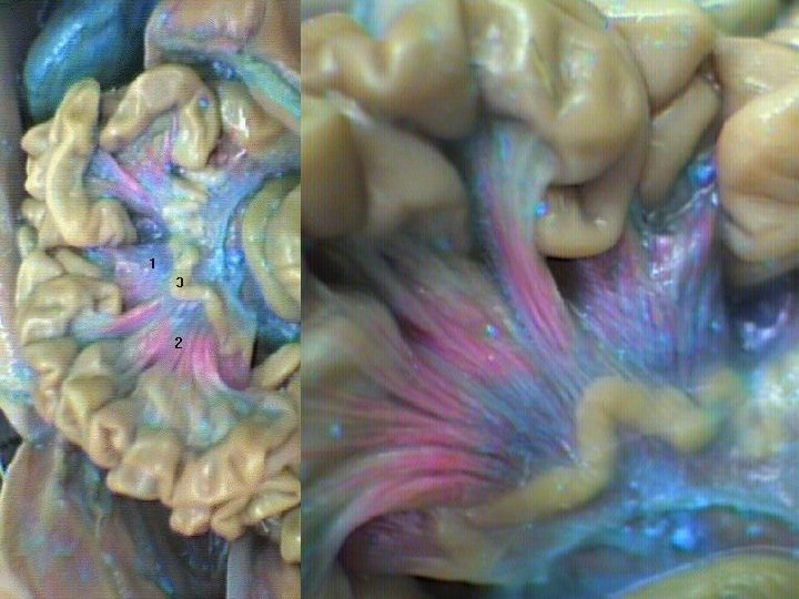
Mesentery �Tissue that supports the intestinal tract. �A double layer of connective tissue in which blood vessels, nerves, lymphatics and other structures are contained.

Thin walls of small intestine with blood supply. Mesentery
.")
Adhesions often form following abdominal surgery or after an abdominal infection (peritonitis).

Peritonitis can also follow a penetrating abdominal injury.

�Made Small Intestine up of three parts: ◦ Duodenum ◦ Jejunum ◦ Ileum
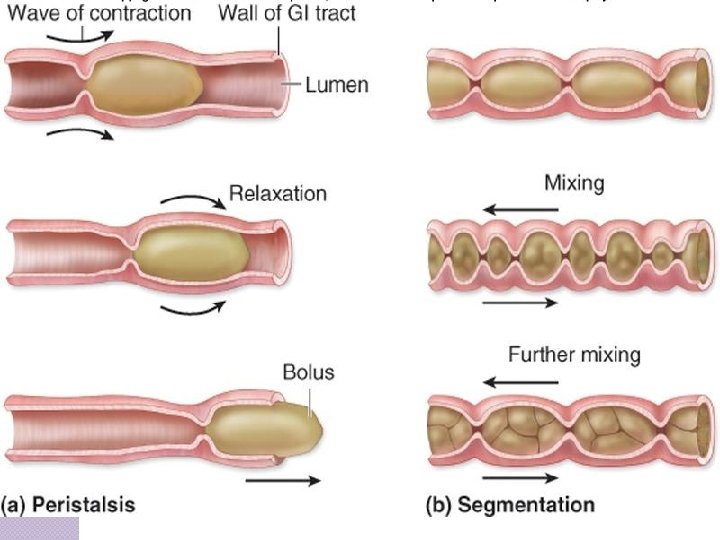

Segmentation �Smooth muscle contractions in both directions that mix and further break down contents of small intestine (chyme) �Is this mechanical or chemical digestion?

Absorption �The small intestine absorbs the majority of nutrients �such as ◦ The breakdown products of �Carbohydrates �Protein �Fats ◦ Vitamins by active and passive mechanisms

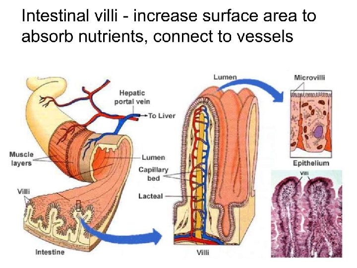
Intestinal villi
. Each villus is covered")
Each villus contains blood capillaries and a lymph capillary (lacteal). Each villus is covered with microvilli.

�Villi – tiny, finger-like projections on the walls of the small intestine �Microvilli – “brush border” further increases surface area

�Within villi structures are blood vessels that carry the absorbed nutrients to the rest of the body �Also contain in the villi are structures called lacteal, which absorbs fats to be delivered into the bloodstream
")
The Colon (AKA large intestine)

Main Functions of the Large Intestine �Water absorption �Absorption of vitamins produced by bacteria �Mass movements (defecation) – removes undigested food
 is digested by enteric bacteria (ex.")
Chemical Digestion �Indigestible food matter (e. g. cellulose) is digested by enteric bacteria (ex. E. Coli) that thrive in the large intestine. �These bacteria produce vitamin K and some B vitamins.

Appendix �Vestigial organ has lost function but retained structure �However, some scientists now think it does serve a function stores good bacteria to help repopulate gut after infection.

http: //www. flickr. com/photos/moonjazz/3530268320/

Endoscopic view of appendix Cecum

Rectum �Connected to the sigmoid colon of the large intestine �Damp Mass of indigestible food remaining in large intestine is called FECES. �Temporarily stores feces before elimination (egestion)
")
Anus �The ending portion of the gastrointestinal tract in which feces (undigested food matter) leaves the body �Anal sphincter controls opening of anus.

Bowel Movement �Receptors in the walls of the large intestine give the central nervous system (brain) signals when a bowel movement is needed. �Some substances stimulate the bowel movements like caffeine.

Defecation to eliminate feces.

Summary

Digestion Time Summary Digestion takes different lengths of time depending on the food being consumed. For healthy adults, it's usually between 24 and 72 hours. ◦ Mouth (20 sec) �Starch is digested to maltose by carbohydrase / amylase. ◦ Esophagus (10 sec) �Muscles squeeze food along the canal. ◦ Stomach (2 -6 hours) �Gastric juice mixes food when stomach churns. ◦ Small intestine (5 hours) �Intestinal juice contains enzymes that complete the digestion process. ◦ Large intestine and anus (min of 24 hours) �Undigested food reaches the large intestine �Lots of water is removed and taken back into the body
 �Bacteria �Food")
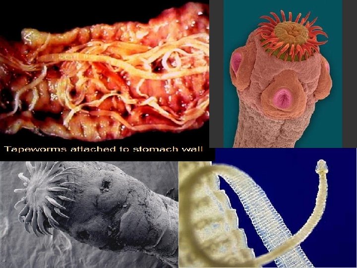
Washing your hands is essential! (Return to regular notes booklet for this) �Bacteria �Food poisoning �Parasites �Worms (Tapeworms) * All can be transferred through the mouth!

The Bacteria in your Gut? �Good, bad �Good √ or ugly? E. coli helps you digest food that you could not digest otherwise. �Bad √ The bacteria that help you out, also produce flatulence as a by-product. Gas is a normal part of digestion. �Ugly √ Some bacteria cause food poisoning, resulting in severe stomach cramps, vomiting and diarrhea.

More important stuff. . . �Our bodies make two hormones that contribute to homeostasis. 1. GASTRIN: Releases gastric juice (HCl) and relaxes gastric sphincter. 2. SECRETIN: Help release bicarbonate ions that neutralize HCl. �Our pancreas also helps with homeostasis in regulating blood glucose.
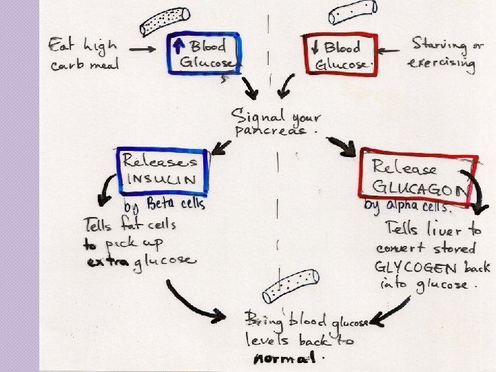

Insulin and Glucagon in Digestive System �Two hormones secreted by pancreas. �Work with liver to control level of glucose in body (blood). �Insulin released after meal allows cells to become permeable to glucose. �Excess glucose stored by liver as glycogen. �Can change back to glucose if blood glucose becomes too low Glucagon
- Slides: 87