Differentiation of the neural tube Development of spinal

Differentiation of the neural tube, Development of spinal cord compiled by András Csillag

Differentiation and early parcellation of neural plate Notochord Neural plate Prechordal plate Neural groove Prosencephalon and mesencephalon Rhombecephalon D D D Spinal cord BMPs (bone morphogenetic proteins) derive from ectoderm, promote migration tendency toward surface ectoderm 1. Planar induction: Primitive node – anti-BMP factors – inhibit migration to surface, neural plate develops 2. Vertical induction: - Rostrally under the effect of prechordal plate (mesendoderm) - Caudally under the effect of notochord (mesoderm) After Larsen, 2003
 Normal development.")
Expression of N-cadherin and E-cadherin adhesion proteins during neurulation in Xenopus. (A) Normal development. In the neural plate stage, N-cadherin is seen in the neural plate, while E-cadherin is seen on the presumptive epidermis. Eventually, the N-cadherin-bearing neural cells separate from the E-cadherin-containing epidermal cells. (The neural crest cells have neither cadherin, and they disperse. ) (B) No separation of the neural tube occurs when one side of the frog embryo is injected with Ncadherin m. RNA, so that N-cadherin is expressed in the epidermal cells as well as in the presumptive neural tube.

Primary neurulation The ‚classic’ mechanism yields three main sets of cells: neural tube, neural crest, epidermis of skin The cells surrounding the neural plate make the neural plate cells proliferate, invaginate, and pinch off from the surface to form a hollow tube Primary neurulation: neural tube formation in the chick embryo. (A, 1) Cells of the neural plate can be distinguished as elongated cells in the dorsal region of the ectoderm. Folding begins as the medial neural hinge point (MHP) cells anchor to notochord and change their shape, while the presumptive epidermal cells move towards the center. (B, 2) The neural folds are elevated as presumptive epidermis continues to move toward the dorsal midline. (C, 3) Convergence of the neural folds occurs as the dorsolateral hinge point (DLHP) cells become wedge-shaped and epidermal cells push toward the center. (D, 4) The neural folds are brought into contact with one another, and the neural crest cells link the neural tube with the epidermis. The neural crest cells then disperse, leaving the neural tube separate from the epidermis. (Photographs courtesy of K. Tosney and G. Schoenwolf; drawings after Smith and Schoenwolf 1997. )
 humans this occurs caudally to somite 35 (Schoenwolf")
Secondary neurulation In mice and (probably) humans this occurs caudally to somite 35 (Schoenwolf 1984; Nievelstein et al. 1993) the neural tube arises from a solid cord of cells which subsequently hollows out (cavitates) to form a tube Secondary neurulation in the caudal region of a 25 -somite chick embryo. (A) The medullary cord forming at the most caudal end of the chick tailbud. (B) The medullary cord at a slightly more anterior position in the tailbud. (C) The neural tube is cavitating and the notochord forming. (D) The lumens coalesce to form the central canal of the neural tube. (From Catala et al. 1995; photographs courtesy of N. M. Le Douarin. )

Rostrocaudal patterning of neural tube Early development of prosencephalon and mesencephalon ANR – anterior neural ridge (crista neuralis anterior, anterior neural crest) Major organizing center, emits rostralizing signals, essential for the development of secondary prosencephalon and its derivatives (telencephalon, optic cup, hypothalamus)

The principal organizing centers determining early regionalization of the brain ANR – anterior neural ridge (determines the rostral limit of neural plate, antagonizes ‘caudalization’ signals, e. g. Wnt 1, and promotes action of prosencephalic factors, e. g. Six 3) ZLI – zona limitans anterior, anterior limiting zone (delineates the prosomeres P 2/P 3) Is. O – isthmic organizer (at the prosencephalon/rhombencephalon boundary, produces factor Fgf 8 toward r 1 cerebellar development - , and Wnt 1 toward the mesencephalon development of colliculi ) Echevarría et al. , 2003
 and Iso (isthmic organizer)")
The isthmic organizer Main organizer regions: anr (ant. neural ridge) and Iso (isthmic organizer) Fgf 8 (fiber growth factor 8) Shh (sonic hedgehog) Otx 2 → caudal Morphogenetic gradient Gbx 2 → rostral Martínez-Ferre és Martínez, Front. Neurosci, 2012. Aroca és Puelles, 2005.

Rostrocaudal subdivisions of the neural tube, schematic illustration Martínez et al. , 2012

Dorsoventral patterning of neural tube Signaling molecules and transcription factors: BMPs from non-neural ectoderm Sonic hedgehog (Shh) from the notochord (N) and the floor plate (F) Retinoic acid from the somites (S) Pax 3, 7 from the roof plate, then from the alar plate Nk 2 (Nkx), and later Pax 6 from the basal plate Sadler, 2007

Dorsoventral patterning of neural tube A – Fusion of neural tube, rostral view B –Shh expression in floor plate C – The four basic components of neural tube (roof plate – RP; alar plate – AP; basal plate – BP; floor plate – FP) Martínez et al. , 2012 D – Shh expression in sagittal section of mouse neural tube Abbreviations: aep – anterior entopenduncular area Di – diencephalon Mes – mesencephalon mge – medial ganglionic eminence Pros – prosencephalon Rh – rhombencephalon Sec. Pros. – secondary prosencephalon p 1 -3 – prosomeres Puelles et al. , 2004

The developing neural tube is divided into 4 plates • Roof plate: signaling center (BMPs and Wnts) • Alar plate: sensory • Basal plate: motor • Floor plate: signaling center (Shh)

The early neural tube is a pseudostratified epithelium • The “apical” portion abuts the central canal • The “basal” portion abuts the surrounding tissue (e. g. somites, notochord, etc. ). • Cell division occurs in the apical portion.

The principal mechanisms of developmental dynamics Histogenesis Proliferation Migration • Radial • Tangential Differentiation

Histogenesis of the central nervous system: proliferation, migration, and differentiation • Replication of DNA in proliferative cells (progenitors) occurs at external limiting membrane • The nucleus of the progenitors migrate to apical portion to undergo either equal or unequal mitosis (depending on orientation of metaphase plate): • “Equal” generates two progenitors that migrate back to ELM to undergo another round of DNA replication • “Unequal” generates a progenitor and cell that will differentiate into a neuron • Appropriate development requires a delicate balance between proliferation and differentiation • Neurons migrate along radial glia to establish layers within the CNS that are very important for its function –disruption of this migration therefore has significant consequences

Neural cell lineages • 1 st major cell fate decision is glial vs. neuronal • If glial, then next decision is O-2 A, 1 A, or radial glia • Radial glia can also generate type 1 astrocytes and ependymal cells

Histogenesis of Neural Tube • Tube differentiates into two concentric rings by day 26: Mantle layer and marginal layer. • Other cells lose contact with the basement membrane and will migrate past the ependymal cells to form a new outer layer of densely packed cells collectively called the: Mantle layer: • Cells that make up the mantle layer are: NEUROBLASTS. • Note that mantle layer is still covered by the external limiting membrane.

Spinal cord 10 mm pig embryo cross-section © 2006 Marshall Andersen

Spinal cord 10 mm pig embryo cross-section © 2006 Marshall Andersen

Histogenesis of Neural Tube • Neuroblasts in the mantle layer will begin to grow processes (axons) that will form a new outer layer: Marginal layer. The marginal layer is also located beneath the external limiting membrane. • The marginal layer will form the white matter of the spinal cord and the brain. • The mantle layer forms the gray matter of the brain and spinal cord (except for the cortices).

Neural Crest: the “ 4 th germ layer” At the time of neurulation, cells at the lateralmost edge of the neural plate are exposed to a unique combination of factors from the adjacent skin, underlying mesoderm, and from the rest of the neural plate and are induced to form neural crest. The neural crest cells downregulate cadherin expression and delaminate from the neuroepithelium, i. e. , they transform from epithelial cells into migratory mesenchymal cells that contribute to forming MANY tissues in the body.

Major Derivatives of the Neural Crest

Trunk Neural Crest: sensory lineage • Migrate along ventrolateral stream and stop just medial to somite • Aggregate into dorsal root ganglia and separate into 2 general lineages: • Sensory neurons (Wnts, NGFs, b. HLHs (Ngn 1, etc. ) • Glia (Schwann cells and satellite cells) via GDNF signaling • Sensory neurons extend processes in two directions: dorsomedially to neural tube and ventrolaterally into growing spinal nerve (established by outgrowth of motor neuron axons from the ventral horn)

Sensory neurons send axons into dorsal horn. Motor neurons send axons out of vental horn. Axons of peripheral nerves are myelinated by Schwann cells

Trunk Neural Crest: sympathoadrenal lineage • Migrate along ventral stream • Dependent on expression of Phox -2 and Mash-1 • BMPs from aorta play further instructive role to specify bipotential progenitors: • Sympathetic ganglion neurons: induced by presence of FGF and NGF in symp. Ganglia • Adrenal chromaffin cells: induced by glucocorticoids in adrenal gland

Circumpharyngeal Neural Crest: vagal crest • Migrate caudal to 6 th arch and then become associated with wall of fore-, mid-, and proximal hindgut • Downregulate expression of Robo, a Slit-2 receptor, which allows them to invade the gut wall. • Hand 2 influences differentiation into cholinergic phenotype

Circumpharyngeal Neural Crest: • • Associated with arches 3, 4, 6 Contribute to thyroid, parathyroid, and thymus Cardiac crest contributes to outflow tract cushions Disturbances (e. g. Di. George syndrome, Hoxa-3 mutation) therefore have multiple effects: craniofacial (jaw) defects, glandular defects, & outflow tract defects

Innervation of the Gut: vagal and lumbosacral crest • Vagal crest: innervate gut wall from foregut to ~proximal 2/3 of transverse colon in hindgut • Sacral crest: innervate hindgut wall from ~distal 1/3 of transverse colon to rectum

Cranial Neural Crest: Contribute to forehead, face, jaw, and pharyngeal arch derivatives

Cranial nerve ganglia arise from neural crest and ectodermal placodes • “proximal” or “superior” ganglia from neural crest • “distal” or “inferior” ganglia from ectodermal placodes • parasympathetic ganglia (ciliary, otic, pterygopalatine, and submandibular) from neural crest

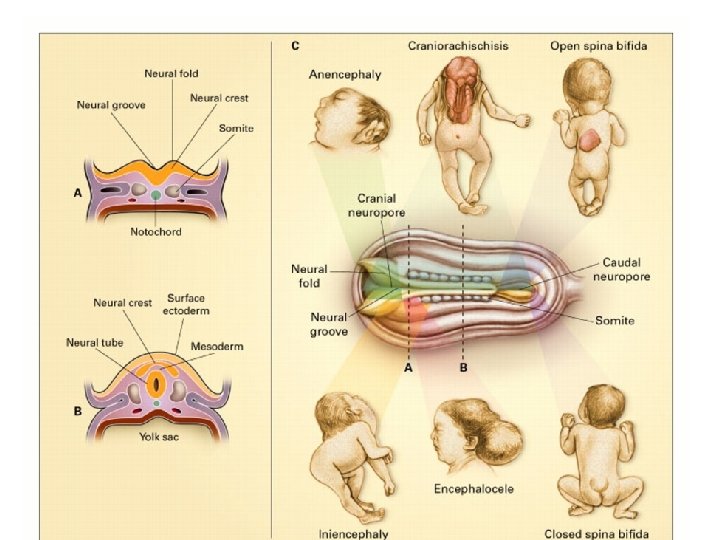
Neural tube defects

Frequent closure defects of the spinal cord and meninges

Tethered cord syndrome Growth of scar tissue Normally, growth of the spine exceeds that of the cord, therefore the adult cord ends at L 2 with a freedom of movement. In tethered cord patients the cord remains adherent to the lower spinal canal, resulting in stretching and reduced blood flow to nerves. Adhesion
- Slides: 34