Differential diagnose in proteinuria and pathologic urine sediment


 ERCP-1 body (capsule and ball); 2 -crimped canaliculus proximal (main Division);")


 Feverish As a result of the deep, vigorous, continuous")
 Stagnant or heart Clubockova, (not more than 1 g/day),")


 In the clinical picture: swelling and pain in the")


 Xp. purulent processes (osteomyelitis, bronhoektaza) Rheumatoid arthritis and other")


 PU Largest small (up to 1. 0 g/day). Proteinuria caused by")


 Allocation of urine red blood cells more physiological norm, namely above 1")


. moderate")


 the cell of origin (human erythrocyte, leukocyte, epithelial) Cylinders")


, HC 400 h.")
- Slides: 68
Differential diagnose in proteinuria and pathologic urine sediment. Tactics of GP. Rakhimova M. E.
PHYSICAL PROPERTIES Ø Ø Ø The amount of urine in 24 hours-800 -1500 ml relative density of the morning portion-1020 -1026 Maximum osmotic concentration-910 mosms/l colour: straw yellow transparent transparency -Reaction-Neutral or slightly acidic p. H of 5. 0 - -6. 0 Protein is missing, tracks (25 -70 mg/day) -0. 070 0. 025 g/d No sugar, traces (up 0. 02 percent)
Proteinuria Hematuria Main manifestations of US Pyuria Cylindria
Proteinuria Selection of protein in the urine, exceeding the physiological norm, namely a -50 30 mg/day. Daily excretion of protein more than 5080 mg always points to the pathology of the kidney
The question, getting up in front of the doctor: first, where the border of the normal and the pathological state, and the second whether any renal proteinuria is a manifestation of kidney failure?
Total protein in the blood 65 -85 g/l Albumin/globulin -1. 6 -2. 8 protein fraction: Albumin-48 -61% Globulin: Alpha 1 -3 -6% -8 -11% alfa 2 beta -11 -15 gamma-16%25%
СТРОЕНИЕ НЕФРОНА (СХЕМА) ERCP-1 body (capsule and ball); 2 -crimped canaliculus proximal (main Division); 3 and 4 -loop (Henle); 5 -crimped canaliculus distal (intercalating Division); 6 -the collecting tubules. In the circles shows the structure of epithelium in different parts of the nephrone.
Классификация протеинурии In connection with diseases By source On the composition of the According size ØFunctional. ØPathological ØPrerenal (overflow); ØRenal (clubockova); ØPostrenal (extrarenal, false) ØSelective; ØNonselective ØMicroalbumin urine test (up to 300 mg/day); Low (traces 1 g/d); Moderate (1 -3 g/d); High or principally nefrotical (> 3 g/day).
Functional proteinuria Reflex Alimentary Emotional Central After the cold with hand forearm immersion in cold water for 2 -5 minutes After eating a hearty protein Under the influence of a strong nervous tension After bouts of epilepsy
Functional proteinuria Pal′patornaâ Voltage (March) Feverish As a result of the deep, vigorous, continuous abdominal palpation of the kidneys. Working does not exceed 1 -2 g/d, occurs after heavy physical exertion, are detected in the first portion of urine disappears when regular physical activity Clubockova, (up to 1 -2 g/day) is observed in febrile States, more often in children and the elderly, disappears after normalization of body temperature.
Functional proteinuria Orthostatic (postural, lordotičeskaâ) Stagnant or heart Clubockova, (not more than 1 g/day), isolated is more common in young persons 13 -20 years asteničeskim physique, disappears in the position of lying PU ≤ 1. 0 -3. 0 g/l is not always regarded as PU functional nature, as the slowing of renal blood flow and hypoxia, may have a deleterious effect on the basal membrane of the renal tubular epithelium and clubockov
Pathological proteinuria By source On the mechanism of occurrence Prerenal′nyj, renal and postrenal′nyj Øglomerulârnuû, tubulârnuû, smešennuû secretory overflow proteinuria, proteinuria ("over-flow" -proteinuria) gisturiû (increase the secretion of protein kanal′cevom epithelium)
Overflow proteinuria Multiple myeloma, makroglobulinemiâ Val′destrema, intravascular hemolysis, rabdomioliz, monocitarnyj leukemia, etc Before light chains of immunoglobulins (protein Bence-Jones), Myoglobin, gemoglobulinom, lizocinom. Usually the overflow PU varies from 0. 1 to 20 g/day. High proteinuria in this case is not a sign of nephrotic syndrome, since it is not accompanied by a who had hypoalbuminaemia and other sign of nephrotic syndrome.
Multiple myeloma ØProducts of pathological immune-globulin one clone. Proteinuria to 20 g/day; Renal tubular cylinder filling consisting of k and �-chains of immunoglobulins and antibodies to protein Thamm. Horsfalla, partly from albumin and Fibrinogen; Acute renal failure. Other clinical symptoms: pain in the bones. spontaneous fractures caused by destructive processes in the bones; Hepato-or splenomegaly; parastezii, anemia, bleeding; In the blood: sharp desproteinemia with mgradient enhanced pathological antibodies; in sternal′nom punctateplasma cells.
Val′destrema Macroglobulinemia Synthesized and accumulates in the blood the pathological Ig. M. kidney damage in this disease is rare (incidence of proteinuria 15 -20%), the most common Other clinical signs: splenogepatomegaliâ, haemorrhagic syndrome; the sharp increase in ERYTHROCYTE SEDIMENTATION RATE; an isolated increase of Ig. M, sometimes there are osteoparoz, mielomopodobnye multiple osteolitičeskie bone defects.
The transfusion of incompatible blood The hemolytic effect of poisons and toxins Drugs, immuno-logical and trauma-metic damage red blood cells Intravascular hemolysis Free hemoglobin binds with plasma gaptoglobulinom. When the saturation of blood hemoglobin level starts to filter gaptoglobinom klubočkami. In the urine: PU, DE, LU, free hemoglobin, grainy cylinders. Развивается клиническая картина ОПН.
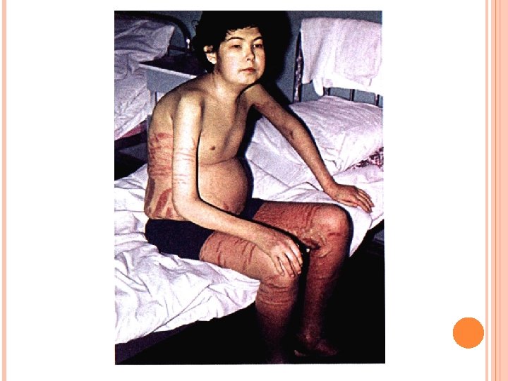
Rhabdomyolysis (a traumatic crash syndrome) In the clinical picture: swelling and pain in the affected muscles, oligo-, anuria with clinical picture of SPD. In the blood increase: aminotrasferazy KLF hyperkalemia, hyperuricemia. PRESCRIBED: proteinuria, myoglobinuria
ØAcute and hraničeskij Glomerulonephritis; Amiloidoze; Diabetic glomeruloskleroz; Thrombosis of the Renal vein Protein fractions: Transferiny Albumin, βmikroglobuliny, Globuoin. The defeat of basement membrane Clubockova proteinuria ( -20 0. 1 g/d)
Isolated urinary syndrome Hypertonic syndrome Chronic Glomerulonephritis Nephrotic syndrome Their combination ХПН
Amyloidosis Chronic infections (tuberculosis, syphilis) Xp. purulent processes (osteomyelitis, bronhoektaza) Rheumatoid arthritis and other diseases. In the 10 -12% of cases develop benign AG In the digestive system (diarrhoea, goiter, syndrome of small suction). In 60 -70% of patients with increased liver, and at 35 -40 percent (gall bladder) In: anemia, blood SEDIMENTATION RATE ≥ 50 mm/s, increased gamma globulin, α 2 and Fibrinogen in serum Decreases the relative density of pee, POO, ZU, sometimes Lou and GU
Сахарный диабет The 10 -12 year disease show signs of severity of Glomerulosclerosis. Most early sign of the disease is somewhat selective proteinuria clubockova. The degree of selectivity is gradually decreasing. The KF-45 m. L/min is kluboč ultrasound and kanaltsevaya nonselective PU. The ad directly correlates with the manifestation of PU. Appearance NS leads to the development of atherosclerosis, accession secondary infection results in CHRONIC RENAL FAILURE With the advent of permanent decline steadily beginning to KF PU with a speed of 1 m. L/min per month.
ØAcute tubular necrosis tubulopathy genetic; Interstitial nephritis; Xp. transplant rejection; Salts of heavy metals poisoning; Toxic effects of drugs. Protein fractions: albumins, β 2 mikroglobuliny, lizocin, light cepy immunoglobulins ØInadequate protein reabsorption in the proximal section of the canaliculus channel protein Overload due to enzyme deficiency Kanaltsevaya proteinuria (2 g/day)
Fanconi syndrome-tubulopatiâ generalized proximal type Includes: -proximal tubular acidosis with bikarbonaturiej; -proteinuria tubulârnogo type; -polyuria; -Renal glycosuria; phosphaturia; -hypophosphatemia; osteomalacia.
Primary Fanconi syndrome-an inherited condition Clinic: delay in physical and mental development, wasting, pain in the bones of the skeleton changes, rahitopodobnye, polyuria, polydipsia, renal glycosuria. Secondary-Fanconi syndrome develops in amiloidoze, giperparatireoze, multiple myeloma, etc. Clinic: PU, deformation of the bones of the skeleton, bone pain, fractures, osteoparoz, polyuria, thirst, gipokaliemičeskaâ male.
Postrenal′naâ (extrarenal, false) PU Largest small (up to 1. 0 g/day). Proteinuria caused by inflammatory exudate rich protein in urine in diseases of the urinary tract (prostatitis), degradation of sperm with the long-term stagnation of urine.
Proteinuria Selective separation of urine protein with low molecular weight, mostly albumin. Histological research on kidney revealed minimal changes. Nonselective-selection of proteins with medium and high molecular weight Demonstrates the serious, sometimes fibroplastičeskom the defeat of kidneys
Minimal proteinuria Loss of protein from 300 mg to 1 g/SUT. Øobstructive uropathy, tubulopathy, nefrolitiaze, polikistoze, intersticial′nom chronic nephritis.
Moderate proteinuria Protein loss in from 1 to 3 g/s Øacute urinary tract infection, cystic-močetočnikovom reflux, kanal′cevom necrosis acute, chronic nephritis with intersticial′nom AG, gepatorenal′nom syndrome, transplant rejection, the primary and secondary glomerulonefritah (excluding NS), amyloidosis, functional stage proteinuričeskoj proteinuriâh.
High or principally nefrotical proteinuria The loss of the protein more than 3 g/day. ØIn the case of the combination of high proteinuria with who had hypoalbuminaemia indicate incomplete nefroticescom syndrome in detecting Hyperlipidemia, gipoalbuminemii, disproteinemii, edema and proteinuria more than 3. 5 g/day of full nefroticescom syndrome.
REMEMBER! ÚGood quality proteinuria should always be considered as a pathological syndrome and in each case, carefully examine the patient to determine the causes of its occurrence.
Proteinuria-Stix-positive test -Test with sulfosalicylic acid-Study utrennoj portions of urine to detect microbial colonies and their sensitivity to antibiotics-Daily proteinuria Proteinuria of less than 200 mg/day normal kidney function normal urine sediment Confirmation Proteinuria over 200 mg/day Ortostatical 200 mg/day Channel Neortostatičeskaâ More than 1 g/d Clubockova Study: Intravenous urography kidney biopsy, test for systemic disease Glomerulonephr itis Histological variants Systemic disease SLE Amyloidosis infekcilonnyj Tubulointersticial′noe disease
Hematuria (èritrocituriâ) Allocation of urine red blood cells more physiological norm, namely above 1 erythrocyte in sight of the microscope in the study of morning urine portion of the toilet. Sample Nechyporenko: more 1 k. red blood cells in 1 ml of urine. Addis-Kakovskogo: more than 1 million. Red for the day.
Mikrogematuria: up to 100 cells in field of view Initial: at the beginning of the Act of urination Hematuria End: at the end of urination Macrogematuria: more than 100 cells in field of view
Causes of hematuria 1. The parenchyma of kidney disease: A. Glomerular Diseases: 1. primary: acute and chronic Glomerulonephritis (including alcohol), Ig. A-jade-Berža disease; 2. secondary: SLE, hemorrhagic Vasculitis, Wegener, granulematoze uzelkovom periarteriite, chronic active hepatitis, etc. Б. Infection (infective endocarditis). В. Renal parenchyma tumor (benign and malignant). Г. Hereditary diseases (Al′porta syndrome).
2. The anomaly development of kidney vessels (bessimtomnaâ, hematuria caused by Renal vein hypertension, possible arteriovenous shunts, Vasculitis). 3. Falling. 4. Vnepočečnye causes of hematuria (blood disease, coagulation defect, DVS-sindrom, overdosage of anticoagulants in the treatment of cytotoxic drugs, as well as systemic disease, hematuria, proteinuria which usually, possible eritrocitariani clindruriâmi).
Hematuria Painful form of hematuria of renal Trauma urolithiasis sickle-cell anemia Polikistoze Without a painful form of Glomerulonephritis hematuria (in this case, it is possible to combine it with PU and TSU); The Tumor.
Pain in the side or upper abdomen usually indicate the origin of hematuria of the upper half of the urinary tract. Irradiruûŝie pain from the abdominal region in the external genitals are when blood clots of ureteral obstruction resulting in bleeding or kidney cancer, sickle-cell anaemia, and papillary necrosis in diabetes mellitus, tuberculosis, etc.
Isolated hematuria may occur if Iga-nephritis in children and adults under the age of 30 years, more often in men is accompanied by a dull pain in the lumbar area and can recur with pharyngitis. In the blood level of Iga. Infective endocarditis in 20% of cases of hematuria due to development of Glomerulonephritis, -60 30%-infarction of kidney. The hematuria is combined with the fever of the correct type, positive gemokul′tura, formation of the heart, thromboembolic complications, splenomegaly, increase Monday. Hereditary nephritis (Al′porta syndrome) is characterized by a combination of gemoturii with reduced vision and hearing, evident in several generations.
Hematuria in conjunction with frequent urges to urinate, urinary incontinence, urinary disorders and fever in young women is often the result of cystitis. Such symptomatology in men of young age usually associated with prostatitis or burns stricture of the urethra, and in older men in consequence of benign hyperplasia or prostate cancer. Cyclic hematuria occurs during menstruation, testifies to the presence of endometriosis of the bladder.
For tumors of the hematuria may be of varying intensity, is not permanent and asymptomatic in the early stages of tumor growth. When the urolitiaze is accompanied by pain-renal colic. Kidney polikostoz clinically manifested "unmotivated" recurrent hematuria, varying in intensity polyuria, blunt pain in the lumbar region, often combined with cysts in the liver, the pancreas. Hematuria of renal tumors, urolitiaze, cysts, trauma and inflammatory diseases of the urinary tract as the rules due to the destruction of the vascular wall.
A. differential diagnosis history physical examination of urinary sediment microscopic examination of the biochemical analysis of blood and urine B. Topographic diagnosis. As a first step, the history of the present illness 3 -õ stakannyj test. urine sediment excretory urography
В качестве второго шага Vascular lesions, tumors Angiography. Echography. Computer tomography Defeat of the urinary tract Urethrocystoscopy. Retrograde pielografia Nephropathy Daily proteinuria. Determination of the erythrocyte cylinders. Kidney biopsy
The excretory kidney size reduction: urogramme
Gemoglobinizirovannye red blood cells when postrenal′noj hematuria, HC 400 h.
Altered renal blood, HC 1000 h.
Pyuria-Excretion in the urine of white blood cells, the amount of which exceeds the norm. PRESCRIBED in more than 56: under review Sample Kakovkogo. Addis-more than 2 -4, 0 h 106/day
Pyuria insignificant (from 8 -10 to 2040 cells in the field of view). moderate (50 -100 in the field of view) pronounced (piuria -leukocytes cover all fields of FPI crowd)
Detection of LOU always indicates the presence of inflammation in the kidneys or urinary tract: tubulointerstitial nephritis, pyelonephritis, rejection reactions kidney transplantirovannoj, urinary tract infection, cystitis, etc. Lejkocituriej accompanied by renal tuberculosis, prostatitis, hydronephrosis, kidney stones and other urinary disorders of the kidneys and urinary tract.
Neutrophils, HC. 250 h
Neutrophils in alkaline urine, HC. 400 h
Neutrophils and eosinophils. Hc. 400 h Èozinofil, HC 1000
Zilindruria This excretion in the urine of cylinders, which are "cast", formed in the lumen of seminiferous tubules of protein or cellular components. Depending on which and how many particles cover the cylinder cast, there are protein
Zilindruria. Hc. 200 x
protein origin (hyaline, granular, voskovidnye) the cell of origin (human erythrocyte, leukocyte, epithelial) Cylinders consisting of breakdown products of red blood cells (pigment) consisting of renal epithelial cells decay products (coarse and fatty, gialinovo-tiny)
hyaline гиалиновые granular зернистые voskovidnye восковидные Цилиндрурия Zilindruria эритроцитарные human erythrocyte Leukocyte
Гиалиновые при всех заболевание почек, сопровождается с протеинурией, состоит из свернувшийся сывороточного белка. Granular formed from epithelial cells of the proximal of degenerated tubules. Voskovidnye formed in the lumen of distal tubules resulting deaths (dystrophy, atrophy) epithelial kanal′cevogo of those divisions. Human Erythrocyte Leukocyte in severe hematuria of different origin. Pigment with piurii in patients with acute, purulent and XP. pyelonephritis in the phase of deterioration, etc. in all gemoglobinuriâh (the impact of toxic substances, etc. ).
The modified kidney cells of the gialinovom cylinder, HC 400 h.
Hyaline cylinder with a renal epithelium, HC 1000 h.
A wide cylinder voskovidnyj Hc 400 h.
Granular cylinder, HC 400 h.
Accumulation of epithelial cylinders, HC 400 h.
KIDNEY EPITHELIUM ON THE SURFACE OF THE CYLINDER HYALINE. PAINTING OF AZUR-EOSIN, HC 1000 H.
SHORT HYALINE CYLINDER. HC 1000 H. Hyaline long cylinder. HC 400 h.
Red blood cylinder Hc. 1000 h
Short cylinders of Leukocyte detritus), HC 400 h.