Different techniques in Immunology Dr G Sunil Babu

Different techniques in Immunology Dr. G. Sunil Babu Assistant Professor, Department of Biotechnology Babasaheb Bhimrao Ambedkar University Rae Bareli Road, Lucknow-226025

PREFACE Antigen-antibody interactions can occur both as well as in vitro LABORATORY ASSAYS Immune reactions Has exquisite specificity & sensitivity. Lower times to perform Possible to detect any molecule (hapten to Ag) Diagnosing the diseases Monitoring the humoral response Identification of biomolecules of interest Both quantitative and qualitative in vivo

Antibody- Central molecule in immune based techniques • Immunoglobulins when taken the role of being antibodies are powerful molecules with exquisite specificities and a myriad of functions. • Antibody molecules have increasingly become indispensable for many applications ranging from Diagnostics, Therapeutics and Research

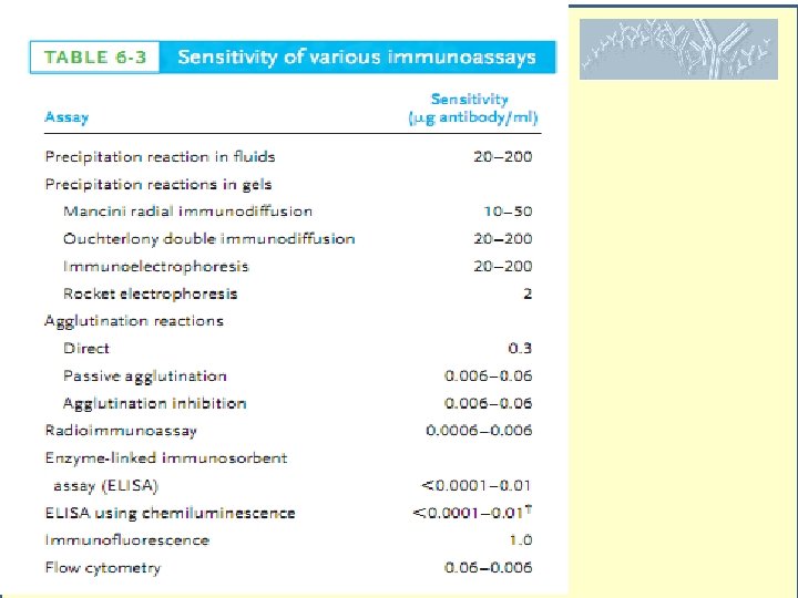
Ab- without tagged molecules Precipitin Reactions Agglutination reactions Ab- with labels Immunofluorescence RIA ELISA FACS Western blotting These assays comes with various formats and sensitivities

Precipitin reactions Principle: Ags and Abs must be bivalent/ polyvalent (monovalent Fab fragments doesn’t form precipitates) Ability to form Lattice Passive diffusion Fluids Double diffusion- Capillary tube precipitation Gels : Radial immunodiffusion (Mancini method) Double immunodiffusion (Ouchterlony method) Active Diffusion: Electrophoresis Immunoelectrophoresis Rocket immunoelectrophoresis Equivalence – Lattice formation

Immunodiffusion Mancini method Ab in gel Ag Ag Diameter 2 Ag Ag Concentration Takes time (24 hrs) Ouchterlony method Ag

Clinical uses Identity A Ag B Ag Ab Partial identity Ag Non-Identity A Ag Ag B Ag A B Ab Ab

Immuno electrophoresis Used to detect presence or absence of proteins in serum. • Determining abnormality of isotypes. • Overproduction of serum proteins such as albumin, transferrin

Rocket Immuno electrophoresis • Quantitaive technique • -vely charged Ag is electrophoresed • The height is proportional to the concentration of Ag Some proteins (Ig. G) not sufficiently charged
 Agglutination reactions + + + Principle: Ab titer Interaction between Ab and particulate")
(control) Agglutination reactions + + + Principle: Ab titer Interaction between Ab and particulate Ag results into visible clumping Efficient isotype is Ig. M followed by Ig. G Agglutination occurs in minutes Heme Agglutination Bacterial Agglutination

Agglutination reactions Passive Agglutination: Soluble Ag can be coated on latex beads Ag reaction measured. Doping tests Agglutination Inhibition Pregnancy kits

Immunofluorescence • 1944 - Albert Coon showed Abs-labelled with flurescent molecules §Direct immunofluorescence §Indirect immunofluorescence Fluorescence is the emission of light by a substance that has absorbed light or other electromagnetic radiation Fluoresceicn- 490(blue)-517 nm(intense yellow-greeen) Rhodamine- 515 -(yellow-green)546(Red) Phycoerythrin red

Radio Immuno Assay Rosalyn Yalow Principle: Competetive binding of radiolabelled and unlabelled Ag to high affinity antibody Ag generally labelled with I 125 and H 3 Step 1: determine the amount of Ab needed to bind 50% or 70% of a fixed quantity of labelled Ag. Ag*+Ab Ag*Ab Precipitated by Antiantibody Protein A Ag*Ab complex Supernatant Ppt Measure radioactivity §Plasma levels of most of our hormones T 3 and T 4 levels §Digitoxin or digoxin in patients receiving these drugs §Certain other drugs §For the Presence of hepatitis B surface antigen (HBs. Ag) in donated blood §Anti-DNA antibodies in systemic lupus erythmatosis Solomon Berson

RIA- Limitations • The expense and hazards of preparing and handling the radioactive Ag • Both 125 I or 131 I emit -radation, requires special counting equipment • Body concentrates iodine atoms- radioactive or not; in the thyroid gland they are incorporated in thyroxine. T 4 • Not able differentiate biologically active or inactive form

ELISA An enzyme conjugated with an antibody reacts with a colourless substrate to generate a coloured reaction product. • Direct • Indirect • Sandwich • Competetive • Multiplex Eva Engvall Peter Perlman Alkaline phosphatase-PNPP (p-Nitrophenyl Phosphate)- yellow water-soluble product ( 405 nm) Horse radish Peroxidase- 1. ABTS (2, 2'-Azinobis [3 -ethylbenzothiazoline-6 -sulfonic acid]- a water-soluble green product ( 410 nm & 650 nm). 2. OPD (o-phenylenediamine dihydrochloride)- yellow-orange ( 492 nm) 3. TMB (3, 3', 5, 5'-tetramethylbenzidine)-a blue color ( 450 nm). TMB is very sensitive and may produce significant background signal if too much protein used. or antibody is

ELISA Variants- Indirect For assaying Antibody, in HIV test. Recombinant HIV Ag coated to the wells HIV specific Abs can be detected generally 6 weeks of infection Diagnostic purpose

ELISA Variants. Sandwich Especially the antigen does not need to be purified prior to use. For example the strain identification of microorganisms or pathogens are generally done with this assay during epidemics. It is useful for quantitation of antigens when antigen concentration is low and/or has higher concentrations of contaminating protein(s).

ELISA Variants- competitive The more Ag present in the sample less colour

Multiplex ELISA Multiplexing means multiple readings from a single sample. For example, using the Q-Plex. TM Cytokine array, with one sample and one well, you can receive data for the following assays: IL 1 alpha, IL-1 beta, IL-2, IL-3, IL-4, IL-5, IL-6, IL-9, IL-10, IL-12, MCP-1, INFgamma, TNFalpha, MIP-1, GMCSF and Rantes. Why Multiplex ELISAs? Not only get more data from one well, but the size of the sample is much less (only 30 ul for 16 cytokines; traditional method require almost 1 ml). The advantages of this are clear in newborn studies where sample draws are very low as well as mouse and other small animal models. Time ; Cost

ELISA – increasing sensitivity CHEMILUMINESCENCE In versions of the ELISA using chemiluminescence, a luxogenic (lightgenerating) substrate takes the place of the chromogenic substrate in conventional ELISA reactions. oxidation of the compound luminol by H 2 O 2 and the enzyme horseradish peroxidase (HRP) produces light: luminol H 2 O 2 Ab-HRP + Ag Limunol+ H 2 O 2 Ab-HRP-Ag light • enhanced sensitivity • detection limit can be increased at least ten-fold • In fact, under ideal conditions, as little as 5 X 10 -18 moles (5 attomoles) of target antigen have been detected.

ELISPOT • Quantitative determination of cells in a population that are producing antibodies specific for a given Ag. • Wells were coated with either Ag specific for Ab specific for Ag. A suspension of cells under study added. • The cells settle on the bottom and secrete molecules specific for Ab, producing a ring • Plate is washed and added enzyme labelled Ab. • Subsequent addition of chromogenic substrate yields a spot where the Ag producing cell is present.

FACS

Western Blotting • Identification of a protein. • Protein mixture is electrophoretically separated by SDS-PAGE • Protein bands are transferred into nitrocellulose membrane • The individual bands were detected through the specific Abs. Use: Confirmatory test for HIV

Western Blotting

Monoclonal Antibodies Hybridoma technology

Thank you
- Slides: 28