Difference Between Prokaryotic And Eukaryotic Cell Cell The

Difference Between Prokaryotic And Eukaryotic Cell

Cell ü The cell Latin cella, meaning "small room”. ü Basic structural, functional and biological unit of all known living organisms. ü Cells are the smallest unit of life that can replicate independently, ü "building blocks of life“ ü There are two types of cells, eukaryotes, which contain a nucleus, and prokaryotes, which do not.

PROKARYOTIC CELLS

Functions of Cells v. A boundary that keeps the cellular contents separate from the external environment but allows for the transfer of some substances into and out of the cell. v. Replication of DNA v. Synthesis of cellular components v. The ability to obtain energy through metabolic processes
 are cellular")
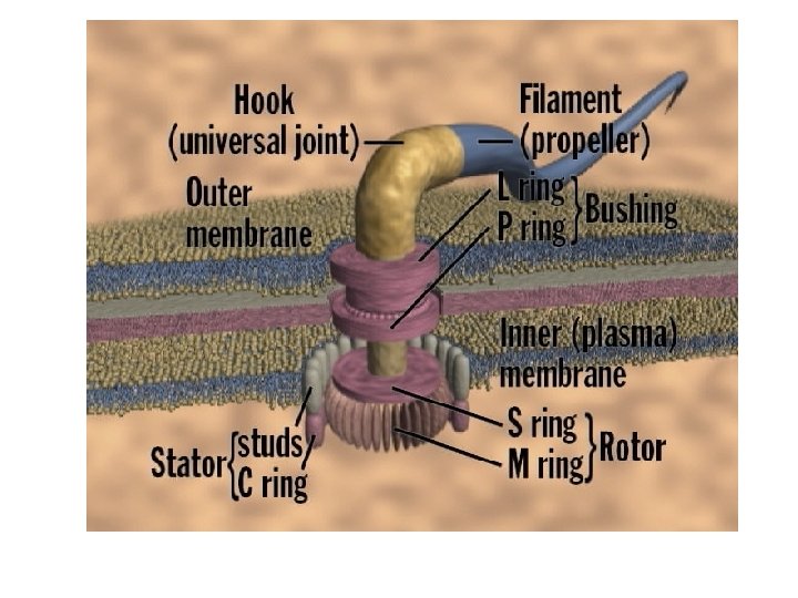
Appendages are basically involved in movement or adhesion Ø Flagella (singular flagellum) are cellular appendages that consist of three parts: 1. A filament that rotates for movement 2. A hook where the filament attaches 3. A basal body that anchors the hook to the cell Ø The arrangement of the hook/basal body articulation allows the hook with its filament to rotate 360 o

Arrangements of Flagella

Axial filaments Ø Modified flagella that occur in spirochetes

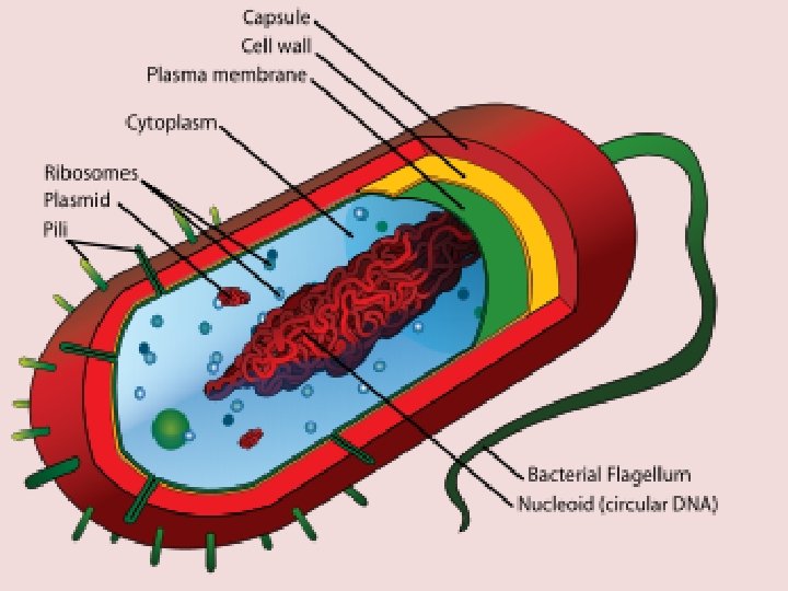
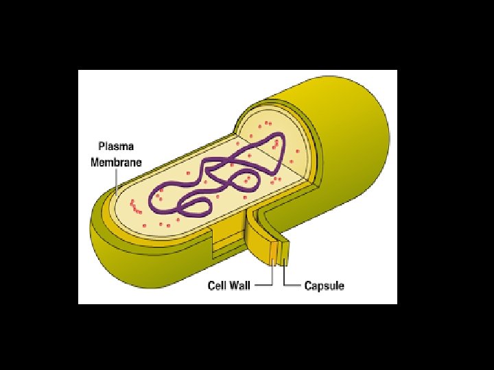
Cell envelopes differ between taxa but they basically consist of three layers: Ø The capsule or slime layer (outermost layer) differs greatly in thickness, organization and chemical composition depending on the bacterial species. Ø Beneath the outer layer lies the cell wall. Ø The cell membrane is a thin flexible sheet that surrounds the contents of the bacterial cell. Its functions include: transport, energy extraction, nutrient processing, and synthesis

The Gram Stain

The protoplasm or cytoplasm o Dense gelatinous solution within the cell membrane o Primary site for the cell’s biochemical and synthetic processes.

Nuclear region Ø chromatin body or the bacterial chromosome Ø Nucleoid or nuclear region of the cell that is associated with the chromatin body

Plasmids Ø Plasmids are tiny circular extra chromosomal strands of DNA Ø Ribosomes are small structures consisting of RNA and proteins that are involved in protein synthesis

Inclusions or granules Ø Inclusions or granules areas where nutrients are concentrated Ø Endospores are dormant structures produced by some species of Bacillus and Clostridium.

Shapes and arrangements of bacteria

FUNGI Ø Fungi are eukaryotic protista; differ from bacteria and other prokaryotes. 1. Cell walls containing chitin (rigidity & support) , other polysaccharides 2. Cytoplasmic membrane contains ergosterols 3. Possess true nuclei with nuclear membrane & paired chromosomes. 4. Divide asexually, sexually or by both 5. Unicellular or multicellular

• • Simplest fungus : - Unicellular budding yeast Hypha : - Elongation of apical cell produces a tubular, thread like structure called hypha Mycelium : - Tangled mass of hyphae is called mycelium. Fungi producing mycelia are called molds or filamentous fungi. Hyphae may be septate or nonseptate

CLASSIFICATION • Depending on cell morphology 1. Yeasts 2. Yeast like fungi 3. Molds 4. Dimorphic fungi

Eukaryotic Cell

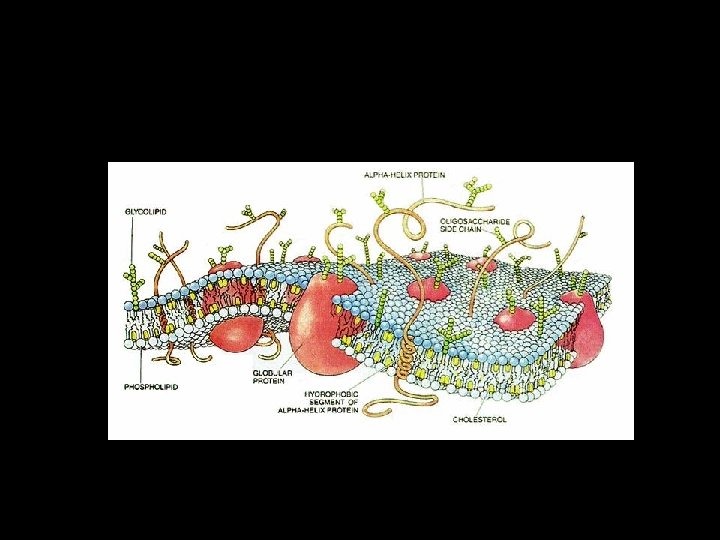
CELL MEMBRANE Structure. Components Arrangement Functions Barrier Transport (know diffusion, osmosis, facilitated diffusion and active transport) Recognition (e. g. , self vs. non-self) Reception (for protein hormones)
 – Nucleolus")
Nucleus Structure and Function – membrane similar to cell membrane (similar function) – Nucleolus (formation of ribosomes) – Chromosomes (gene expression) – Nucleoplasm (matrix)

Ribosomes Ø Structure – r. RNA – Proteins Ø Function – Site of protein formation (translation) Ø Found in both prokaryotes and eukaryotes (different structurally)

Endoplasmic Reticulum Ø Structure membranous system of tunnels and sacs – Rough – with ribosomes on surface – Smooth- no ribosomes on surface Ø Function – Rough – protein synthesis – Smooth- lipid synthesis

Golgi Apparatus Ø Structure also membranous, kind of like a stack of pancakes Ø Function processing of lipids and proteins

LYSOSOMES Ø Structure Membrane bound sac containing hydrolytic enzymes Ø Function Digestion

Mitochondria q Structure – cigarshaped, double membrane-bound organelle q Function – Energy transfer by ATP synthesis

Chloroplast v. Structure Also cigar or spindle shaped, double membrane-bound, green v. Function photosynthesis

Other structure • • Cell walls, not in animal cells Vacuoles Cytoskeleton Cytoplasm

Differences Between Prokaryotic and Eukaryotic cells

Features No 1. Occurrence Prokaryotic These are cells are the are found in all, characteristic animals and of bacteria plants, except and blue green algae and bacteria.

2. Size Mostly 10 -100 μm 1 -10 μm 3. Multicellular Rare forms Common, with extensive tissue formation

4. Cell wall Present in most but not The animal in all cells. In Bacteria, cells lack cell wall is made up cell wall, murein, polysaccharides, but plants lipid and proteins. cell wall is made up of cellulose and chitinous cell wall is present in fungi.

5. Plasma membrane Present 6 Nucleus Present

7. Nuclear Membranes Absent 8. Chromatin with Absent histone 9. Number of chromosomes Present Number of Each cell chromosomes Has only per cell one depends chromosome upon the type of organism.

10 Chromosome 11. Genetic material The Chromosome is circular ring lacking a centromere. Each chromosome is linear having a centromere Linear double Circular or stranded DNA: linear, double genes frequently by stranded DNA: interrupted intron sequences only exons are especially in higher present eukaryotes (called as split genes).

12 Nucleoli and Absent Mitotic apparatus Present 13. Nucleolus Present (for the synthesis and organization of ribosomes) Absent

14. Plasmid Commonly present 15 Mesosomes Absent perform the function of Golgi bodies and mitochondria, and also help in the separation of chromosomes during cell division. Rare

16. Cell organelles Absent Mitochondria Present Endoplasmic Reticulum Absent Present Vacuoles Absent Present

Lysosomes Absent Present Chloroplast Absent Present Centrioles Present Absent Ribosomes Only 70 S type of ribosomes are present which lie free in cytoplasm, or are engaged in protein synthesis. The cytoplasm has 80 S type of ribosomes; and plastid and mitochondria have 70 S

Microtubules Flagellae Absent Present Complex Simple structure 9+2 Composed of the Structure protein Flagellin. of tubulin and other protein.

17 Respiration Many strict anaerobes Photosynthetic Bound to plasma 18 Enzymes membrane as All aerobic, but some facultative Anaerobes By secondary modifications. Enzymes packed in plastids composite bound chromatophore by membrane

Metabolic Great Variations Patterns 19 20 Sexual System Rare: If present one way (and usually forming partial diploids or merozygotes) All share cytochrome electron transport chains, Krebs cycle oxidation, glycolysis. Both sexes involved in sexual participation and entire genomes transferred

21 22 Cyclosis There are no streaming movements of cytoplasm Protein Transcription and translation Synthesis take place in cytoplasm. 23 Duration of cell cycle Cell cycle is short, takes 20 -60 minutes to complete. Cytoplasm shows streaming movements Transcription occurs in nucleus and translation takes place in cytoplasm. Cell cycle is long, takes 12 -24 hours to complete.

CONCLUSION Ø Cell are basic unit of organization or structure of all living matter Ø There are two types of cell, that are prokaryotic and eukaryotic cell Ø There are so many difference in between them include occurance, size, cell wall, nucleus , nuclear membrane, cell organelles respiration, sexual system , protein synthesis.

REFERENCE Ø Prescott LM Harley JP and Klein DAMicrobiology Ø John Webster-Introduction to fungi Ø Voet and voet Ø Tortora-Microbiology an introduction Ø Pelczar Jr. MJ Chan, Ecs and Kreig. Microbiology Ø Lehninger’s principle of biochemistry
- Slides: 49