Die angeborenen Defecte der Kammerscheidewand des Herzens Z

‘Die angeborenen Defecte der Kammerscheidewand des Herzens’ Z. klin. Med. , 32, Suppl. - Heft, 1 -28, 1897.
![� Victor Eisenmenger � [1864 -1932] � Austrian � 1897 Physician](http://slidetodoc.com/presentation_image/3b9088df83bb810d1d9da5d77d0aff9b/image-2.jpg "� Victor Eisenmenger � [1864 -1932] � Austrian � 1897 Physician")
� Victor Eisenmenger � [1864 -1932] � Austrian � 1897 Physician

The time frame � 1888 -Etienne � 1944 - Louis Arthur Fallot First BTT shunt surgery

The case of V Eisenmenger 32/M, powerfully built man � H/o cyanosis+ � Moderate breathlessness since infancy � Jan 1894 worsening dyspnoea � Aug 1894 Frank HF � O/E cyanosis, clubbing, polycythemia � Distended liver, extensive edema � Loud systolic murmur 3 rd & 4 th LICS � Basal diastolic murmur from pulm incompetence � Improved with rest & digitalis � Nov 13 massive hemoptysis sudden collapse & death �

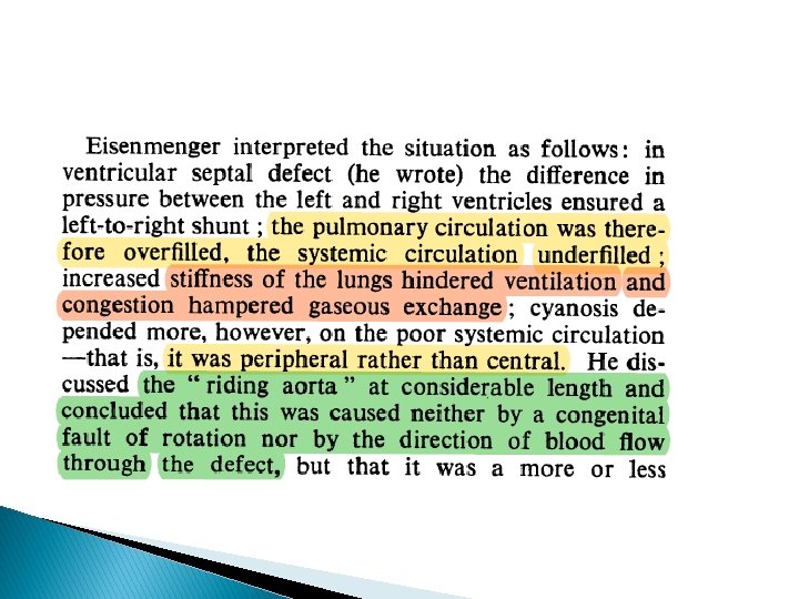
Pitfalls � Cyanosis was peripheral rather central � SM to left right shunt � Attributed the rise in pulmonary vascular resistance to multiple thrombosis and pulmonary infarction –Lt UL & Rt LL � Reasons for misinterprentation: ◦ Cyanosis since birth couldn’t attribute it to reversed shunt due to PH ◦ Misleading JVP findings

Conflicting Hypothesis � 1924 - Abbot & Dawson � 1947 - H Taussig � 1950 - Brown
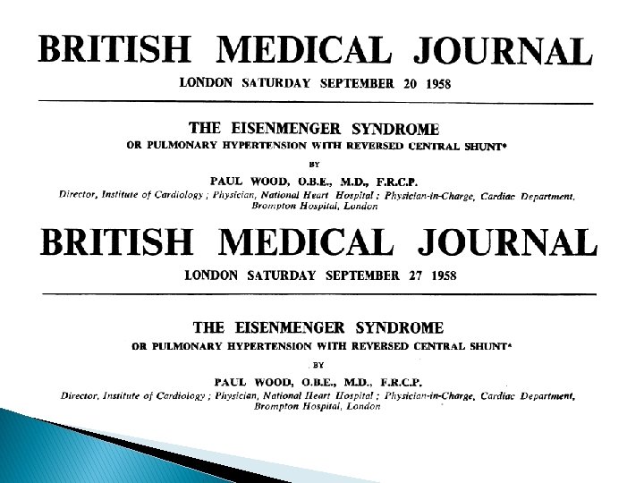

Paul Wood 3 � 1907 � Born in India � Died in 1962 � AWMI � Coined the term “Eisenmenger Syndrome”

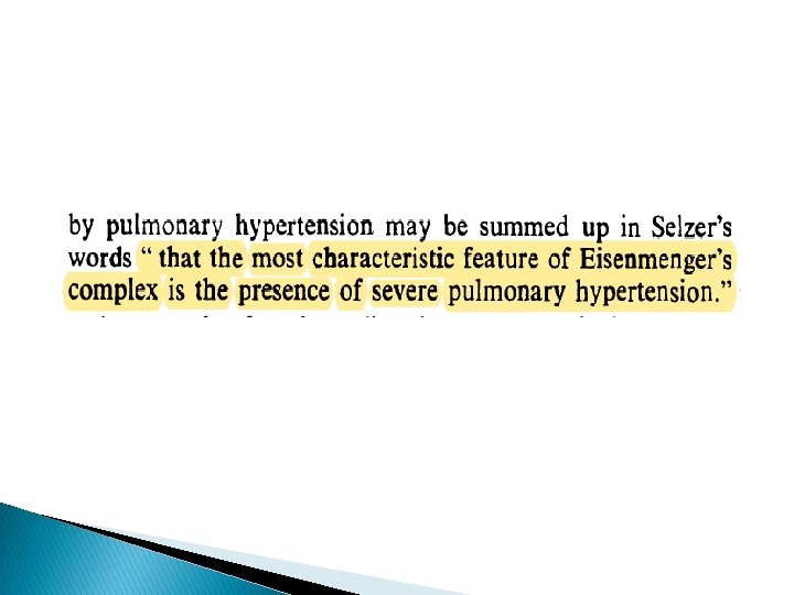
� 1947 - Bing et al ◦ 5 cases ◦ Pulm pressures at systemic level ◦ Bidirectional shunt � 1950 - Burchell � 1950, 1952 - Wood � 1951 – Selzer � 1953 - Soulie

The syndrome described is Pulm HTN with Reversed or Bidirectional Shunt It matters very little where the shunt happens to be
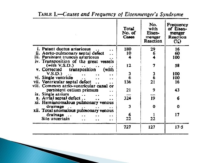

� Total period -11 years � Total cases – 127 � Death – 18 � Autopsy – 15 � 38 autopsy results from Britain � Total – 53 cases for study

Age

Sex

Onset of ES

Symptoms � Effort intolerance + � PDA- ES - best tolerated � Angina, Syncope, Hemoptysis, CHF

Hemoptysis � Eisenmenger Complex ◦ 33% ◦ Never before 24 yrs ◦ 100% by 40 yrs � 25% of ASD-ES � 4% of Pri PAH � Large hemoptysis – imm death -29% of 42 deaths

5 Facets of Arterial desaturation � Cyanosis � Clubbing � Polycythaemia � Differential � Squatting Cyanosis –seen in 50% of PDA-ES ◦ Uncommon, when + usually a/w VSD

� Jugular Venous Pressure & Pulse ◦ Commonly normal ◦ Small dominant a wave – 20 -25% of each subtype ◦ Giant a wave seen in 18% of ASD-ES � Right atrial Gallop ◦ 38% of ASD-ES ◦ 2 -3% of others � S 2 – single in 55% of VSD-ES ◦ Close split S 2 - indeterminate ◦ Obvious or wide split – favoured ASD-PAH -86% of cases
 ◦ 20% of ASD-ES ◦ None in")
ECG � Paroxysmal AT or flutter(rarely fibrillation) ◦ 20% of ASD-ES ◦ None in VSD-ES ◦ 5% of PDA-ES � RV dominance seen in 80% of ASD-ES ◦ 44% when shunt elsewhere ◦ Some evidence of LV enlargement in addition to RV with large biphasic clx in chest leads PDA-ES �Q in V 5, V 6 ◦ Present in 82% of PDA-ES ◦ Absent in 87% of ASD-ES

CXR � Rt Aortic Arch - 16% of VSD-ES � Rounded shadow overlying the aortic knuckle [double edged app representing the duct itself] � Ductal calcification � Cardiomegaly � Diminished – MC with ASD-ES 85% Pulm Vascular markings beyond the dilated main hilar branches >60% all grps

Eisenmenger Syndrome-Definition

Eisenmenger Complex � “Maude Abbott” coined the term � VSD with reversed shunt in the absence of Pulmonary stenosis � Rt to Lt shunt ◦ Initially attributed to the over riding aorta ◦ Later found to be d/t increased PVR

Dye dilution Curve

Size of the defect � PDA <5 mm � VSD <1 cm � ASD ~ 3 cm

Elastic arteries � In fetus / neonates: ◦ Elastic fibers in PA – long & densely packed ~ Aorta � Fall in PVR Fibers break, shorten & loosen up � PA in VSD & PDA ◦ Resembled foetal (aortic ) pattern � In ASD & all acquired PAH ◦ Normal PA appearance ◦ - Heath[1958]

� Pulm HTN in Eisenmenger Syndrome ◦ VSD & PDA – from birth ◦ ASD – must be acquired

� Muscular Arteries: ◦ <1 mm diameter ◦ Persistence of fetal type of muscular artery ◦ Contributed to the high pulmonary vascular resistance � Arterioles: ◦ ◦ <100 u in diameter endothelium, single elastic layer & adventitia not supplied by muscle Thin walled & dilated

Overriding of Aorta � � � VSD - 1 of 12 ◦ may lead to high PVR & reversed shunt ◦ An overriding aorta or its physiological equivalent - absent in 7 of these conditions PVR – High Sp. O 2 ~ 80% Eisenmenger complex –classical ◦ Defect in muscular IVS ◦ There can be NO OVERRIDING OF AORTA

Experimental Pathophysiology

Course & Prognosis � � ES begins @ birth or during the first 2 yrs when the shunt is post tricuspid ES – typically acquired when the shunt is inter atrial Cyanosis or Dyspnoea – no appreciable change till adolescence Hemoptysis signal for the downward trend

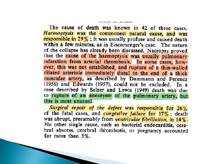
Mode of Death � Cause of death- known in 42 cases � MCC- Hemoptysis -29% ◦ Pulmonary infarction from arterial thrombosis ◦ Rupture of a thin walled dilated arteriole immediately distal to end of a thick muscular artery ◦ Rupture of aneurysm of PA (Most unusual)
![� Of the 18 + 38 deaths, [avg age of death] ◦ 33 yrs](http://slidetodoc.com/presentation_image/3b9088df83bb810d1d9da5d77d0aff9b/image-38.jpg "� Of the 18 + 38 deaths, [avg age of death] ◦ 33 yrs")
� Of the 18 + 38 deaths, [avg age of death] ◦ 33 yrs for both Aortopulmonary & VSD ◦ 36 yrs for ASD � 14% - SCD presumed to be VF � 26% - died from surgical attempt � 17% - CHF

Secret of Eisenmenger
![� 1. ◦ Alan Burton, 1958 �Resistance – Kinked vessels �Gnarled appearance[rough& twisted] �](http://slidetodoc.com/presentation_image/3b9088df83bb810d1d9da5d77d0aff9b/image-40.jpg "� 1. ◦ Alan Burton, 1958 �Resistance – Kinked vessels �Gnarled appearance[rough& twisted] �")
� 1. ◦ Alan Burton, 1958 �Resistance – Kinked vessels �Gnarled appearance[rough& twisted] � 2. ◦ Williams �Kinks get straightened – lowers resistance ◦ Jaykka �Blood fills up the vascular bed- expansion ◦ Dawes et al �Only when gaseous filling PVR decreases � Hypoxic vasoconstriction

� 3. ◦ Dawes �Fn of thick muscular arteries �Well developed in fetal life in relation to systemic pressures �Are in a state of vasoconstriction – can be released by Ach � 4. ◦ Bayliss �Slowly tone in muscular arteries decreases as pressure decreases ◦ Williams �Resistance decreases Flow increases Vessels dilate Further fall in resistance �As pressure falls muscular arteries atrophy decrease the resistance

Pulm HTN in ASD � During the critical period of involution and relaxation of muscular arteries no interatrial shunt � Before the shunt operates, the filling resistance needs to be considered � How soon the RV involutes sufficient enough to permit an interatrial shunt- unknown � It is slow, slower than involution of muscular arteries � Nadas ◦ Pulm SM characteristic of increased flow not heard until the child is well past the neonatal period � Difficult for ES to set in later, pulmonary vessels are fully involuted and increased PBF follows as in others

Summary � Defn: - PH d/t high PVR with reversed or bidirectional shunt @ A-P or V or A level � 12 diff condtions � Large inter circulatory connections ◦ Exceeding >0. 7 cm in A-P ◦ >1. 5 cm when interventricular ◦ >3 cm when interatrial � Obliterative pulm vascular disease d/t “endateritis fibrosa” � Thrombo-obstructive lesions are late

ASD VSD SEX RATIO 1: 3 DOE GRADE 3 GRADE 2 ONSET LATE EARLY CENTRAL CYANOSIS CLUBBING, POLYCYTHEMIA DIFFERENTIAL CYANOSIS 75% -- 1: 1 90% --- DOMINANT a OR LARGE V in JVP 1/3 RD RV LIFT CONSIDERABLE ( NEVER ABSENT) SLIGHT OR MODERATE (ABSENT IN 10%) S 2 OBVIOUSLY SPLIT SINGLE OR CLOSE SPLIT ECG-P PULMONALE RVH Q IN V 5, V 6 XRAY – RAE RT SIDED AORTA LEFT SVC CALCIFIED DUCT PROMINENT AORTIC KN. >50% 2/3 RD -60% ----- RARE <50% 1/3 RD 15% 16% 8% -SEEN PDA 1: 2 GRADE 2 EARLY 30% 50% UNUSUAL SLIGHT OR MOD. (ABSENT IN 10%) CLOSE SPLIT UNUSUAL 1/3 RD 50% 15% --RARE SEEN

� 188 patients � Median follow up period - 31 years � 64 males, 124 females � Mean age - [33 +/- 12. 8 yrs] � CHD ◦ Simple – 128 �ASD-21, VSD-71, PDA-36 ◦ Complex – 60 �TGA+VSD-8, AVCD-23, DORV-4, SINGLE VENTRICLE- 9, TRUNCUS- 11, CCTGA+VSD -4
 atleast")
Results � Complex CHD ◦ earlier deterioration, shorter survival � 20. 2% (38) atleast one episode of hemoptysis between 20 -40 years � 13. 2% (25) had pulm thromboembolism � 7. 9% (15) had stroke � 3. 7% (7) had cerebral abscess

� Cerebral complications - impaired the QOL � Venesection ◦ Target Hb <18 g/dl ◦ did not reduce cerebral complications ◦ 20% caused IDA � Non cardiac surgery – 23. 5% of deaths

� Pregnancy ◦ ◦ ◦ ◦ 39 Pregnancies in 22 pts 15 pregnancies - terminated 14 pregnancies - Spontaneous abortion – (35. 8%) Only 10 continued All delivered before 37 wks – 9 LSCS, 1 NVD Clinical deterioration in all post partum 3 deaths – � 2 cases - 3 rd & 14 th day post LSCS � 1 case - 10 days after NVD ◦ 10 babies survived – all SGA – one PS, one COA

� Total deaths- 61 ◦ Sudden death – 29. 5% �SCD MORE D/T ARRYTHMIAS THAN HEMOPTYSIS ◦ Heart failure – 22. 9% ◦ Hemoptysis – 11. 4% � 8 cases – Heart Lung TX ◦ 5 died between 1 week and 4 years

� 1976 -92 � Total - 201 pts � Mean age of presentation - 19 yr � 12 anatomic lesions ◦ MC - VSD(33. 3%), ASD(29. 85%), PDA (14. 3%) � 20 deaths in follow up period of 54 +/-54 mths � SCD (30%), CHF(25%) & HAEMOPTYSIS(15%) � 5 YR, 10 YR, 15 YR SURVIVAL was 86. 95%, 79. 6%&76. 9% � Level of shunt didn’t influence survival � Prognostic factors identified were ◦ Syncope, elevated RAP >8 mm. Hg, Sp. O 2 < 85%

THANK YOU
- Slides: 53