Diagnostiek en beleid bij sterk gevasculariseerde zwangerschapsresten Thierry

Diagnostiek en beleid bij sterk gevasculariseerde zwangerschapsresten Thierry Van den Bosch VVOG jaarcongres Gent Donderdag 2 oktober 2014

Geschiedenis Color Doppler
 171 -178 Case")
European Journal of Obstetrics & Gynecology and Reproductive Biology 92 (2000) 171 -178 Case series n=9 Selective embolisation: 1
 171 -178 Case")
European Journal of Obstetrics & Gynecology and Reproductive Biology 92 (2000) 171 -178 Case series n=9 Cross-sectional series n = 385 Enhanced myometrial vascularity (8. 3%) ~ retained products of conception
 171 -178 Case")
European Journal of Obstetrics & Gynecology and Reproductive Biology 92 (2000) 171 -178 Case series n=9 Cross-sectional series n = 385 Prospective n = 93 52% EMV = involution of placental bed Williams JW. Am J Obstet Gynecol 1931; 22: 661 - 4%
 171 -178 Case")
European Journal of Obstetrics & Gynecology and Reproductive Biology 92 (2000) 171 -178 Case series n=9 Cross-sectional series n = 385 Prospective n = 93 J Ultrasound Med 2008; 27: 343 -347 Prospective n = 1070 RPOC in 6, 3% EMV in 77, 3%
 ~ PSV (> 60 cm/sec)")
Case series n = 30 8 embolisations (27%) ~ PSV (> 60 cm/sec)

Complications after embolisation … § § § thrombo-embolic events uterine perforation vesicovaginal fistula perforation transverse colon uteroenteric fistula buttock necrosis labial necrosis septic shock affect ovarian reserve cause synechiae infertility • • • • Acharya J, Bancroft K, Lay J. Perforation of Transverse Colon: A Catastrophic Complication of Uterine Artery Embolization for Fibroids. Cardiovasc Intervent Radiol 2012; 35: 1524 -1527. Aungst M, Wilson M, Vournas K, Mc. Carthy S. Necrotic leiomyoma and gram -negative sepsis eight weeks after uterine artery embolization. Obstet Gynecol 2004; 104: 1161 -1164. Denen SB, Mani NB, Zuckerman DA, Thaker PH. Uteroenteric fistula after uterine artery embolization. Obstet Gynecol 2011; 118: 434 -436. Dietz DM, Stahlfeld KR, Bansal SK, Christopherson WA. Buttock necrosis after uterine artery embolization. Obstet Gynecol 2004; 104: 1159 -1161. Diop AN, Bros S, Chabrot P, Gallot D, Boyer L. Placenta percreta: urologic complication after successful conservative management by uterinearterial embolization: a case report. Am J Obstet Gynecol 2009; 201: e 7 -8. Dutton S, Hirst A, Mc. Pherson K, Nicholson T, Maresh M. A UK multicentre retrospective cohort study comparing hysterectomy and uterine artery embolisation for the treatment of symptomatic uterine fibroids (HOPEFUL study): main results on medium-term safety and efficacy. BJOG 2007; 114: 1340 -1351. Gonsalves C, Franciosa SV, Shah S, Bonn J, Wu C. Patient presentation and management of labial ulceration following uterine artery embolization. Cardiovasc Intervent Radiol 2007; 30: 1263 -1266. Imoto S, Takeda A, Koyama K, Taguchi S, Horibe K, Nakamura H. Late occurrence of severe hyponatremia followed by extrapontine osmotic demyelination syndrome after successful conservative management of postpartum hemorrhage due to placenta accreta by uterine artery embolization. J Matern Fetal Neonatal Med 2010; 23: 742 -746. Maassen MS, Lambers MD, Tutein Nolthenius RP, van der Valk PH, Elgersma OE. Complications and failure of uterine artery embolisation for intractable postpartum haemorrhage. BJOG 2009; 116: 5561. Maheux-Lacroix S, Lemyre M, Laberge PY, Lamarre A, Bujold E. Uterine artery embolization complicated by uterine perforation at the site of previous myomectomy. J Minim Invasive Gynecol 2012; 19: 128 -130. Pandey B, Sunanda GV, Crowe P. Enterouterine fistula: a rare and unusual complication of uterine artery embolisation. J Obstet Gynaecol 2012; 32: 32 -33. Yeagley TJ, Goldberg J, Klein TA, Bonn J. Labial necrosis after uterine artery embolization for leiomyomata. Obstet Gynecol 2002; 100: 881 -882. Hehenkamp WJ, Volkers NA, Broekmans FJ, de Jong FH, Themmen AP, Birnie E, Reekers JA, Ankum WM. Loss of ovarian reserve after uterine artery embolization: a randomized comparison with hysterectomy. Hum Reprod 2007; 22: 1996 -2005. Berkane N, Moutafoff-Borie C. Impact of previous uterine artery embolization on fertility. Curr Opin Obstet Gynecol 2010; 22: 242 -247. Jang J. Uterine rupture following uterine artery embolisation due to retained placenta. Ultrasound Obstet Gynecol 2014; 44 (Suppl 1): 335.

Beleid bij sterk gevasculariseerde zwangerschapsresten ? D&C: Bloedings gevaar Embolisatie complicaties

Study design PREOP ECHOGRAFIE PSV > 60 cm/sec BLOEDVERLIES Heelkundige verwijdering onder echogeleide POSTOP ECHOGRAFIE PSV ? FOLLOW-UP HISTOLOGIE

Patients’ characteristics N = 18 5 5 1 7 term mid-trim TOP 1 st-trim TOP RPOC confirmed on histology in all patients

Results • BEFORE surgery Mean Min Max PSV 105 cm/sec 61 cm/sec 153 cm/sec EBL 202 ml 20 ml 1000 ml • AFTER surgery – In 17 cases: • PSV ≤ 30 cm/sec • No residual trophoblastic tissue – In 1 case: • PSV = 74 cm/sec (pre-surgery 98 cm/sec) • Incomplete removal (second procedure needed)

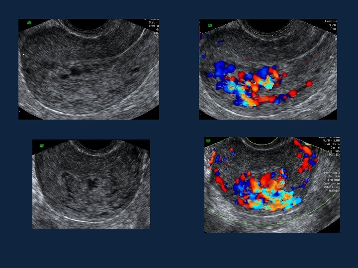
26 jaar - P 0 G 1 4 weken na spontaan miskraam op 11 weken amenorrhoea EBL ~ 300 ml

As Time Goes By … ὕβρις
 1200 1000 800 600 N E GE 400 200")
Bloedverlies versus PSV EBL (ml) 1200 1000 800 600 N E GE 400 200 0 50 70 90 ! e i t la e r r co 110 130 150 PSV (cm/sec)

Bloedverlies versus laatste zwangerschap

PSV versus laatste zwangerschap

33 jaar 6 weeks na AAP op 8 weken S/ aanslepende spotting Spiraal arteries PSV 103, 8 cm/sec Decidua Chorionic villi

Besluit EMV ≈ trofoblastrest Volledig verwijderen rest stoppen bloeding verdwijnen EMV PSV ≠ predictor voor hoeveelheid bloedverlies
 Chirurgische")
Besluit Echografie met kleurendoppler - diagnose trofoblastrest - risico perop. bloeding (informeer anesthesist) Chirurgische verwijdering onder echogeleide (onder hysteroscopie) Embolisatie ? … meestal niet Bedankt !
- Slides: 21