Diagnostic methods in pulmonology RASOUL ALIANNEJAD Diagnostic methods


")
")


 Expiratory flow rate L/sec TLC FVC Inspiratory")


 Reduced peak flow,")


")
 Pa. O 2 Pa. CO 2 PH HCO 3")


- Slides: 69
Diagnostic methods in pulmonology RASOUL ALIANNEJAD
Diagnostic methods in pulmonology
Diagnosis Prognosis Treatment
Diagnostic Methods Tools/tes t process
Symptoms Signs disease
Hypothesis Test Diagnosis
• Hypothesis DDx Physical ex • Hypothesis History • Hypothesis generation/revis ion Problem list • Testing hypothesis Diagnostic tests Final diagnostic
Data Hypothesis generation Diagnosis Patient Questions Data gathering Hypothesis evaluation
History & PHx Anatomical investigation functional investigation Infectious test Integrative test : CPET, PSG • Radiology imaging • Bronchoscopy • Ventilation tests • Perfusion tests • Diffusion tests • Sputum based • Blood samples based
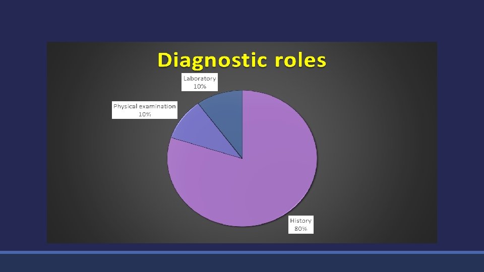
Taking history
Physical examination
Anatomical investigation
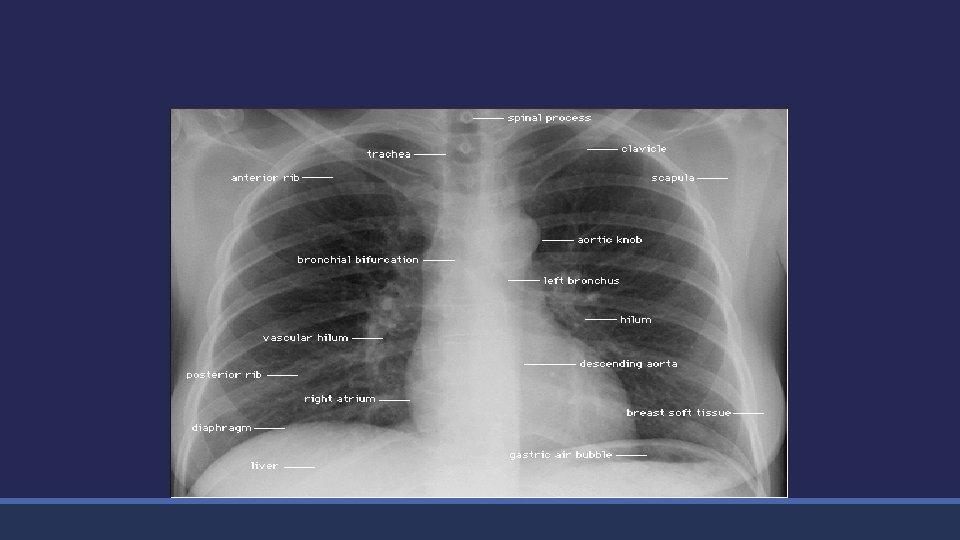
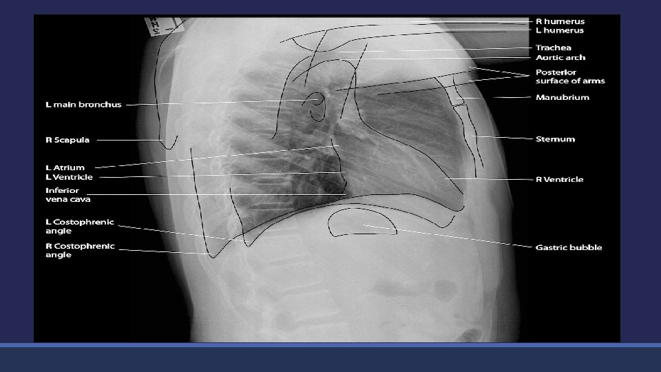
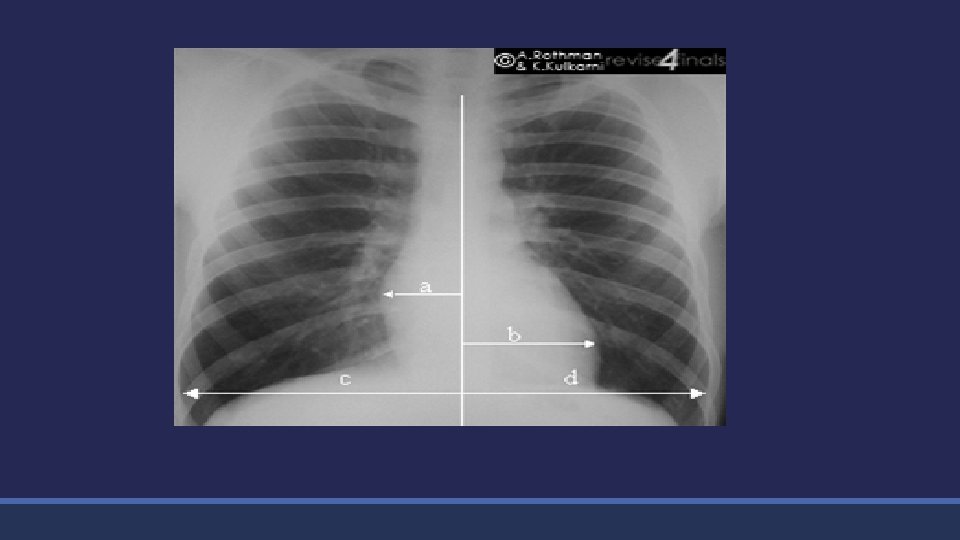
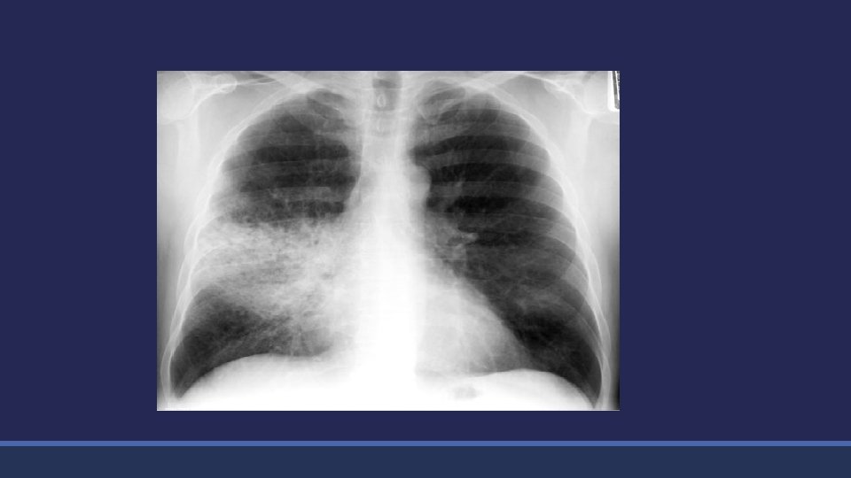
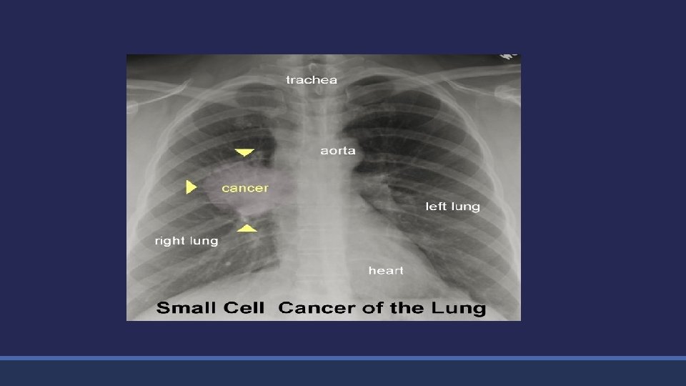
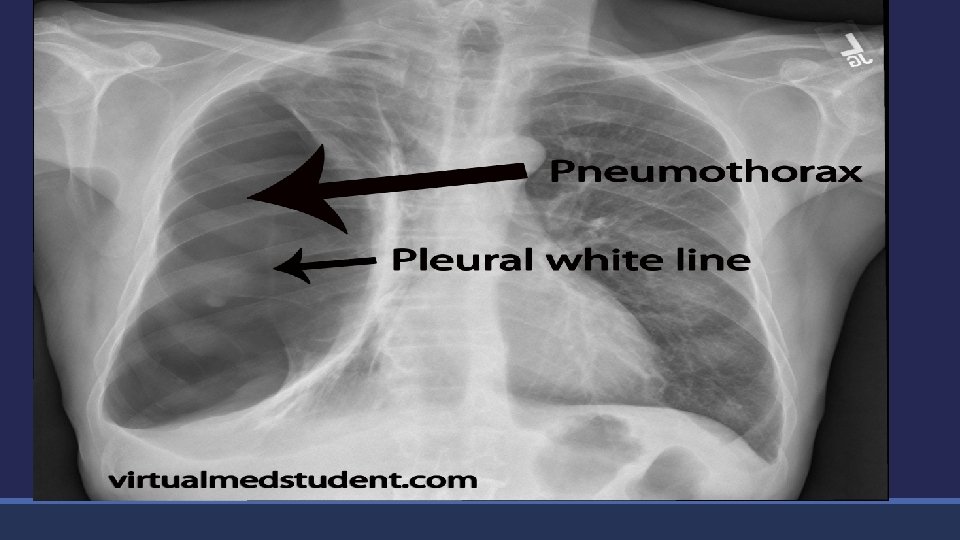
Chest X-ray
Chest CT scan
CHEST CT Angiography
Ventilation/perfusion scan
CHEST PET/CT scan
Fiberoptic bronchoscopy
Fiberoptic bronchoscopy(FOB)
Endobronchial ultrasound(EBUS)
Thoracentesis
Plural biopsy
Medical Thoracoscopy
Mediastinoscopy
Functional investigation
History & PHx • Radiology imaging Anatomical investigation • Bronchoscopy functional investigation Infectious test Integrative test : CPET, PSG • Ventilation tests • Perfusion tests • Diffusion tests • Sputum based • Blood samples based
What is Spirometry? Spirometry is a method of assessing lung function by measuring the volume of air the patient can expel from the lungs after a maximal expiration.
Why Perform Spirometry? Confirm presence of airway obstruction Assess severity of airflow obstruction Monitor disease progression Assess response to therapy Assess prognosis (FEV 1)
Flow Measuring Spirometer
Small Hand-held Spirometers
muscles of respiration inspiratory muscles neck musculature expiratory muscles internal intercostal abdominal musculature external intercostal diaphragm
maximal deep inspiration sternocleidomastoideus scalenis
the limits of breathing maximal inspiration maximal expiration
mechanics during spirometry volume neck muscles elasticity diaphragm elasticity & abdominal muscles diaphragm abdominal muscles time
maximal movements of the chest “vital capacity” maximal inspiration TLC-niveau maximal expiration RV-niveau
limits of the vital capacity volume deep inspiration _ VC visceral pleura VC deep expiration + time _ 0 _ + + intrathoracic pressure airway closure
trumpet or funnel model sum of the transverse areas of each branching generation amount crosssection total area trachea 0 convection 3 cm 2 segmental bronchi 3 diffusion 1 0 flow speed 20 0, 15 cm 2 6 cm 2 respiratory bronchioli 18 2. 105 0, 00195 cm 2 390 cm 2
airflow PPAlv VV PAtm Poiseuille’s law alveolar pressure muscular strength airflow is dependent on: elasticity resistance airway collapse airway obstruction
alveolar and intrathoracic pressure PAlv Palv = Pint. th = Pcw + Pmusc + Plung
expiratory airway collaps PAlv pressure PAtm transmural pressure Plung Pcw intra-thoracic pressure Equal Pressure Point Pmusc alveolus airway mouth
airways and lung parenchyma connective tissue cartilage smooth muscle alveolar walls loss alveolar septa
airway mechanics in lung emphysema flow + Plung + P volume expiration Plung Pcw Pmusc alveolus airway mouth F-V curve in lung emphysema inspiration
Lung Volume Terminology Inspiratory reserve volume Total lung capacity Inspiratory capacity Tidal volume Expiratory reserve volume Residual volume Vital capacity
Normal Trace Showing FEV 1 and FVC Volume, liters 5 4 FEV 1 = 4 L 3 FVC = 5 L 2 FEV 1/FVC = 0. 8 1 1 2 3 4 5 Time, seconds 6
Flow Volume Curve Maximum expiratory flow (PEF) Expiratory flow rate L/sec TLC FVC Inspiratory flow rate L/sec Volume (L) RV
FEV 1 FVC • Forced expiratory volume in one second • Forced vital capacity: • The total volume of air that can be forcibly exhaled in one breat FEV 1/FVC • The fraction of air exhaled in the first second relative to the total volume exhaled MEFR • Derived from the mid portion of the flow volume curve VC • A volume of a full breath exhaled in the patient’s own time and not forced. Often slightly greater than the FVC, particularly in COPD
Criteria for Normal Post-bronchodilator Spirometry • FEV 1: % predicted > 80% • FVC: % predicted > 80% • FEV 1/FVC: > 0. 7
Normal Trace Showing FEV 1 and FVC Volume, liters 5 4 FEV 1 = 4 L 3 FVC = 5 L 2 FEV 1/FVC = 0. 8 1 1 2 3 4 5 Time, seconds 6
Spirometry: Obstructive Disease Volume, liters 5 4 Normal 3 FEV 1 = 1. 8 L 2 FVC = 3. 2 L 1 FEV 1/FVC = 0. 56 1 2 3 4 5 Time, seconds 6 Obstructive
Criteria: Restrictive Disease • FEV 1: % predicted < 80% • FVC: % predicted < 80% • FEV 1/FVC: > 0. 7
Spirometry: Restrictive Disease Normal Volume, liters 5 4 3 Restrictive FEV 1 = 1. 9 L 2 FVC = 2. 0 L 1 FEV 1/FVC = 0. 95 1 2 3 4 5 Time, seconds 6
Flow Volume Curve Patterns Obstructive and Restrictive Severe obstructive Volume (L) Reduced peak flow, scooped out midcurve Restrictive Expiratory flow rate Obstructive Volume (L) Steeple pattern, reduced peak flow, rapid fall off Volume (L) Normal shape, normal peak flow, reduced volume
Spirometry: Abnormal Patterns Restrictive Time Slow rise, reduced volume expired; prolonged time to full expiration Mixed e Volume Obstructive Time Fast rise to plateau at reduced maximum volume Time Slow rise to reduced maximum volume; measure static lung volumes and full PFT’s to confirm
Body Plethysmography TGV =FRC TLC RV Raw
Lung diffusion capacity(DLco)
Arterial blood gas(ABG) Pa. O 2 Pa. CO 2 PH HCO 3
Pulse oximetry
Ergospirometry VO 2 VCO 2 pulse Breathing reserve EQ CO 2 EQ O 2 ET CO 2
Polysomnography
Sputum examination
Sputum culture