Diagnostic Imaging of Horses LACP Chapter 7 Pages


--Extended Lateromedial (Flexed lateral)--Flexed Dorsopalmar (DP) Dorsolateral-palmaromedial oblique (DLPMO)")


 away from the patient.")


 Lateral Medial Dorsal")


- Slides: 75
Diagnostic Imaging of Horses LACP Chapter 7 Pages 147 -175
Diagnostic Radiology Very similar to small animals Principles are the same as in small animals Large animal radiology is very unique due to the conformation of the patient and the fact that most radiographs are performed on a standing, awake patient. The temperament of the animal can hinder the ability to position a patient for ideal film studies.
Safety: “Fear Factors” Injured horses-painful Strange noises Strange things touching the horses; for example, ideally the film cassette should be placed to contact the patient’s skin. Personnel and equipment are in vulnerable positions with a patient that is often suspicious of the situation.
Safety: Personnel Involved Move slowly and speak in a low calm voice Do not make loud crashing noises Let the horse know that you are getting ready to do something to him/her. Gently rub the area of interest-touch it lightly if it is not injured. PPE-Wear these items! What are they?
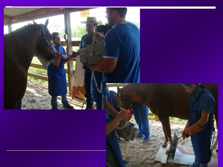
Just how many people does it take to take a radiograph? One person to tend to the horse’s head- your restrainer One person to operate the radiograph machine One person to hold/position the film cassette That’s right-this equals 3
Anatomic Direction • Femur tibia/fibula – Cranial - Caudal • Tarsus - digits – Dorsal Plantar
The five Routine views Lateromedial (Lateral)--Extended Lateromedial (Flexed lateral)--Flexed Dorsopalmar (DP) Dorsolateral-palmaromedial oblique (DLPMO) Dorsomedial-palmarolateral oblique (DMPLO)
Radiograph labeling Standard convention Marker is placed Cranial / Dorsal Lateral Be sure to “flash” the cassette Permanently identifies film as belonging to a certain patient Owners name Patient name / number Name of clinic / Location Date of exam Limb examined (RF, LF, RH, LH)
The Equipment: Radiograph machine Caliper Radiograph cassette holder Cassette/film Play-doh or putty Positioning blocks Processor to develop the film Chemical Sedation/Restraint devices PPE Time and patience!
Radiograph Cassette holder
Film cassette in cassette holder
Portable radiograph machine
Portable radiograph machine
Portable radiograph machine with a light collimator
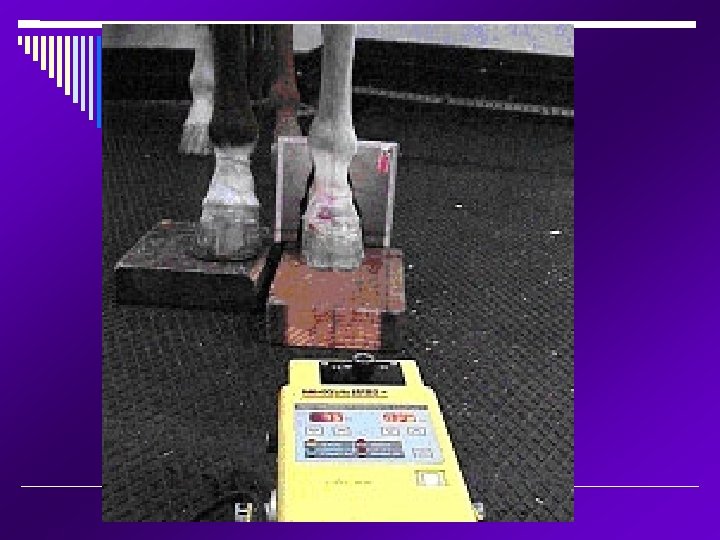
Wood and Plexiglas positioning block for the equine foot.
Position of the cassette for stand-on radiographic views
Position of the foot and cassette for stand-on views of the foot.
Slots provide additional support for film cassettes.
Packing of the grooves of the sole to prevent artifacts created by the air pockets of the hoof. These artifacts can sometimes mimic fracture lines and may obscure true lesions. Do not over pack. Always be sure to clean the hoof wall, sole and recesses of the frog. Hoof picks, soap, water and a brush may be needed.
Here’s the setup! Without the horse of course…
Generally you are about 30 -40 inches (80 cm. ) away from the patient.
Removal of the shoe is recommended but is expensive to remove and place back on the horse. Many owners will object to this. Always obtain owner consent before removing shoes!
Horseshoes interfere with interpretation of radiographs
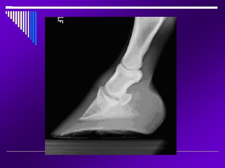
Hoof anatomy
Lateral • Horse is weight bearing • Cassette holder is used • X-ray beam is perpendicular to the middle carpal joint
Flexed Lateral • Foot is elevated • Carpus held in flexion • X-ray beam is centered at middle carpal joint • Tendency to shift carpus toward midline • Hands are close enough for scatter radiation-use gloves!
Dorsopalmar • Weight bearing • X-ray beam centered at middle carpal joint • Cassette parallel to the palmar aspect of the limb
DLPMO • Weight bearing • Foot of interest placed slightly cranial • Centered at middle carpal joint • Beam is 60 degrees lateral off a straight dorsal-palmar
DMPLO • Same as previous, BUT, beam comes from 60° MEDIAL from a straight DP • Since beam comes across the front of the horse, have a cooperative horse Sedation if needed
A Review… DORSAL LATERAL MEDIAL PALMAR
http: //www. vetmed. wisc. edu/Dr__Adams _938 -675. 220. 1. html
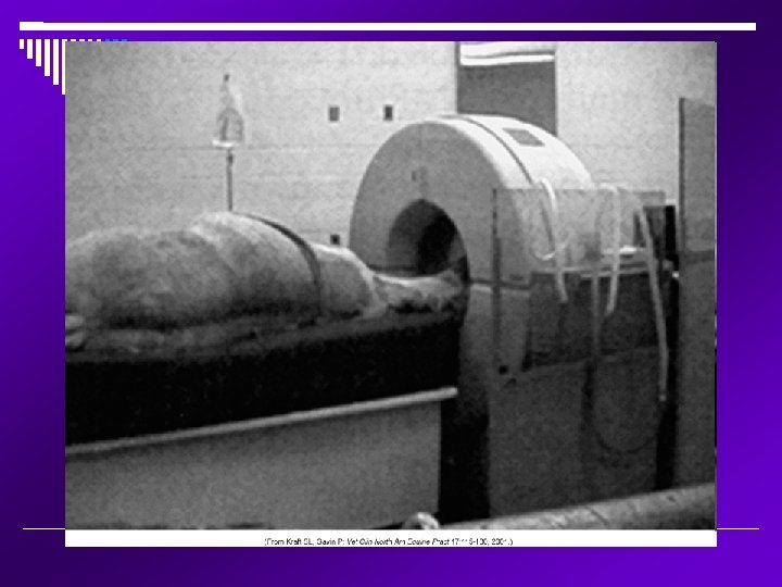
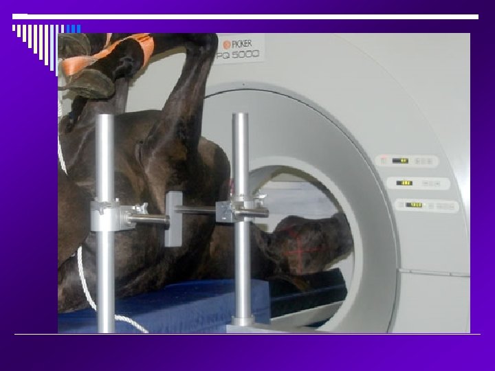
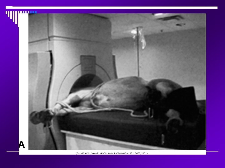
CT or CAT Scan Computed tomography has just recently become available for large animal patients. This equipment is very expensive and limited to specialized facilities. Use is restricted in adult horses to the examination of the head, cranial cervical spine and distal limbs. Patient must be under anesthesia. Must be injected with radioisotope and is absorbed by bony areas Used as a last resort in diagnosing.
CT scan of the horse’s nasal passages
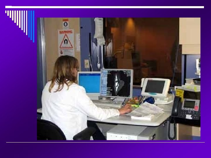
This is the CT control room. The animal patient must be anesthetized because you can not remain in the room with them. There can be no motion or movement involved.
“By scanning foals as they mature, one is able to directly quantify the rate of bone development in the distal limb. At this stage the foal must be anesthetized for a short time so there is no movement artifact. ”
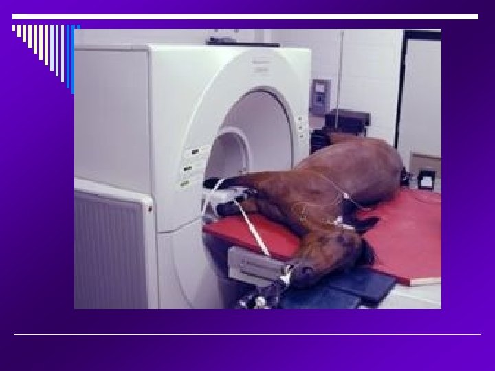
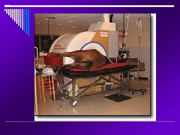
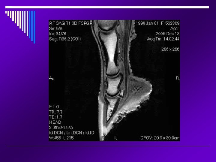
MRI Magnetic Resonance Imaging; the area being imaged is placed within a strong magnetic field and stimulated by radio-frequency pulses. These radio-frequency signals are collected analyzed by computers to form the image. Very, very costly and not widely available Anesthesia is required MRI tend to be superior to CT for soft tissue imaging The head, cervical spinal cord and lower legs can be imaged in an adult animal Precise and focal imaging tool that produces images of all tissue types; bone, tendons, ligaments, and fluid. Mainly utilized in equine lameness.
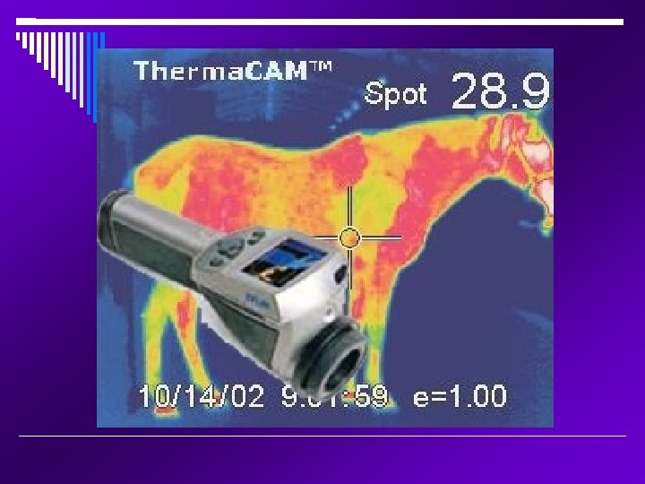
Thermography Uses a heat camera to scan the body surface temperature of the patient. Very popular because it is non-invasive, equipment is affordable and portable. Primarily used to locate “hot spots” which may indicate inflammation near the body surface. Deeper locations can not be detected, such as within the thorax or abdomen.
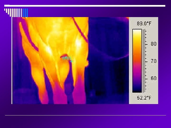
Stifles: the right stifle shows a “hot spot” over the medial femorotibial joint.
IR PROx Thermal Infrared Camera
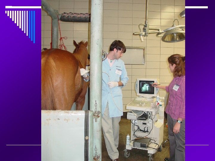
Ultrasound Operates on high frequency sound waves beyond our hearing. Uses sound waves to detect differences in tissue density Generally superior to standard radiographs for visualizing soft tissues. Radiographs are superior for imaging bony structures.
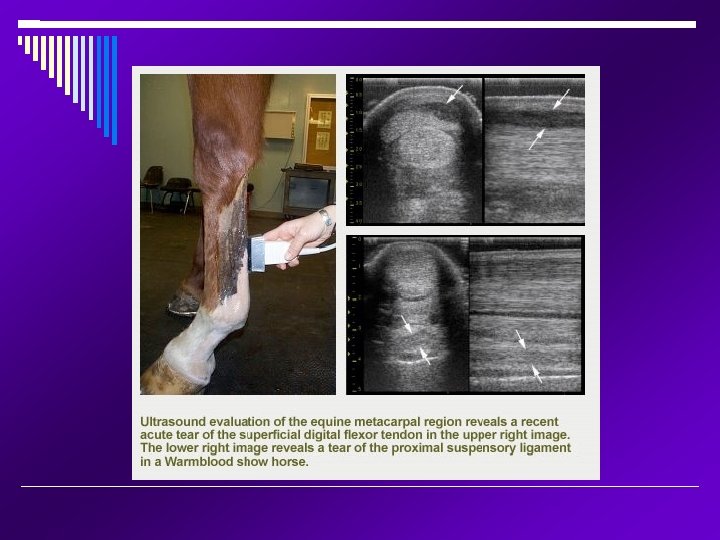
Common uses of Ultrasound Visualize kidneys and capable of ultrasound guided biopsy Lungs for pneumonia diagnosis and treatment GI for colics - small intestinal distension, large colon wall thickness, peritoneal fluid, diagnosis of abcesses and tumors Foals - GI disease, umbilical structures (diagnose umbilical infections), ruptured bladder Liver - ultrasound guided biopsy Assist with lameness diagnosis, including the extent of tendon and ligament damage Eyes
Common uses of Ultrasound cont… Monitor the mare's reproductive tract and optimize the time for breeding The genital tract in stallions Early detection of pregnancy Early detection of problem pregnancies, including fetal abnormalities Cardiovascular Ultrasound can be used to image the heart, lungs, kidneys, liver, spleen, and intestines, even during colic episodes.
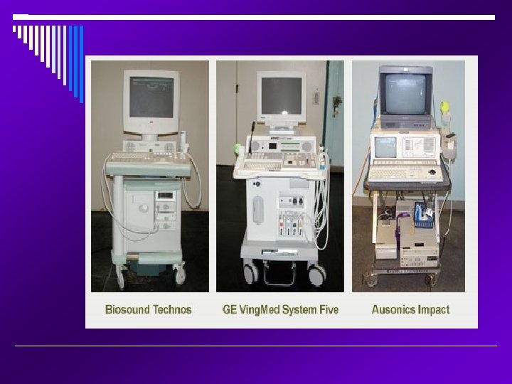
Portable veterinary ultrasound
Brief Introduction to Ultrasound • Transducer produces sound waves and also • • receives reflected sound waves. Sound waves travel in a plane through tissue. Sound waves are transmitted, absorbed or reflected by tissues. Computer forms image – in shades of gray. Bone appears white, fluids appear black From: Zagzebski, JA; Essentials of Ultrasound Physics, Mosby, © 1996
Musculoskeletal Palmar distal extremity Infected umbilicus Reproduction Uterine cysts Echocardiography Pericardial effusion Abdomen Liver - cholelithiasis Thorax Pleuropneumonia
Patient Preparation Clip the area Clean area Coupling medium alcohol commercial gel
Imaging technique Systematic organized approach – must be familiar with normal Scan from proximal to distal Evaluate structures individually Transducer perpendicular to structure Limb should be weight bearing
Imaging Techniques Label images - patient info, directions and location Two methods for location 1. Zones 2. Reference points - cm distal to standard point accessory carpal bone point of hock point of ergot
Transverse images Palmar (skin surface) Lateral Medial Dorsal
Rectal ultrasound examination of a fetus
A 16 -day pregnancy, visualized rectally with a 5 -m. Hz probe. Video. .
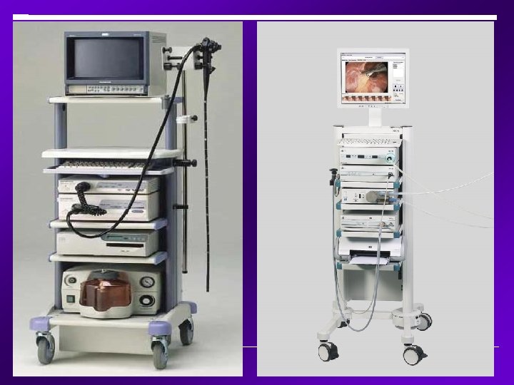
Endoscopy
Endoscopy Endoscopes come in two basic varieties; rigid and flexible. A thin tube that contains a fiberoptic camera and a tool at the end allowing samples to be taken from various locations in the body. It is passed through virtually any open cavity on the body. Usually takes 2 -3 people to operate
Control handpieces
Air/Water channel Illumination lens Instrument channel Viewing lens Illumination lens
Control handpiece and insertion tube of the flexible endoscope.
Endoscope examination of the male urethra. Notice how many people are involved.
Maintenance of the Equipment You will be utilizing some if not all of the previously mentioned equipment. Take care of the equipment and it will take care of you in the long run. Please follow ALL procedures in your hospital/clinical pertaining maintenance and trouble shooting equipment. Follow ALL cleaning procedures as well. Read, read, and read! Ask questions!
The End