Diagnosis Postsurgical hypoparathyroidism Bilateral central neck operations including


Diagnosis?

Postsurgical hypoparathyroidism
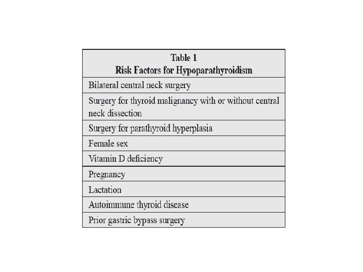

• Bilateral central neck operations, including total thyroidectomy, bilateral central neck dissection, and total laryngectomy can result in hypoparathyroidism.

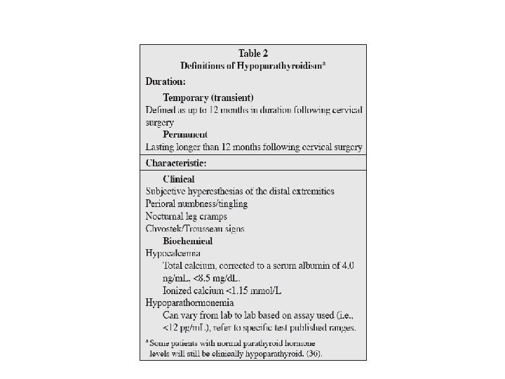
Diagnosis • The diagnosis of postsurgical hypoparathyroidism is usually obvious in a patient who has acute onset of symptoms of hypocalcemia immediately following neck surgery.

• Transient hypoparathyroidism occurs in up to 20 percent of patients after surgery for thyroid cancer and permanent hypoparathyroidism occurs in 0. 8 to 3 percent of patients after total thyroidectomy and is more common when the goiter is extensive.
 : with recovery")
DEFINITIONS OF HYPOPARATHYROIDISM • Hypoparathyroidism following surgery : Ø Temporary (transient) : with recovery in days, weeks or months. Ø Permanent: is defined when a medical regimen is required for longer than 12 months. • Transient hypoparathyroidism may be due to manipulation of the blood supply to or removal of one or more parathyroid glands during surgery.

• Hypomagnesemia may also accompany hypocalcemia and should be diagnosed and corrected as indicated as magnesium is required for full PTH secretion and action.

MITIGATION OF POSTOPERATIVE HYPOPARATHYROIDISM q Patients can be tested for 25 OHD blood levels preoperatively: ü vitamin D deficient (25 OHD<20 ng/m. L), then aggressive treatment with high-dose vitamin D should be considered. ü If 25 OHD is between 20 and 30 ng/m. L, less aggressive replacement is sufficient. Typically, 50, 000 IU vitamin D 3 (cholecalciferol) is given by mouth weekly. q Intraoperative preservation of the parathyroid glands with their blood supply intact, typically from the inferior thyroid artery, is paramount for preventing hypoparathyroidism. Every effort should be made to visually identify each parathyroid close to the thyroid gland, assessing the location of the vascular pedicle to the parathyroid.

• Pathologists routinely indicate the presence or absence of microscopically identified parathyroids in the final pathology report. Because parathyroids are quite small and may resemble thyroid tissue, they may be missed on gross exam. Unfortunately, requesting meticulous pathologic dissection of the thyroid for large multinodular goiters is not practical.
 refers to blood specimens drawn during and shortly after")
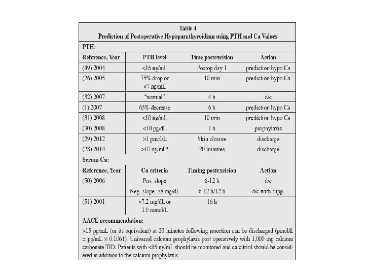
• Intraoperative PTH (IOPTH) refers to blood specimens drawn during and shortly after central neck surgery (i. e. , 5, 10, and 20 minutes after thyroidectomy is completed). The results may become available while the patient is still in the operating room or once she/he has arrived in recovery. This data can expedite same-day discharge or predict the need for observation and postoperative calcium management. • Patients with a PTH value >15 ng/m. L measured 20 minutes or longer after surgery can be discharged home on prophylactic calcium. • Patients with <15 ng/m. L PTH should be started on calcitriol (0. 5 mcg BID) in addition to calcium (and possibly magnesium) and observed overnight. • i. PTH level <10 to 15 pg/m. L is predictive of later hypocalcemia.

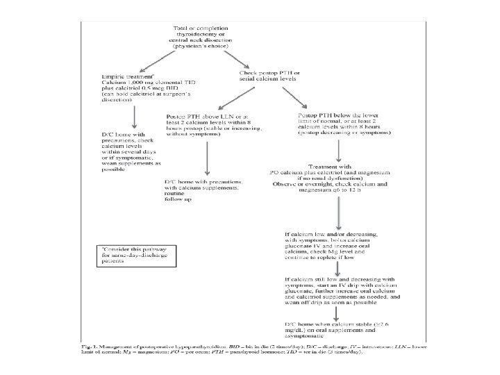
q Postoperative calcium testing : Ø The lag time for calcium changes is greater than that of PTH, and a calcium nadir may not occur for 24 to 72 hours following surgery. Ø Calcium levels, when used, are drawn the evening after surgery, the following morning, and every 6 to 12 hours thereafter as indicated. Ø If calcium levels are stable or increase over an observation period, discharge is generally considered safe. Ø If calcium levels continue to decline despite medical treatment, these patients require optimization of calcium replacement therapy (and possibly magnesium) and ongoing observation until calcium stability or increase is observed.
 Ø Prophylactic approach to preventing postoperative hypocalcemia is to routinely initiate")
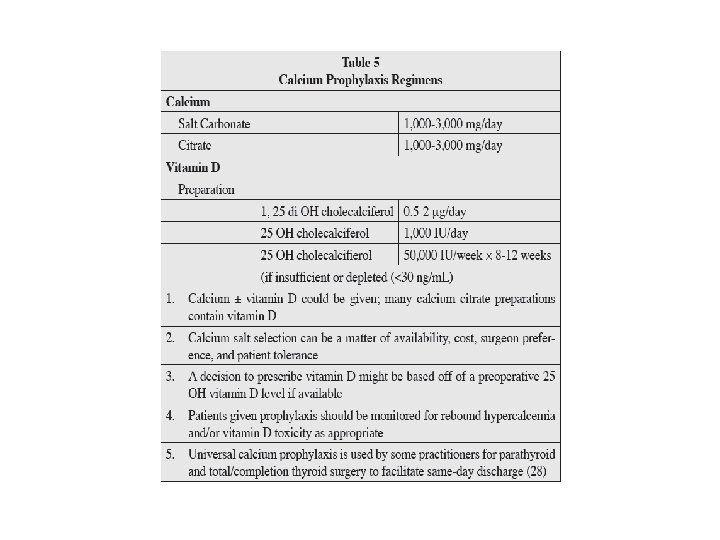
TREATMENT( Postoperative Prophylaxis) Ø Prophylactic approach to preventing postoperative hypocalcemia is to routinely initiate treatment with oral calcium with or without calcitriol.

• Routine oral calcium has been demonstrated to reduce postoperative hypocalcemia to approximately 10%. Wang TS, Cheung K, Roman SA, Sosa JA. To supplement or not to supplement: a cost-utility analysis of calcium and vitamin D repletion in patients after thyroidectomy. Ann Surg Oncol. 2011; 18: 1293 -1299.

• A prospective, randomized study after total thyroidectomy showed that 1, 500 mg oral calcium plus 1 mcg calcitriol BID was superior to 0. 5 mcg of calcitriol BID or no calcitriol. Routine use of calcitriol is more expensive than calcium alone but is much less costly than hospital admission. Karakas E, Osei-Agyemang T, Schlosser K, et al. The impact of parathyroid auto transplantation during bilateral surgery for Graves’ disease on postoperative hypocalcemia. Endocr Regul. 2008; 42: 39 -44.

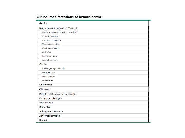
• Acute Management of Hypoparathyroidism: • Calcium levels should be monitored at least every 12 hours and more frequently if total calcium levels are <7 mg/d. L, or if symptoms of hypocalcemia (e. g. , perioral numbness/tingling, positive Chvostek or Trousseau sign, or carpopedal spasm) should occur.
 or a serum")
• Patients with symptomatic hypocalcemia (circumoral and distal extremity paresthesias) or a serum calcium concentration below 7. 8 mg/d. L should be treated with calcitriol 0. 5 mcg twice daily and calcium carbonate 500 mg four times daily. • Patients with more severe symptoms (muscle cramps) or a serum calcium concentration below 7. 5 mg/d. L should also receive calcium gluconate by continuous intravenous drip for 24 to 36 hours or until the serum calcium concentration rises and is maintained above 8. 0 mg/d. L. • Emergency therapy is indicated in patients with tetany, seizures, laryngospasm or markedly prolonged QT intervals on the electrocardiogram. Treatment is initiated with the intravenous administration of one 10 m. L ampule of 10 percent calcium gluconate over 5 to 10 minutes, followed by the intravenous infusion.

ü If severe hypocalcemia develops with symptoms: IV calcium should be administered as a bolus (1 to 2 g of calcium gluconate over 20 minutes) or infusion of a solution composed of 11 g calcium gluconate added to normal saline or 5% dextrose water to provide a final volume of 1, 000 m. L administered at 50 m. L/hour (equivalent to 50 mg/hour) and adjusted to maintain the calcium level in the low normal range. ü Higher doses of oral calcium (3 to 4 grams of elemental calcium daily given in 3 to 4 doses) should be initiated as soon as the patient can swallow oral medication. ü Vitamin D analogs such as calcitriol should be employed but may take up to 72 hours to be effective.

• Calcitriol markedly increases the intestinal absorption of calcium and also liberates calcium from bone. • Calcitriol has a relatively short half-life, 5 to 8 hours, but this can double in patients with renal failure. • In contrast, fat-soluble vitamin D 3, cholecalciferol, has a halflife of weeks to months. Therefore, toxicity from excessive calcitriol can be reversed in days, whereas vitamin D 3 toxicity can last for weeks (with the potential for soft tissue calcification, renal stones, or permanent renal failure).

• After discharge, calcium levels should be monitored at least twice weekly, anticipating a reduction in calcitriol after steady state is achieved in approximately 1 week.

Long-term Management of Hypoparathyroidism • The primary goal of chronic management : maintain serum calcium within an asymptomatic range and to avoid significant hypo- or hypercalcemia. • To reduce the risk of symptoms, kidney stones, and ectopic soft tissue calcification, it is recommended that serum calcium and phosphorus be maintained in the low and high normal reference ranges, respectively. • Some authors have specifically recommended keeping the 24 hour urine calcium excretion <7. 5 mmol/day and the calciumphosphorus product <55 mg 2/d. L 2. • This long-term patient management is usually best accomplished under the care of an endocrinologist.

• Calcium is typically provided as calcium carbonate or calcium citrate. Most patients requiring 1, 500 mg elemental calcium daily. Dosing is divided into 2 or 3 split doses to maximize absorption. • Calcitriol improves intestinal calcium absorption and is almost always required. Doses range from 0. 125 to 4. 0 mcg/day, with most patients requiring 0. 25 mcg daily. • The doses are typically divided when 1 mcg or more per day is required. • Vitamin D 2 (ergocalciferol) or vitamin D 3 (cholecalciferol) are occasionally used along with the activated metabolite of vitamin D (calcitriol) and may help to provide smoother control of calcium levels.

q Thiazide diuretics can be added to the regimen when calcium control is difficult or hypercalcuria (>150 mg/24 h) is aproblem. • Thiazide diuretics enhance distal renal tubular calcium reabsorption, thereby increasing serum calcium and reducing urinary calcium excretion. • Hydrochlorothiazide (12. 5 -50 mg daily) can be effective. q Vitamin D therapy has the unwanted effect of increasing intestinal phosphate absorption. When severe hyperphosphatemia is problematic, intestinal phosphate binders may be necessary.

• Calcium levels may rapidly become unstable in the setting of acute gastrointestinal disease or acute kidney injury because it can alter volume status, serum magnesium levels, or calcium/vitamin D absorption.

• The long-term consequences of permanent hypoparathyroidism include nephrolithiasis, nephrocalcinosis, basal ganglia calcifications, ectopic soft tissue calcification, cataracts, potential defects in bone metabolism, and an impaired quality of life. • Periodic measurement of 24 -hour urine calcium is probably prudent, and some physicians perform periodic renal ultrasound monitoring. • Bone mineral content tends to be increased in hypoparathyroidism, but cancellous bone microarchitecture in hypoparathyroidism is abnormal, and the effect on fracture risk is uncertain. Hypoparathyroidism may induce greater bone stiffness that could make the skeleton more predisposed to develop micro fractures when loaded. • Dual-energy X-ray absorptiometry testing is not likely to help assess this risk. • One area particularly prone to ectopic calcification is the basal ganglion area of the brain, but there are currently no formal recommendations for routine head computed tomography scanning. • Additionally, patients with hypoparathyroidism often report increased anxiety and decreased sense of well-being compared to controls. These symptoms are not improved with current therapies.

• A new option for the treatment of hypoparathyroidism recently approved by the U. S. FDA in January 2015, is recombinant human PTH (1 -84), which is identical in structure to the fulllength endogenous hormone. The quality of life for these patients can be poor, requiring daily medication, frequent physician visits, and many episodes of breakthrough hypocalcemia symptoms. Mannstadt M, Clarke BL, Vokes T, et al. Efficacy and safety of recombinant human parathyroid hormone (1 -84) in hypoparathyroidism (REPLACE): a double-blind placebo-controlled, randomized, phase 3 study. Lancet Diabetes Endocrinol. 2013; 1: 275 -283.

• Recombinant PTH was studied in the REPLACE trial. When injected subcutaneously into the thigh once daily, 53% of patients were able to reduce their calcium and vitamin D requirements by more than 50%, and 43% were able to achieve independence of vitamin D and reduction in calcium to <500 mg daily. • Importantly, urinary calcium and serum phosphorus levels were also reduced. The hope with these findings is there might be a reduction in renal calculi. • Recombinant PTH may also restore normal bone metabolism and improve bone microarchitecture.

• It is only recommended for patients who cannot be wellcontrolled on calcium supplements and active forms of vitamin D alone. • It was not studied in patients with acute postsurgical hypoparathyroidism. • The label carries a warning for potential risk of osteosarcoma, although this was only observed in rats that received the drug at higher levels than those used in humans. • The drug is initiated at a dose of 50 mcg once daily and can be titrated to doses of 25, 50, 75, or 100 mcg. Serum calcium must be monitored 3 to 7 days after starting the drug or adjusting doses.
 double during a normal pregnancy.")
pregnancy • Serum concentrations of 1, 25 D (calcitriol) double during a normal pregnancy. However, intact PTH concentrations remain low to normal, suggesting that PTH does not mediate the late partum rise in 1, 25 D production. The increase in serum 1, 25 D may be regulated by other pregnancy hormones, which are normal in hypoparathyroid women, such as PTHrelated protein (PTHr. P), prolactin, estrogen, and placental growth hormone. • During pregnancy, calcitriol requirements may increase or decrease.

lactation • During lactation, calcitriol requirements may abruptly decrease. If the calcitriol dose is not reduced, the combination of elevated serum 1, 25 D and PTHr. P can lead to increases in intestinal absorption and bone resorption and hypercalcemia. • Thus, serum calcium concentrations should be measured frequently during late pregnancy and lactation in women with hypoparathyroidism, who may have a rise in serum calcium requiring a decrease in calcitriol dose. • The requirement for calcitriol will return to antepartum levels with cessation of lactation.
- Slides: 37