
DIAGNOSIS OF PREGNANCY Department of ObGyn Kathmandu Medical

DIAGNOSIS OF PREGNANCY Department of Ob/Gyn Kathmandu Medical College
The reproductive period of a woman begins at menarche and ends in menopause. It usually extends from 13 – 45 years. Lina Medina in Lima, Peru was the youngest one, delivery by caesarean section when she was only 5 years 7 months old and the oldest one at 57 years and 4 months.

DURATION OF PREGNANCY Menstrual age or Gestational Age: 9 calendar months and 7 days or 280 days or 40 weeks calculated from the first day of the last menstrual period. Fertilization or Ovulatory age: The true gestation period is to be calculated by subtracting 14 days from 280 days i. e. 266 days.
First trimester (0 -12 weeks) Second trimester (13 – 28 weeks) Third trimester (29 – 42 weeks)
SUBJECTIVE SYMPTOMS Ø Amennorrhoea Pregnancy, however, may occur in women who are previously amenorrhoeic – during lactation and puberty Ø Morning sickness Ø Frequency of micturation Ø Breast discomfort Ø Fatigue
OBJECTIVE SIGNS: Breast changes Evident between 6 -8 weeks. Enlargement with vascular engorgement evidenced by the delicate veins visible under the skin. The nipple and the areola become more pigmented specially in dark women. Montgomery’s tubercles are prominent. Thick yellowish secretion can be expressed as early as 12 th week.

OBJECTIVE SIGNS Per abdomen – Uterus remains a pelvic organ until 12 th week, it may be just felt per abdomen as a suprapubic bulge. Pelvic signs Jacquemier’s or Chadwick’s sign Vaginal sign Cervical signs Uterine signs Hegar’s sign Palmer’s sign
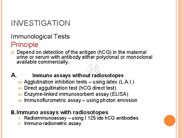
INVESTIGATION Immunological Tests Principle Depend on detection of the antigen (h. CG) in the maternal urine or serum with antibody either polyclonal or monoclonal available commercially. A. Immuno assays without radiosotopes Agglutination inhibition tests – using latex (L. A. I. ) Direct aggultination test (h. CG direct test) Enzyme-linked immunosorbent assay (ELISA) Immunoflurometric assay – using photon emission B. Immuno assays with radiosotopes § § Radioimmunoassay – using I 125 ido h. CG antibodies Immuno-radiometric assay
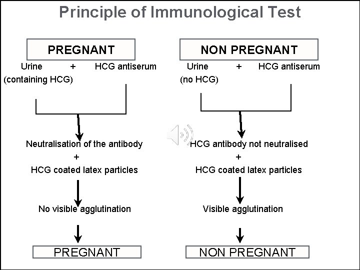
Principle of Immunological Test PREGNANT Urine + (containing HCG) HCG antiserum Neutralisation of the antibody + HCG coated latex particles No visible agglutination PREGNANT NON PREGNANT Urine (no HCG) + HCG antiserum HCG antibody not neutralised + HCG coated latex particles Visible agglutination NON PREGNANT
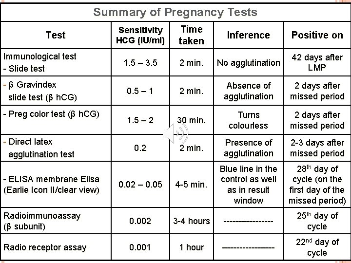
Summary of Pregnancy Tests Sensitivity HCG (IU/ml) Time taken Inference Positive on Immunological test - Slide test 1. 5 – 3. 5 2 min. No agglutination 42 days after LMP - β Gravindex slide test (β h. CG) 0. 5 – 1 2 min. Absence of agglutination 2 days after missed period 1. 5 – 2 30 min. Turns colourless 2 days after missed period 0. 2 2 min. Presence of agglutination 2 -3 days after missed period 4 -5 min. Blue line in the control as well as in result window 28 th day of cycle (on the first day of the missed period) --------- 25 th day of cycle --------- 22 nd day of cycle Test - Preg color test (β h. CG) - Direct latex agglutination test - ELISA membrane Elisa (Earlie Icon II/clear view) Radioimmunoassay (β subunit) Radio receptor assay 0. 02 – 0. 05 0. 002 0. 001 3 -4 hours 1 hour
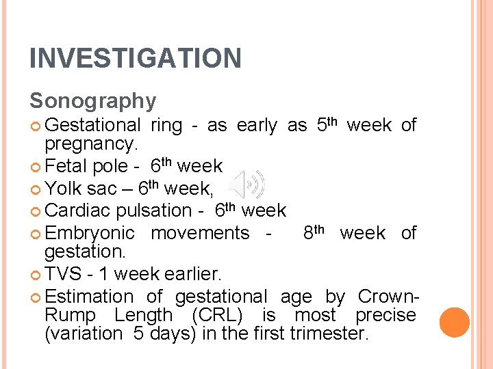
INVESTIGATION Sonography Gestational ring - as early as 5 th week of pregnancy. Fetal pole - 6 th week Yolk sac – 6 th week, Cardiac pulsation - 6 th week Embryonic movements 8 th week of gestation. TVS - 1 week earlier. Estimation of gestational age by Crown. Rump Length (CRL) is most precise (variation 5 days) in the first trimester.
SECOND TRIMESTER Symptoms: The subjective symptoms – such as nausea, vomiting and frequency of micturition usually subside, while amenorrhoea continues “Quickening” (feeling of life) denotes the perception of the first active fetal movement by the women. Progressive enlargement of the lower abdomen by a mass
OBJECTIVE SIGNS Chloasma Breast changes Breasts are more enlarged with prominent veins under the skin Secondary areola specially demarcated in primigravidae, usually appears at about 20 th week Montgomery’s tubercles are prominent and extend to the secondary areola Colostrum becomes thick and yellowish by 16 th week Variable degree of striae may be visible with advancing weeks
OBJECTIVE SIGNS Abdominal Examination Linear nigra Striae (both pink and white) of varying degree are visible in the lower abdomen, more towards the flanks. Palpation – fundal height is increased with progressive enlargement of the uterus.
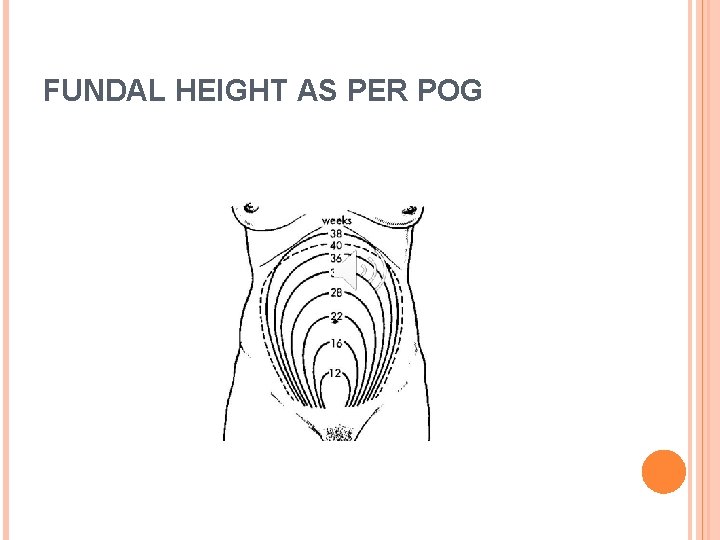
FUNDAL HEIGHT AS PER POG
Abdominal examination § § § The uterus feels soft and elastic Braxton-Hicks contractions Palpation of fetal parts Active fetal movements – it not only gives positive evidence of pregnancy but of a live fetus. External ballottement
Fetal heart sound (FHS) is the most conclusive clinical sign of pregnancy Normal – 110 -160 per minute Uterine soufflé - is a soft blowing and systolic murmur, synchronous with the maternal pulse Funic or fetal soufflé - rush of blood through the umbilical arteries, synchronous with the fetal heart sounds
INVESTIGATIONS Sonography Routine Sonography at 18 -20 weeks permits a detailed survey of fetal anatomy, placental site and the integrity of the cervical canal Gestational age is determined by biparietal diameter and femur length with variation of ± 10 days. Radiologic evidence of fetal skeletal shadow may be visible as early as 16 th week.
THIRD TRIMESTER Subjective Symptoms Amenorrhoea Enlargement of the abdomen Lightening Frequency of micturition Fetal movements
OBJECTIVE SIGNS Cutaneous changes Uterine shape Braxton-Hicks contractions are more evident Fetal movements are easily felt Palpation of the fetal parts FHS may not be audible in cases of maternal obesity

INVESTIGATION Sonography Fetal growth assessment can be made provided accurate dating scan has been done in first or second trimester
DIFFERENTIAL DIAGNOSIS OF PREGNANCY Uterine fibroid Cystic ovarian tumour Encysted tubercular peritonitis Haematometra Distended urinary bladder. Pseudocyesis- it is a psychological disorder where the woman has the false but firm belief that she is pregnant although no pregnancy exists.
SUMMARY Positive or absolute signs Manual appreciation of fetal parts and perception of active fetal movements by the examiner at about 20 th week. Fetal heart sounds Ultrasonic evidence of embryo as early as 6 th week and later on the fetus Radiological demonstration of the fetal skeleton at 16 th week and onwards.
SUMMARY Presumptive symptoms and signs Amenorrhoea Frequency of micturation Morning sickness Fatigue Breast changes Skin changes Quickening
SUMMARY Probable signs Ø Ø Ø Ø Ø Abdominal enlargement Braxton-Hicks contractions External ballottement Changes in the size, shape and consistency of the uterus Jacquemier’s sign Softening of the cervix Osiander’s sign Internal ballottement Immunological tests

ESTIMATION OF GESTATIONAL AGE AND PREDICTION OF EXPECTED DATE OF DELIVERY Gestational age is about 280 days calculated from the first day of the last normal menstrual period (LMP) Naegele’s formula if the interval of cycles is longer, the extra days are to be added and if the interval is shorter, the lesser days are to be subtracted to get the EDD. But in significant cases (20 -30%), the patients either fail to remember the LMP or report inaccurately.
Date of the single fruitful coitus - accuracy of 50% within 7 days on either side 266 days are to be added to the date of the single fruitful coitus to calculate the expected date Date of quickening – a rough idea about the probable date of delivery can be reduced by adding 22 weeks in primigravidae and 24 weeks in multiparae to the date of quickening.
Previous records Clinical Size of the uterus Palpation of fetal parts Auscultation of FHR Investigation records Recording of positive pregnancy test Ultrasonographic findings at the earliest are: Gestation sac – at 5 weeks Measurement of crown rump length detected at 7 weeks, approximates 10 mm. (CRL in cm + 6. 5 = weeks of pregnancy
Sonographic estimation Estimation of gestational age by Crown-Rump Length is most precise, variation: ± 5 days Second trimester – gestational age is determined by BPD and FL (variation: ± 10 days) Third trimester: Gestational age estimation by BPD, FL, AC and HC are less accurate (variation: ± 3 weeks)

Thank You!
- Slides: 30