
Diagnosis Of Periodontal Diseases EXAMINATION ON CLINICAL BASES
Diagnosis Of Periodontal Diseases ﺭﺍﺋﺪ ﻋﺰﻳﺰ. ﺩ. ﺃ
EXAMINATION ON CLINICAL BASES: Color & texture alteration of the gingiva. Increased bleeding in gingival sulcus & pocket area. Increased pocket depth. Tooth mobility or change in its position. EXAMINATION ON RADIOGRAPHICAL BASES: Horizontal & or vertical loss in alveolar bone. Widening of P. L space. EXAMINATION ON HISTOLOGICAL BASES: Inflammatory cells infiltration within CT. Obvious loss and degradation of collagen. Loss of C. T attachment in more severe cases. Apical migration of J. E.
Conventional clinical diagnostic tools: Visual diagnosis. Changes in color and contour of the gingiva are pathologic changes. Periodontal probing: is used to determine pocket depth and clinical Attachment levels. Bleeding on probing: is indicative of periodontal inflammation Radiographic image: . to estimate the severity of bone loss (amount, distribution, pattern of distruction).
First visit • Medical History -The possible role systemic diseases, may play in the cause of periodontal disease. • The presence of systemic diseases may require special precautions. • The possibility that oral infections may influence a variety of systemic diseases & conditions. Dental History Loose teeth (spacing), foul taste, sensitivity when chewing, sensitivity to hot & cold. •
Second Visit Oral Hygiene. Oral Examination • The extent of accumulated plaque , food debris, & stains. • Examination includes each surface of the present teeth this can be achieved using PLI Oral Malodor. (halitosis) • Poor OH , Caries & PD Diseases • It may indicate patient has systemic diseases (liver disease, , DM, tonsillitis. . etc. )
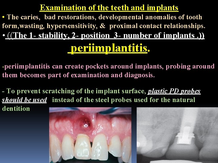
Examination of the teeth and implants • The caries, bad restorations, developmental anomalies of tooth form, wasting, hypersensitivity, & proximal contact relationships. • ((The 1 - stability, 2 - position 3 - number of implants. )) periimplantitis. -periimplantitis can create pockets around implants, probing around them becomes part of examination and diagnosis. - To prevent scratching of the implant surface, plastic PD probes should be used instead of the steel probes used for the natural dentition
Plaque and Calculus. • The presence of supragingival plaque and calculus can be directly observed. • Warm air may be used to deflect the gingiva and aid in visualization of the subgingival calculus. Wasting Disease of the teeth. Wasting is defined as any gradual loss of tooth substance causes formation of smooth, Polished surfaces,
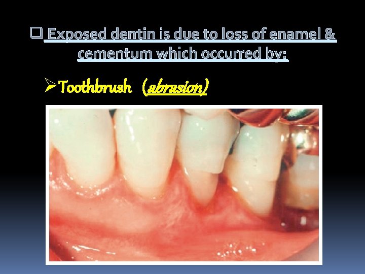
q Exposed dentin is due to loss of enamel & cementum which occurred by: ØToothbrush (abrasion)
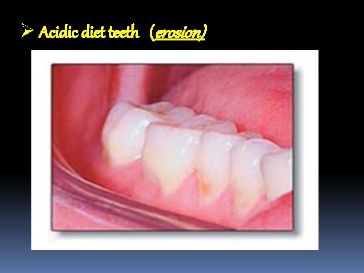
Ø Acidic diet teeth (erosion)
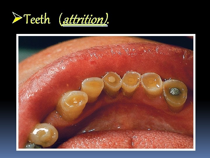
ØTeeth (attrition).
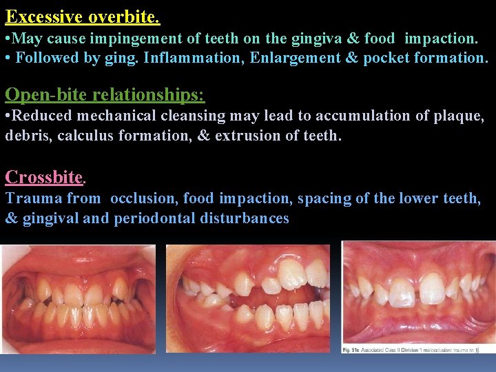
Excessive overbite. • May cause impingement of teeth on the gingiva & food impaction. • Followed by ging. Inflammation, Enlargement & pocket formation. Open-bite relationships: • Reduced mechanical cleansing may lead to accumulation of plaque, debris, calculus formation, & extrusion of teeth. Crossbite. Trauma from occlusion, food impaction, spacing of the lower teeth, & gingival and periodontal disturbances
Periodontal Pockets. • Is the distance between the base of the pocket & gingival margin. • Type of pockets @- (suprabony or infrabony). @- (True and false pockets)
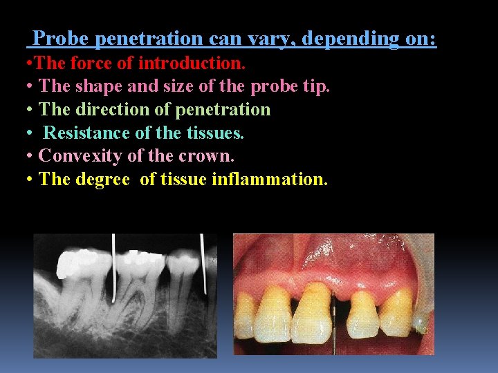
Probe penetration can vary, depending on: • The force of introduction. • The shape and size of the probe tip. • The direction of penetration • Resistance of the tissues. • Convexity of the crown. • The degree of tissue inflammation.
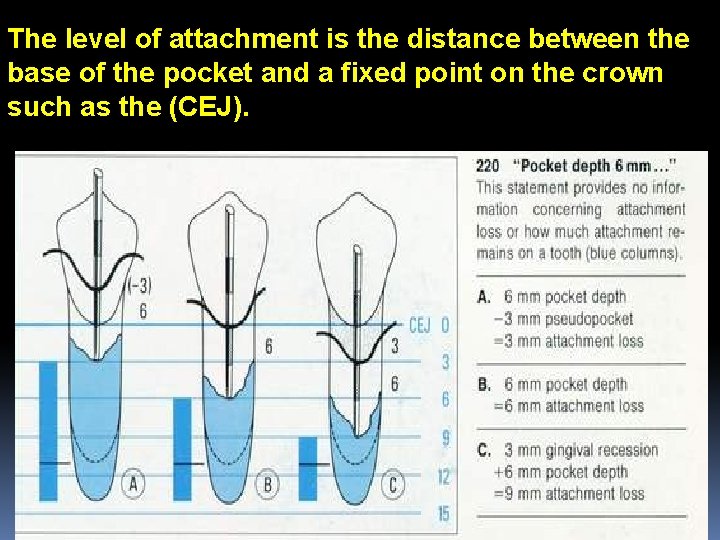
The level of attachment is the distance between the base of the pocket and a fixed point on the crown such as the (CEJ).
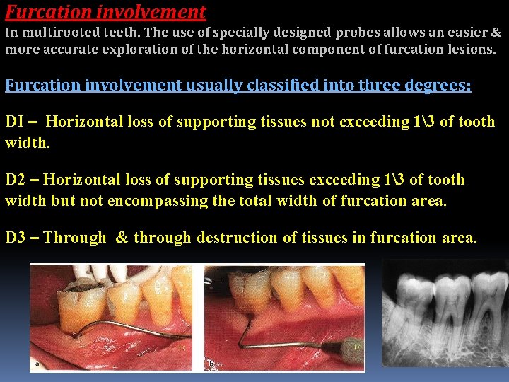
Furcation involvement In multirooted teeth. The use of specially designed probes allows an easier & more accurate exploration of the horizontal component of furcation lesions. Furcation involvement usually classified into three degrees: DI – Horizontal loss of supporting tissues not exceeding 13 of tooth width. D 2 – Horizontal loss of supporting tissues exceeding 13 of tooth width but not encompassing the total width of furcation area. D 3 – Through & through destruction of tissues in furcation area.
Abnormal mobility most often occurs faciolingually. Mobility is graded according to the ease and extent of tooth movement as follows: - • D 1 - Mobility of the crown of the tooth in horizontal direction (0. 2 -1 mm). • D 2 - Mobility of the crown of the tooth in horizontal exceeding 1 mm. • D 3 - Mobility of the crown in vertical as well as horizontal direction.
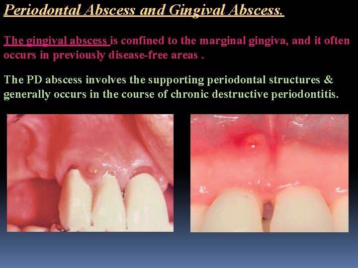
Periodontal Abscess and Gingival Abscess. The gingival abscess is confined to the marginal gingiva, and it often occurs in previously disease-free areas. The PD abscess involves the supporting periodontal structures & generally occurs in the course of chronic destructive periodontitis.
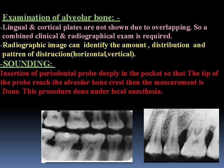
Examination of alveolar bone: -Lingual & cortical plates are not shown due to overlapping. So a combined clinical & radiographical exam is required. -Radiographic image can identify the amount , distribution and pattren of distruction(horizontal, vertical). -SOUNDING: Insertion of periodontal probe deeply in the pocket so that The tip of the probe reach the alveolar bone crest then the measurement is Done. This procedure done under local anesthesia.

THANK YOU FOR YOUR KIND ATTENTION
- Slides: 19