DIAGNOSIS OF OSTEOPOROSIS Mahtab Niroomand M D Assistant

DIAGNOSIS OF OSTEOPOROSIS Mahtab Niroomand M. D. Assistant Professor of Endocrinology SBUMS

Lecture outline Epidemiology of osteoporosis Definition of osteoporosis Clinical manifestations of osteoporosis Diagnosis of osteoporosis Bone densitometry Fracture risk assessment

Epidemiology Osteoporosis is a systemic skeletal disorder that is characterized by the loss of bone tissue, disruption of bone architecture, and bone fragility, leading to an increased risk of fractures Bone loss and low bone mass are asymptomatic until fractures occur The burden of suffering associated with osteoporosis is related to the increased incidence of fractures in individuals with low bone mass and microarchitectural deterioration. Osteoporosis is the most common metabolic bone disease and the most common cause of fractures in older adults in the United States

Epidemiology Fractures of the hip and spine are associated with: � Increased mortality rate (10 -20%) � Limitation of ambulation � Depression � Loss of independency � Chronic pain

Definition Osteoporosis: Low bone mass � Microarchitectural disruption � Skeletal fragility, resulting an increased risk of fracture �
: fracture that occur with a fall from")
Definition Osteoporotic fracture (fragility fracture, low-trauma fracture): fracture that occur with a fall from standing height or less or with no trauma
 Bone mass density Bone Strength Degree of")
Bone geometry (size and shape of bone) Bone mass density Bone Strength Degree of mineralization Bone turnover microarchitectur e

Bone strength = Bone quality + Bone quantity Micro-architecture increases bone strength Normal Loss of Quantity + Quantity Architecture Loss of Architecture
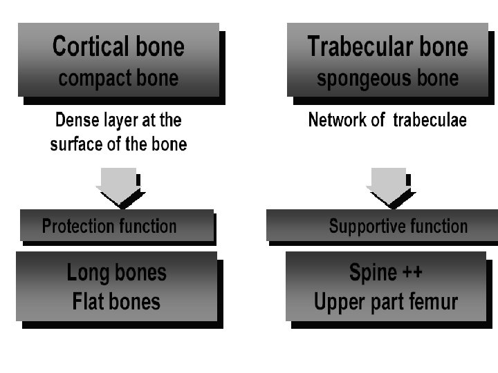

AP spine Lateral spine Femoral neck Ward Trochantric Mid radial Distal Ultra Total Trabecular % Cortical % 66 34 ++++ 25 75 ++++ 50 50 1 99 20 80 66 34 20 80

Clinical manifestation Osteoporosis has no clinical manifestation until there is a fracture. Complication of fractures include: � Pain, deformity, disability , loss of height Vertebral fracture: the most common clinical manifestation of osteoporosis: Asymptomatic (two-third): incidental finding on CXR or Abdominal X-ray � Symptomatic( one-third): height loss � Other fractures: hip fractures(15% of women and 5% of men by 80 years old) � Colles fracture (distal radius fractures) �

Age-specific incidence rates for Hip , Vertebral and distal Forearm fractures in men and women
 : � Bone biopsy")
Diagnosis Assessment of microarchitecture (not routinly used in clinical practice) : � Bone biopsy � Micro. CT � Micro MRI BMD assessment is the gold standard to diagnose osteoporosis Clinical diagnose of osteoporosis may be made in the presence of a fragility fracture, without BMD

Bone Mass Densitometry BMD testing is a widely available clinical tool to: � Diagnose osteoporosis � Predict fracture risk � Monitor response to therapy

Osteoporosis Measurement Plain film, Subjective, Radiogrammetry, Osteogram SPA DXA QCT US MRI
 Quantitative computed tomography (QCT) Peripheral")
Osteoporosis Measurement Central Skeleton Dual Energy X-ray absorptiometry (DXA) Quantitative computed tomography (QCT) Peripheral Skeleton Quantitative ultrasound Digital X-ray Radiogrammetry (DXR) Single X-ray Absorptiometry (SXA) Peripheral DXA (p. DXA) Peripheral QCT (p. QCT)

The WHO criteria for the diagnosis of osteoporosis are based upon BMD measured by DXA, and therefore do not apply to these other technologies. However, these technologies can be used to predict fracture

Techniques for BMD Technique Site Precision, percent Accuracy, percent Examinatio n time, min Absorbed radiation dose, mrem Single – photon absorptiomet ry Proximal and distal radius, calcaneous 1 -3 5 15 10 -20 Dual – energy Spine, hip, photon total body absorptiomet ry 2 -4 4 -10 20 -40 5 0. 5 -2 3 -5 3 -7 1 -3 2 -5 5 -20 10 -15 100 -1000 DXA Spine, hip, total, body Quantitative CT Spine

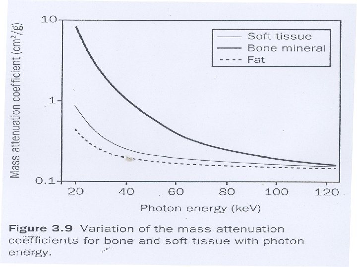
DXA: Why Dual Energy X-rays? Differential absorption by bone and soft tissue � � Low energy (30 -50 ke. V): Bone mineral attenuation >> Soft tissue High energy (>70 ke. V): Bone mineral attenuation ≈ Soft tissue allows to suppress impact of overlying soft tissue Partially corrects for presence of varying amount of fat
 Very low")
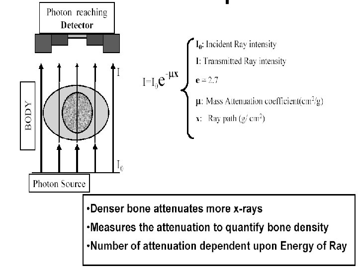
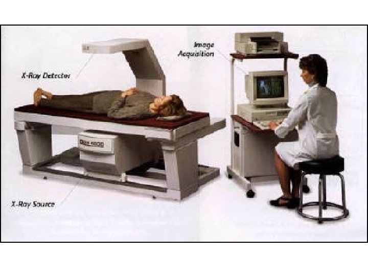
DXA Technology Detector (detects 2 tissue types - bone and soft tissue) Very low radiation to patient (similar magnitude to daily background radiation) C-arm Patient Photons Very little scatter radiation to technologist (no shielding of the technologist or the room is necessary) Collimator (pinhole for pencil beam, slit for fan beam) X-ray Source (produces 2 photon energies with different attenuation profiles)

DXA Biomechanical studies: � Strong correlation between mechanical strength and BMD measured by DXA Prospective cohort studies: � Strong relationship between fracture risk and BMD measured by DXA RCTs: � Relationship between decreased fracture risk with drug therapy and increases in BMD measured by DXA
 �")
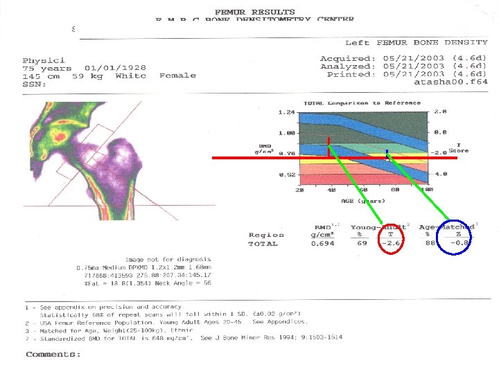
What DXA Really Measures: DXA measure: � Bone mineral content (BMC, in grams) � Bone area (BA, in square centimeters) Then calculates “areal” BMD in g/cm 2 BMD (g/cm 2) = BMC (gr) /BA (cm 2)
 Z – Score : Checked with Age & Sex Matched (2)")
Standard Scores (1) Z – Score : Checked with Age & Sex Matched (2) T - Score : Checked with Young Adult

T-score The value used for diagnosis of osteoporosis Patient’s BMD – Young-Adult Mean T-Score= BMD 1 SD of Young-Adult Mean BMD

Z-score Patient’s BMD – Age-Matched Mean Z-sore= BMD 1 SD of Age-Matched Mean BMD in g/cm 2 Low Z-score (less than -2. 0) may suggest increased likelihood of secondary osteoporosis, however. . . • This is not validated in clinical trials • High index of suspicion for secondary causes of osteoporosis is suggested in all patients

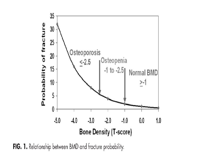
WHO Criteria for Diagnosis of Bone Status Diagnostic criteria Classification • T is above or equal to -1 • Normal • T is between -1 and -2. 5 • Osteopenia • T is -2. 5 or lower • Osteoporosis • T is -2. 5 or lower + fragility Fx • Severe or stablisshed osteoporosis

Why

Why are bone mass measurements performed? Diagnosis of osteopenia or osteoporosis Fracture risk prediction Identification of individuals for treatment Serial assessment of bone mass

Who Should be selected For BMD Measurement ? Targeted Case Finding VS Mass Screening

Indication of bone densitometry There are several strategies for incorporating BMD measurements into osteoporosis screening program: � Screening all individuals over a certain age (when fracture risk increases) � Screening only those women and men over a certain age with clinical risk factors for fracture � Screening those with clinical risk factors who are close to an intervention threshold and in whom the selective addition of BMD testing may result in intervention
 have the highest risk of fracture There")
Screening Individual with T-score below 2. 5(osteoporosis) have the highest risk of fracture There are greater number of individuals with osteopenia than osteoporosis Absolute number of fractures in subject with osteopenia (according BMD) is greater than subjects with osteoporosis.
 Thus, treatment strategies relying solely on BMD testing will miss many patients")
Screening (cont’d) Thus, treatment strategies relying solely on BMD testing will miss many patients at risk for fracture Therefore, assessment of risk factors that are independent of BMD is important for fracture prediction
 Screening for osteoporosis involves: Fracture risk assessment � Measurement of BMD �")
Screening (cont’d) Screening for osteoporosis involves: Fracture risk assessment � Measurement of BMD �
: � Increased the sensitivity of fracture")
Fracture Risk Assessment Non-BMD factors (clinical risk factors): � Increased the sensitivity of fracture risk assessment � Improves treatment intervention strategies � Including: Major risk factors Minor risk factors BMD

Major risk factors Age > 65 years • Vertebral compression fracture Fragility fracture after age 40 yrs Family history of fracture (especially maternal hip fracture > 50 yrs) Systemic glucocorticoid therapy (>3 months duration) Malabsorption syndrome • Primary hyperparathyroidis m • Propensity to fall • Osteopenia apparent on x-ray • Hypogonadism • Early menopause (before age 45)

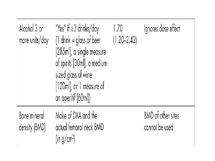
Minor risk factors Rheumatoid arthritis Past history of clinical hyperthyroidism Chronic anticonvulsant therapy Low dietary calcium intake Excessive alcohol intake Excessive caffeine intake Weight < 57 kg Weight loss > 10% of weight at age 25 Chronic heparin therapy

Identifying those at high risk The OSC recommends that � all postmenopausal women and � men over 50 years of age be assessed for the presence of risk factors for osteoporosis

Clinical Risk Factors Validated risk factors that are independent of BMD include the following: Advanced age � Previous fracture � Long-term glucocorticoid therapy � Low body weight (less than 58 kg [127 lb]) � Family history of hip fracture � Cigarette smoking � Excess alcohol intake �

Strongest non-BMD predictors Age Prior fragility fracture Kanis JA; Borgstrom F; De Laet C; Johansson H; Johnell O; Jonsson B; Oden A; Zethraeus N; Pfleger B; Khaltaev N SO - Osteoporos Int 2005 Jun; 16(6): 581 -9

Clinical risk factors are : � predictive of bone density and fracture risk � But, may not be as useful for predicting response to antiresorptive therapy

Several osteoporosis risk assessment instruments have been developed to improve the selection of women and men for measurement of BMD None of the tools was very specific, and most were not validated in other cohorts

Risk Assessment Questionnaires

Risk Assessment Questionnaires
 Introduced by WHO in 2008 Estimates the 10 -year probability")
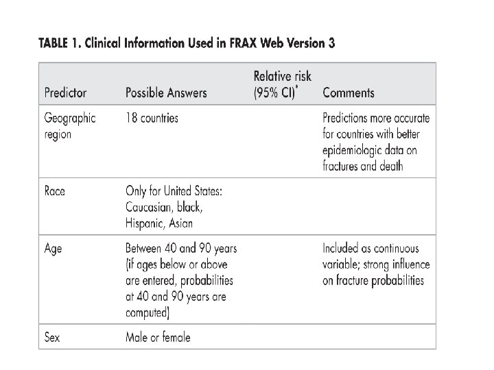
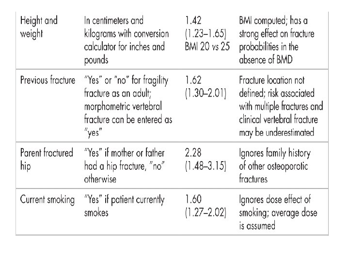
Fracture risk assessment tool(FRAX) Introduced by WHO in 2008 Estimates the 10 -year probability of hip fracture and major osteoporotic fracture for untreated patients between age 40 -90 years according: � Clinical risk factors for fracture � Femoral neck BMD(g/cm 2) Base upon data from large prospective observational studies of men and women of different ethnicities and different world regions in which clinical risk factors, BMD and fractures were evaluated

High sensitivity and low specificity Most of these instruments have high sensitivity and are able to identify patients with low BMD in the osteopenic and osteoporotic ranges. However, few are able to accurately identify patients with normal BMD who could be spared the expense of DXA testing.

WHO Absolute Fracture Risk Weighted Probability Calculation Age Sex Previous Fractures Parental Hip Fracture Rheumatoid Arthritis Secondary Osteoporosis Height Weight Current Smoking Glucocorticoid Use Alcohol > 3 unit/day Femoral Neck BMD Treat when probability of: Hip Fracture > 3% Major Osteoporosis Fracture > 20% www. shef. ac. uk/FRAX

BMD Measurement appropriate for: for all women age 65 and older because of the high risk of osteoporosis and fracture after that age and Targeted case-finding among people under age 65.
 Testing Women aged 65 and older Postmenopausal women")
Indications For Bone Mineral Density (BMD) Testing Women aged 65 and older Postmenopausal women under age 65 with risk factors for fracture. Women during the menopausal transition with clinical risk factors for fracture, such as low body weight, prior fracture, or high-risk medication use. Men aged 70 and older Men under age 70 with clinical risk factors for fracture Adults with a fragility fracture.
 Testing (Cont’d) Adults with a disease or condition")
Indications For Bone Mineral Density (BMD) Testing (Cont’d) Adults with a disease or condition associated with low bone mass or bone loss Adults taking medications associated with low bone mass or bone loss Anyone being considered for pharmacologic therapy Anyone being treated, to monitor treatment effect Anyone not receiving therapy in whom evidence of bone loss would lead to treatment Women discontinuing estrogen should be considered for bone density testing according to the indications listed above

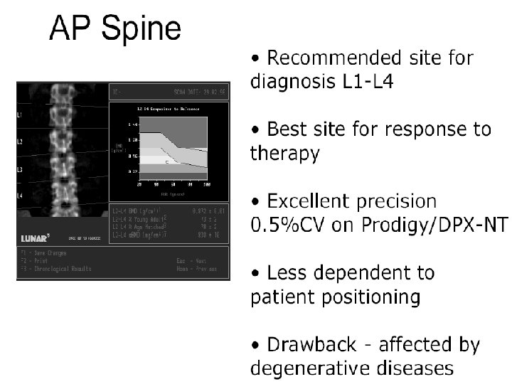
Skeletal site to measure The risk for fracture at a particular skeletal site is best estimated by measuring BMD at that skeletal site Hip fracture is often associated with significant morbidity and mortality compared with other fractures, DXA of the hip is generally regarded as the best site for diagnosis of osteoporosis. In contrast, the lumbar spine is often considered the best skeletal site to monitor because: � Less variability � Detect responses to therapy earlier than hip BMD

Skeletal site to measure The best site to measure for osteoporosis screening is controversial. Measurements from different sites (spine, hip, forearm) are often discordant with regard to the WHO diagnostic classification When measurements of different skeletal sites are performed, the diagnosis of osteoporosis is based upon the lowest T-score for BMD assessed at one of these sites. Assessment of multiple sites does not appear to improve fracture prediction The use of multiple sites for the diagnosis of osteoporosis, osteoporos Int 2006

Skeletal site to measure Younger postmenopausal women with risk factors for fracture: � DXA measurements of both the spine and hip because: early menopause is associated with greater BMD loss at the spine than the hip Aging women and men (spinal osteophytes are common) � interference from osteophytes and vascular calcifications on the spine measurement is usually minimal in that age. measurement of hip BMD alone could be sufficient. If pharmacologic therapy is planned, measurement of spine BMD might be useful for monitoring response to therapy. In the presence of degenerative changes of the spine, bone mineral density can be monitored at another skeletal site, such as the hip or radius, although the sensitivity for detecting changes at these locations is

Which Skeletal Sites Should Be Measured? Some patients Every patients Spine � L 1 -L 4 Forearm (33% Radius) Hip � If hip or spine cannot � Total Proximal Femur be measured � Hyperparathyroidism � Very obese � Femoral Neck � Trochanter Use lowest T-score of these sites

Contraindication of DXA should not be done in: � women who are pregnant or may be pregnant (because ionizing radiation, albeit it in very small doses) � DXA should be postponed until pregnancy is completed. � Unless the results are likely to play a role in the management of the patient. It may not be possible to do a DXA of the hip and spine in some patients due to inability to get on the table
 Bone mass density Bone Strength Degree of")
Bone geometry (size and shape of bone) Bone mass density Bone Strength Degree of mineralization Bone turnover microarchitectur e
 Understanding the mechanism of action of therapeutic agents (in clinical")
Bone Turnover Markers (BTM) Understanding the mechanism of action of therapeutic agents (in clinical trial) Role in the care of individual patients is not well established. Potential roles of BTMs in clinical practice: � prediction of fracture risk � monitoring response to therapy � improving compliance with therapy

Bone turnover markers Bone formation markers Octeocalcin Bone specific alkaline phosphataze P 1 NP: N-terminal propeptide of type 1 procollagen P 1 CP: C-terminal propeptide of type 1 procollagen Bone resorption markers NTX: cross-linked Ntelopeptide of type 1 collagen CTX: cross-linked C- telopeptide of type 1 collagen D-PYR: Free deoxypyridinoline PYD: Free pyridinoline HYP: Hydroxyproline TRACP 5 b: isoform 5 b of tartrate resistant acid phosphatase.

Biochemical markers of bone remodeling BMD and bone turnover markers are independent predictors of hip fracture risk urinary C-terminal collagen crosslink excretion (CTX) or free deoxypyridinoline excretion (D/Pyr)

High bone turnover is predictive of rapid bone loss DPD OC PYD BAP

BTMs Several studies have demonstrated that markers of bone turnover may be useful in predicting rates of bone loss. The values in normal subjects and patients with osteoporosis overlap substantially (biochemical markers are not useful in making the diagnosis of osteoporosis) There is currently no consensus on a role for BTMs in determining candidates for osteoporosis therapy. The decision to treat such patients should be based upon fracture risk assessment using: BMD � clinical risk factors �

Diagnosis of Osteoporosis • Technique – • Site – • DXA is the gold standard Hip (Neck, Total, or both) Reference range – NHANES III data (women 20 -29 yrs)

Discrepancies in T score Determination Different Reference database Different Site of measurement Different technologies

Key point The WHO criteria were never intended to be applied to healthy premenopausal women.

Recommendations regarding selection of patients for DXA

 : � Measures volumetric bone density of")
Bone densitometry Quantitative computerized tomography (q. CT) : � Measures volumetric bone density of the spine and hip � Analyze cortical and trabecular bone separately � Useful in clinical research and may be used in individual patients to follow therapeutic responses particularly to intermittent parathyroid hormone (teriparatide) therapy, where large changes may be observed � it is not recommended for screening, because : The application of T-scores to predict the risk of fracture has not been validated with q. CT This method is more costly Results in greater exposure to radiation than DXA
, x-ray absorptiometry, peripheral quantitative computed")
Peripheral measurements: � Ultrasound, peripheral DXA (p. DXA), x-ray absorptiometry, peripheral quantitative computed tomography (p. QCT) of the heel, radius, or hand � The WHO criteria for the diagnosis of osteoporosis are based upon BMD measured by DXA, and therefore do not apply to these other technologies

There is no consensus about which patients to assess who are younger than 65 years
- Slides: 77