Diagnosis of Fish Diseases Principles of disease diagnosis


 for soluble antigens • Soluble antigen and antibody are")


 • Detection of antigen or antibody • Chemical conjugation")


- Slides: 40
Diagnosis of Fish Diseases
Principles of disease diagnosis • Diagnosis of the aetiological agent is the most important aspect of the health management of any animal against any of the infectious diseases. • Identification of causative agent of infection as well to identify subclinical infections if any.
Investigation of the possible disease problems • Level I - changes in feeding or swimming behaviour followed by appropriate sampling and analysis • Level II - traditional diagnostic techniques like histopathology, bacteriology, virology and water analysis • Level III - Advanced techniques like PCR using molecular biological methods for fast and confirmatory diagnosis of infections
Case history and clinical sign in diagnosis Case history would give a presumptive idea about the nature of problem that caused the infection. Look for clinical signs indicative of diseased condition Clinical signs not giving direct indication of aetiology go for confirmatory diagnosis using various diagnostic tests. Behavioural changes in fish Reduced or stop feeding Viral, bacterial, parasitic and environmental Lethargic swimming Viral, bacterial, parasitic, fungal and water quality Spinning, erratic swimming Viral, parasitic or toxicants.
Normally found external signs in fish and causes • Gills necrotic – Bacterial, parasitic, fungal • Gills with excess mucus – Bacterial, parasitic, environmental or nutritional • Gills pale – Viral, bacterial, nutritional • Skin with excess mucus – Parasitic, environmental • Red pigmented areas in skin – Bacterial or parasitic • Dark skin pigmentation – Viral, bacterial, nutritional, eye parasite • Haemorrhage, erythraemia – viral, bacterial, parasitic, toxicants
Contd…. . • Frayed, eroded, erythraemia in fins – Bacterial, Parasite, mechanical/Physiological disorder • Exophthalmia, haemorrhaged opaque eyes – Viral, Bacterial, parasitic, gas super saturation • Ulceration, necrotisation – Bacterial, parasitic • Hydropsy – Bacterial, viral, metazoan parasite. • Enlarged abdomen (fluid accumulation) – Viral, bacterial, parasitic • Growth, nodules, raised ativite spot on skin – Viral, parasitic neoplasmic, fungal.
Clinical signs observed in shrimps • Major behavioural changes that can be observed in shrimp are: • reduced escape reflexes • abnormal swimming near pond edges or at the surfaces associated with lethargy • unusual aggregations • reduced preening activity and • increased feed consumption followed by anorexia.
Morphological abnormalities observed in shrimps • surface fouling of appendages and cuticle for filamentous bacteria and protozoans. • oral region for bacterial "plaques" (presumptive for Vibrio spp. ). • larval mycosis - diagnosed by demonstration of hyphae, discharge tube, and motile zoospores.
Contd…. . • muscle atrophy in abdomen, especially in 6 th abdominal segment (muscle should fill half of space available). • hepatopancreas atrophy, vacuolisation, lipid droplets and BP inclusion bodies. • cuticular deformities: bent or broken setae or spines • melanized appendages tips or foci.
Histological techniques • Histology: - Microscopic examination of thin, stained tissue sections in order to study their structure and function. • determine changes that occur in the tissues due to pathogens and disease • Histopathology can be used as diagnostic tool for identifying a variety of pathological conditions associated with many of the disease conditions. • Immunohistochemical methods are also used to detect specific pathogens in tissue sections.
Direct microscopical observation of the pathogen in the tissue sample • Fresh preparations are made from different fish tissues and observed microscopically • Many of the parasites are detected primarily by their movement. • The preparations of skin scrapings and gill are examined under the microscope and examined for the presence of for parasites.
Direct culture of the infectious agents and identification • Aseptically collected samples of tissues are used for the isolation and culture of aetiological agents such as bacteria, fungi or viruses using appropriate culture techniques. • Using standard biophysical characteristics, biochemical tests, immunological or nucleic acid based diagnostic techniques these pathogen can be identified.
Immunodiagnostic techniques • Make use of the basic principle of the specificity of antigen and Antibody • Simplest form –agglutination test – for particulate antigens- bacterial identification • Improved techniques like ELISA are now available
NATURE OF ANTIGEN-ANTIBODY REACTIONS • A. Lock and Key Concept Fab portion - constructed from the hypervariable regions of the heavy and light chains • B. Non-covalent Bonds The bonds are non-covalent - hydrogen bonds, electrostatic bonds, Van der Waals forces and hydrophobic bonds. • C. Reversibility Since antigen-antibody reactions occur via non-covalent bonds, they are reversible.
Factors affecting measurement of antigen-antibody reactions • 1. Affinity • 2. Avidity • 3. Antigen to antibody ratio • 4. Physical form of the antigen
Slide Agglutination • Simple form of Ag-Ab reaction • Microscopic slides • rapid and convenient method for determining the presence of agglutinating antibodies. • Positive - granulation or flocculation
Precipitation • when the antigen is soluble • when antigen and antibody are mixed under correct conditions • quantitative precipitation is done in a series of test tubes. • tubes where most precipitate appears contain optimal proportions of antigen and antibody.
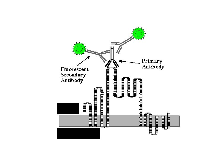
Immunofluorescence • For localisation of antigen at tissue level • Fluorescent compounds covalently attached to antibody molecules without impairing the biological activity • fluorescein isothyocyanate (FITC) forms covalent bonds with free amino groups on antibody molecules • antibody molecules highly fluorescent - can be seen with fluorescent microscope • used to identify antigens. • direct and the indirect methods
Direct and indirect methods • direct method - fluorescein tagged antibodies contact with antigens in a smear on a slide • Ag bind the tagged antibody • slide is examined with a fluorescent microscope. • indirect method - for detecting specific antibodies or identifying antigens • A secondary labeled antibody for detecting primary antibodies • Examine through microscope
Immunoperoxidase test Glucagon α cells Insulin β cells
Haemagglutination assays • Erythrocytes- microparticulate-agglutinate with antibodies against the antigenic determinants on them. • Passive haemagglutination- when erythrocytes are carrier particles for antigens. • Commonly used sheep erythrocytes (SRBC)-treat the SRBC with tannic acid. • Antigen coating on SRBC. • Antigen coated SRBC will be made to react with the antibody which will lead to agglutination.
Haemagglutination
Passive haemagglutination
Detection of bacterial toxins Latex agglutination test • Polystyrene latex particles sensitized with antibody (antiserum) raised against the toxin • Agglutination done with the test sample and the latex particles sensitized with antiserum • Positive reaction –clumps of agglutinated latex particles settle giving irregular appearance Negative reaction- latex particles settle as a button • Best example – Test kits for cholera toxin – Heat labile enterotoxin of E. coli
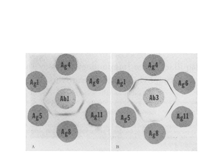
Gel diffusion techniques • Ouchterlony(1949) for soluble antigens • Soluble antigen and antibody are allowed to diffuse passively in a solid agar gel medium • When the reactants meet at the optimal concentration, visible insoluble complex is formed due to precipitation • Simple rapid and needs only small volumes of reactants
Radial immunodiffusion • The antibody or mono/poly-specific antiserum incorporated into the agarose, and the antigen is applied into the well made inside the agarose (Mancini et al, 1965) • Antigen diffuses into the gel radially around the well and a precipitin ring is formed around the well when the concentration is optimum • Reaction can be done with antigen in the gel and antibody in the well- but less sensitive.
Immunoelectrophoresis • Combines the principle of electrophoresis and immunoprecipitation • Partial separation of mixed antigens • Negatively charged molecules migrate rapidly in an antibody gel. • Can be used for serum immunoglobulin identification • Bottleneck: - Large volume of soluble antigen is required
Immunoelectrophoresis
Counter current electrophoresis • Active migration of antibody and antigen in an agar medium under the influence of an electrical field. • the antigens with higher negative charge move towards the anode and meet the antibodies moving towards the cathode • Facilitate precipitation
Counter current electrophoresis
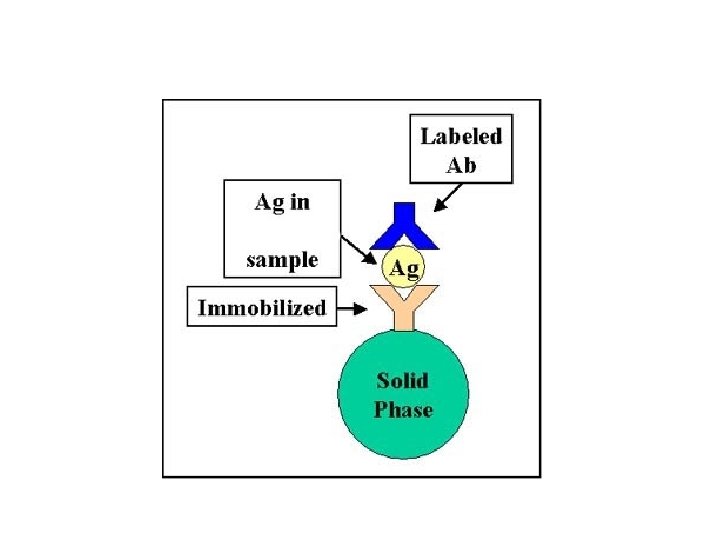
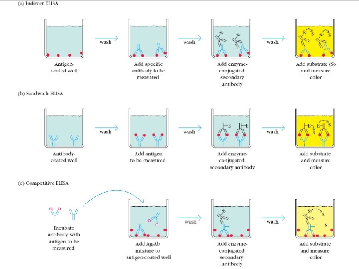
Enzyme linked immunosorbent Assay (ELISA) • Detection of antigen or antibody • Chemical conjugation of an enzyme to the primary or secondary antibody • Allows the detection of immune complex formed on a solid surface • Enzyme will react with its substrate and give rise to a colored product • Visualized and measured by optical density
Sandwich Elisa protocol • • Coating with capture antibody Blocking with BSA Addition of antigen Addition of labeled antibody Addition of substrate Conversion of chromogen Addition of stop solution Reading in ELISA reader at 450 nm
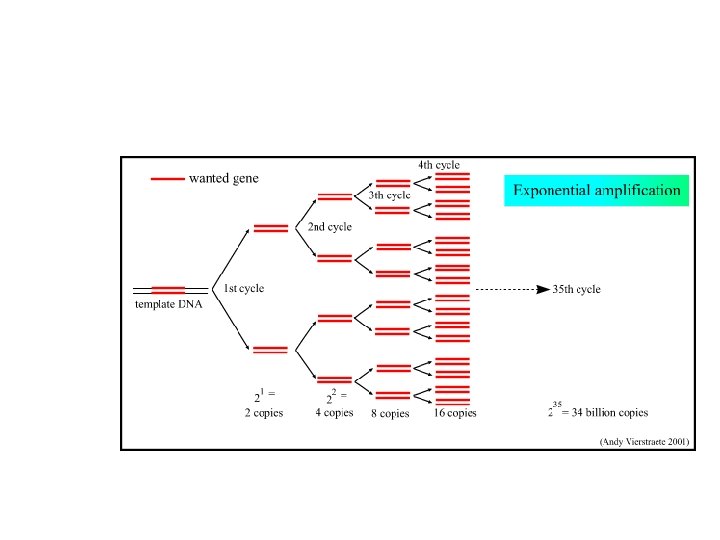
Nucleic acid based rapid diagnostics • Recent advances in molecular biology has revolutionised the diagnostic capabilities against many infectious agents. • The most efficiently used nucleic acid based techniques at present include PCR (Polymerase Chain Reaction) and DNA probes.
38
Thank you for your kind attention