Diagnosis Malignant melanoma of anterior ciliary body and
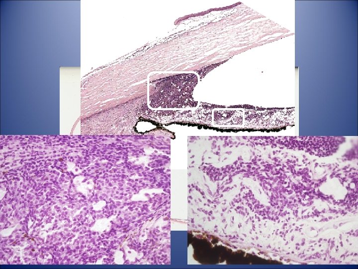
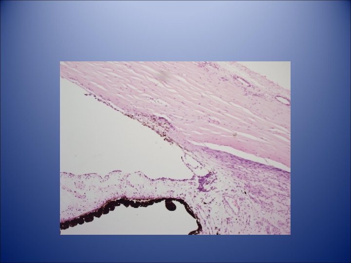
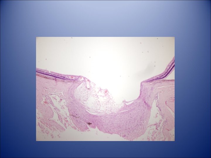

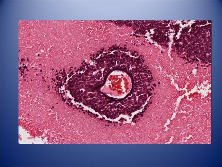
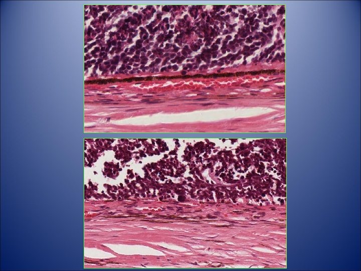
• Diagnosis – Malignant melanoma of anterior ciliary body and iris root, spindle cell type with invasion of Schlemm’s canal and collector channels – Secondary glaucoma

Iris melanoma Most common primary neoplasm of the iris Constitutes 5% to 8% of uveal melanoma Arise from the anterior border layer tissue Most iris melanomas are composed of spindle cells, therefore relatively benign. • Mortality rate is 4 to 5% • •

Retina Cases

Case 6

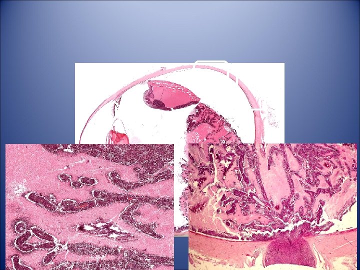
Case History • Two days prior to enucleation, a 6 -yo Caucasian girl was first noted to have left leukocoria. • Left eye was blind and was enucleated because of the clinical diagnosis of retinoblastoma. • Right eye examination and family history were unremarkable.
 • 23 x 22 mm with 11 mm of optic nerve")
Case History (Gross) • 23 x 22 mm with 11 mm of optic nerve attached. • Anterior half of the optic nerve stump exhibited a fusiform swelling with a maximum diameter of 6 mm. • Posterior to this, expansion the nerve stump was 3 mm in diameter. • Posterior segment of the eye transmitted light poorly.
 • Eye was opened horizontally. • Cornea was clear and the")
Case History (Gross) • Eye was opened horizontally. • Cornea was clear and the anterior chamber deep. • Chamber angle was open. • Densely yellow lens was in place and intact.
 • Vitreous was displaced anteriorly by a large tumor of cottage-cheese")
Case History (Gross) • Vitreous was displaced anteriorly by a large tumor of cottage-cheese consistency that occupied most of the posterior segment, sending ragged extensions anteriorly to the ora serrata. • Extravasated blood was seen within the center of the tumor.
 • Retina was identified with difficulty. • Disc was obscured by")
Case History (Gross) • Retina was identified with difficulty. • Disc was obscured by the mass. • Choroid, sclera, and episcleral tissue were unremarkable.
- Slides: 15