
DIAGNOSIS Jafar Naghshbandi D D S M S
DIAGNOSIS Jafar Naghshbandi D. D. S; M. S Diplomate of the American Board of Periodontology

Periodontal Diagnosis ØDefinition: Recognizing a departure from normal in the periodontium and distinguishing one disease from another. ØBased on information acquired from: I. Medical and dental histories II. Clinical and radiographic examination III. laboratory findings
Periodontal Diagnosis ØProper diagnosis is essential for accurate treatment ØPeriodontal diagnosis steps: 1) Determine whether disease is present 2) Identify its type, extent, distribution, and severity 3) Identify its causes and understand the underlying pathologic processes
CLINICAL DIAGNOSIS ØMedical history ØDental history ØExtra-oral exam ØIntra-oral exam ◦ Soft tissue ◦ Hard tissue ØRadiographic Assessment ØPeriodontal Survey
Medical History ◦ Systemic disease ◦ Antibiotic prophylaxis ◦ Pregnancy ◦ Drug habit
Dental History ◦ Chief complaint ◦ Oral habits ◦ History ◦ Extra-oral Exam ◦ Lymph nodes
Intra-oral Exam ØSoft Tissue ◦ evaluate for appearances an ØHard Tissue ◦ Malpositioned teeth ◦ Caries ◦ Restorations ◦ Habits ◦ Abfractions atypical or abnormal
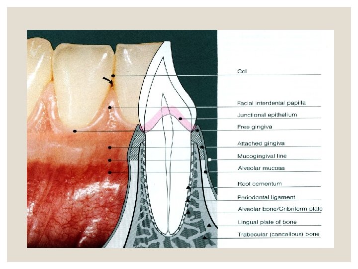
Examination of the Periodontium ◦ plaque and calculus ◦ clinical attachment loss ◦ periodontal pockets ◦ amount of attached gingiva ◦ furcation ◦ Class I, III ◦ mobility ◦ Grade I, III ◦ periodontal abscess ◦ suppuration
Examination of the Periodontium • The periodontal examination should be systematic. ◦ start in the molar region in either maxilla or mandible and proceeding around the arch. ◦ It is important to detect the earliest signs of gingival and periodontal disease. • Periodontal charts: Is used to record periodontal and associated findings. ◦ They provide a guide for a thorough examination and record of the patient's periodontal condition. ◦ They are used for evaluating response to treatment and for comparison during recall visits.
Periodontal examination ◦ The diagnostic procedures are: A- Clinical intra- and extra -oral examination B- Radiographic examination C- Occlusal examination D- Tests of tooth mobility E- Evaluation of tooth vitality F- Periodontal examination Biopsy, if necessary ◦ Supplemental diagnostics
Gingiva ◦ The gingiva must be dried before evaluation. ◦ Light reflection from moist gingiva obscures detail. ◦ In addition to visual examination, exploration with instruments is essential. ◦ Firm but gentle palpation should be used for detecting pathologic alterations in normal resilience and for locating areas of pus formation
Gingiva The following features of the gingiva should be considered: ◦ Color ◦ Size ◦ Contour ◦ Consistency ◦ surface texture ◦ Position ◦ ease of bleeding ◦ pain
Gingival inflammation cause two basic types of tissue response: ◦ 1. edematous ◦ 2. fibrotic. Edematous tissue response is characterized by: smooth, glossy, soft, red gingiva Fibrotic tissue response is characterized by: ◦ Some of the characteristics of normality persist ◦ The gingiva is more firm, stippled, and opaque ◦ It is usually thicker and its margin appears rounded
Periodontal Examination Pocket Probing: ◦ Six surfaces are probed on each tooth and depth is recorded in mm. These surfaces are; Disto-buccal, mesio-buccal, disto-lingual, lingual and mesio-lingual.
Periodontal pockets Examination for periodontal pockets must include : • presence and distribution on each tooth surface • pocket depth • level of attachment on the root • type of pocket (suprabony or intrabony)
periodontal pockets Detection of periodontal Pockets • The only accurate method of detecting and measuring periodontal pockets is careful exploration with a periodontal probe • Pockets are not detected by radiographic examination • The periodontal pocket is a soft tissue change • Radiographs indicate areas of bone loss where pockets may be suspected but important to understand: 1) they do not show pocket presence or depth 2)They show no difference before or after pocket elimination unless bone has been modified
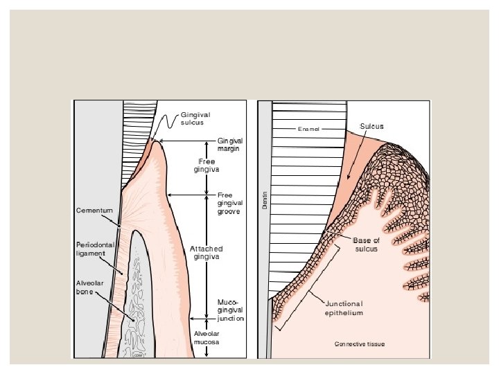
Pocket Probing The two different pocket depths are: A) Biologic or histologic pocket depth B) Probing or clinical pocket depth • Biologic depth is the distance between the gingival margin and the base of the pocket • Probing depth is the distance to which a probe penetrates into the pocket
Pocket Probing The depth of penetration of a probe in a pocket depends on factors: ◦ Size of the probe ◦ Force with which it is introduced ◦ Direction of penetration ◦ Resistance of the tissues ◦ Convexity of the crown
Clinical Attachment Level ( CAL) versus Pocket Depth(PD) ◦ • Pocket depth is the distance between the base of the pocket and the gingival margin ◦ • It may change from time to time even in untreated periodontal disease owing to changes in the position of the gingival margin so it may be unrelated to the existing attachment of the tooth. ◦ • The Clinical Attachment Level ( CAL) is the distance between cementoenamel junction and the base of the pocket ◦ • Changes in the CAL can be due only to gain or loss of attachment and is a better indication for the degree of periodontal destruction or regeneration
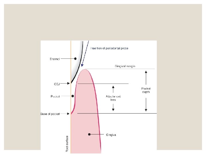
Clinical Attachment Levels (CAL) ◦ The assessment of clinical attachment levels (CAL) provides information relating to the gain or loss of connective tissue attachment to the root surface ◦ It is the most practical method of determining that the disease is progressive (active) when a significant loss of attachment has occurred over time.
Bleeding on Probing • The insertion of a probe to the bottom of the pocket elicits bleeding if the gingiva is inflamed and the pocket epithelium is atrophic or ulcerated. Non inflamed sites rarely bleed. • In most cases, bleeding on probing is an earlier sign of inflammation than gingival color changes. • Sometimes color changes are found with no bleeding on probing.
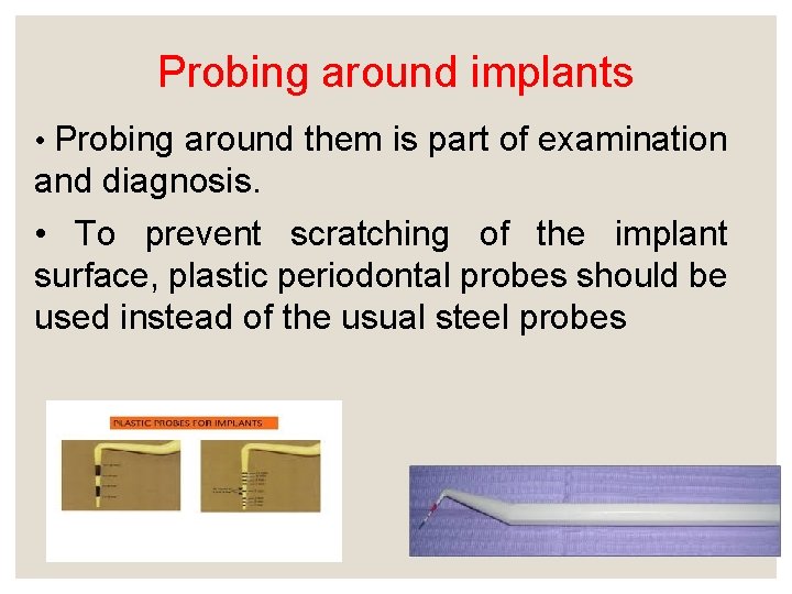
Probing around implants • Probing around them is part of examination and diagnosis. • To prevent scratching of the implant surface, plastic periodontal probes should be used instead of the usual steel probes
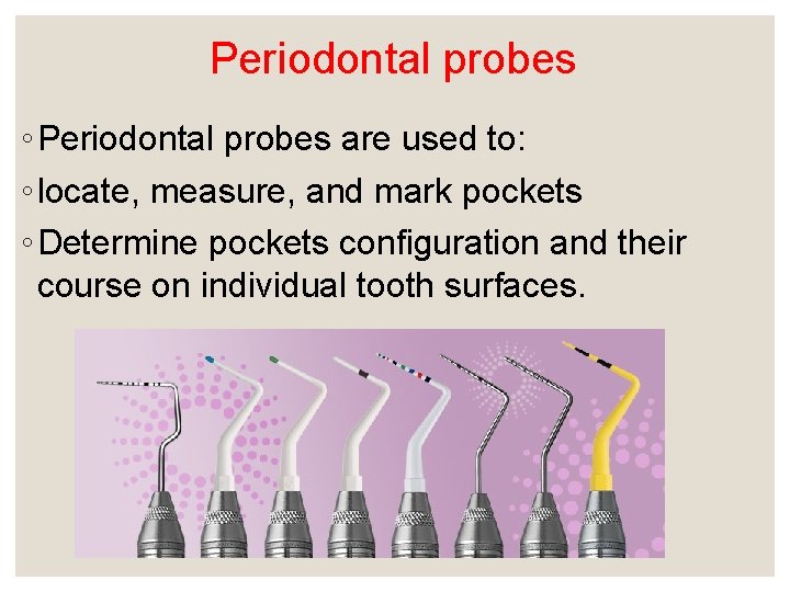
Periodontal probes ◦ Periodontal probes are used to: ◦ locate, measure, and mark pockets ◦ Determine pockets configuration and their course on individual tooth surfaces.
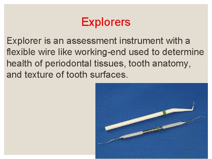
Explorers Explorer is an assessment instrument with a flexible wire like working-end used to determine health of periodontal tissues, tooth anatomy, and texture of tooth surfaces.
Function of explorers ◦ Explorers are used to: 1. Detect, by tactile means, the texture and character of tooth surfaces, before, during and after periodontal debridement to assess the progress and completeness of instrumentation. 2. To examine tooth surfaces for calculus, decalcified and carious lesions, dental anomalies, and anatomic features such as grooves, curvatures, or root furcations.
Types of Periodontal Probes ◦ There are three types of periodontal probes: 1. Calibrated periodontal probes 2. Naber’s furcation probe 3. Computer assisted probes
Radiographic Assessment ◦ Radiographic examination and interpretation ◦ It is essential that all radiographic studies be of diagnostic quality ◦ Sometimes X-ray fails to show bone defects as probing will. In cases of interproximal two wall craters the buccal and lingual intact bone shows clearly on X-ray. ◦ Furcation defects may be better determined by furcation probing
Radiographic Assessment ◦ Radiographs do not Show periodontal pockets ◦ Distinguish between successfully treated and untreated cases ◦ Show structures on buccal, lingual, and labial aspect of tooth ◦ Record tooth mobilty
Radiographic Assessment full mouth series ◦ Look At: ◦ lamina dura ◦ crown: root ratio ◦ interproximal bone ◦ hypercementosis ◦ furcation involvement ◦ periapical pathology ◦ overhangs ◦ calculus ◦ resoption
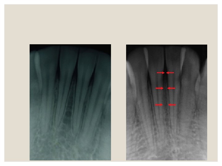
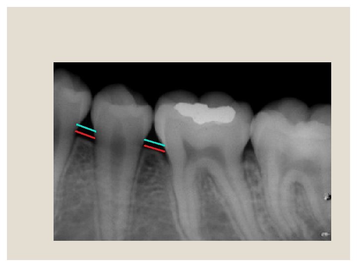
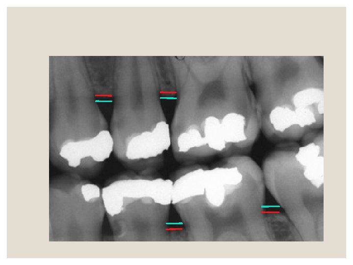
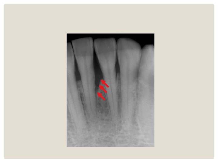
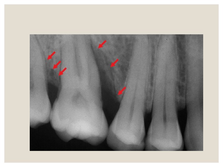
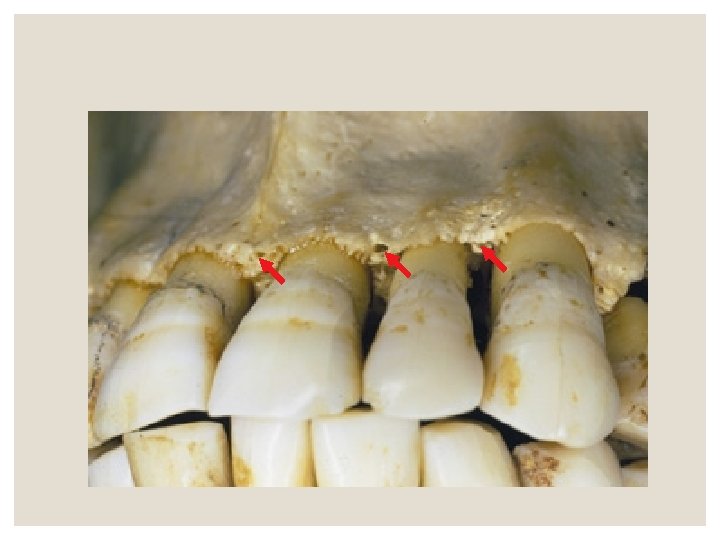
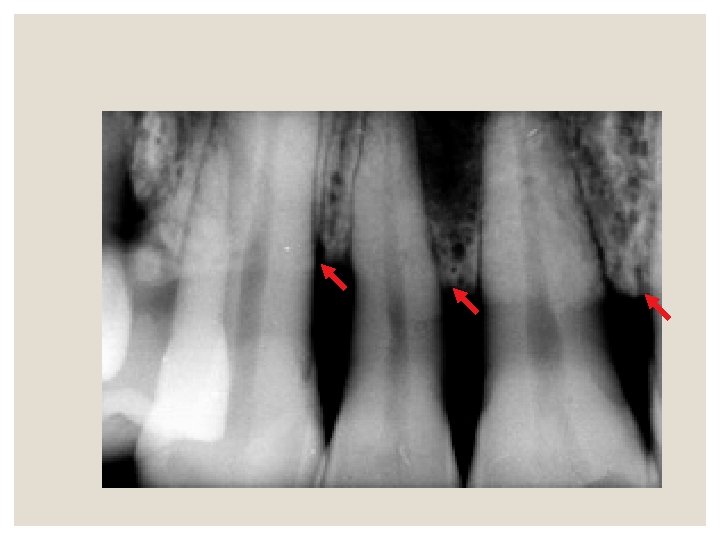
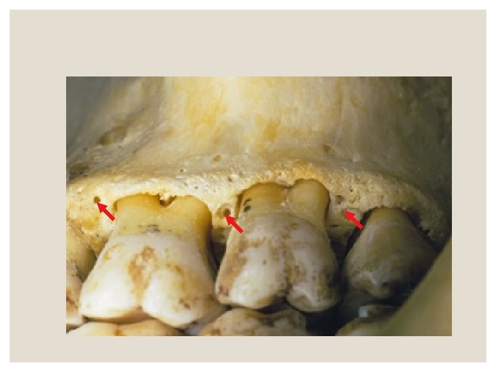
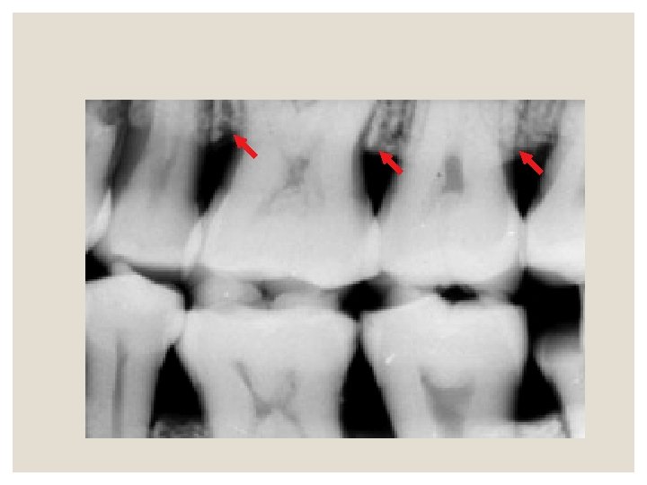
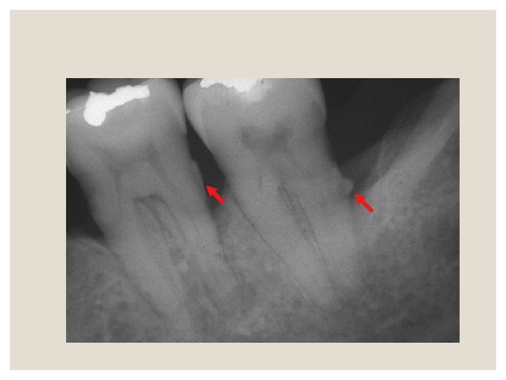
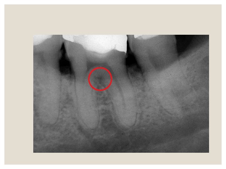
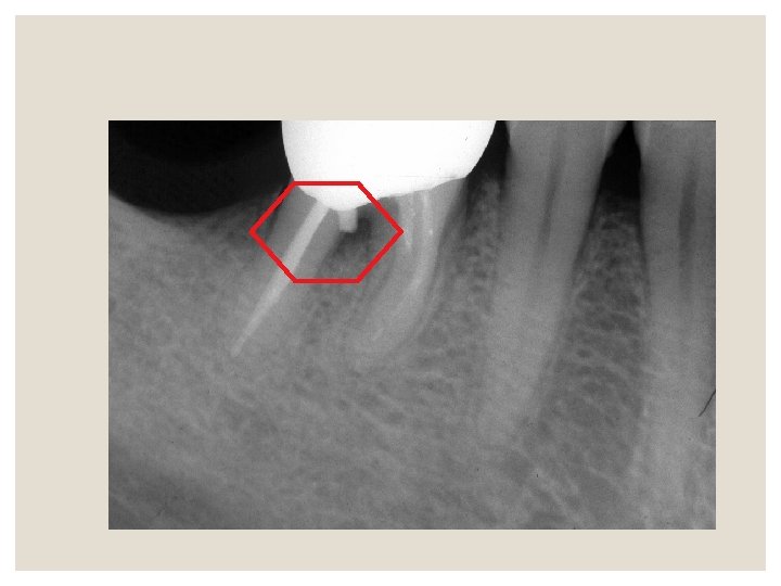
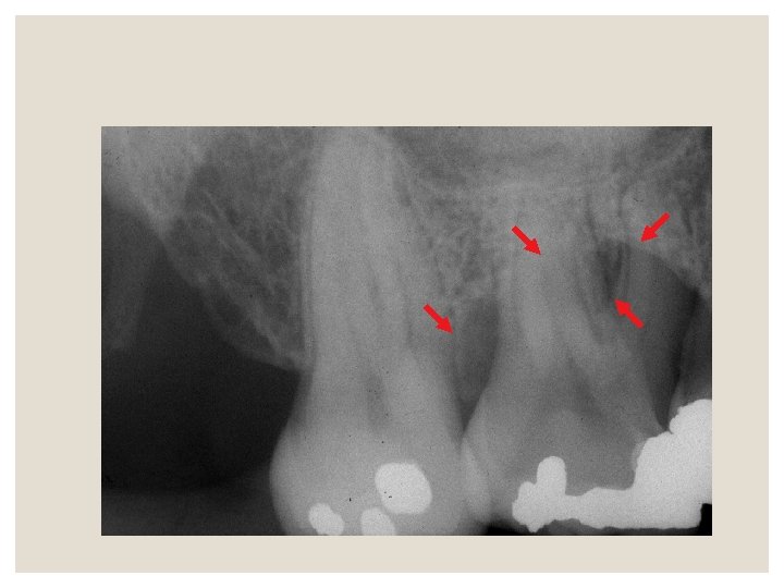
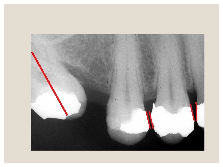
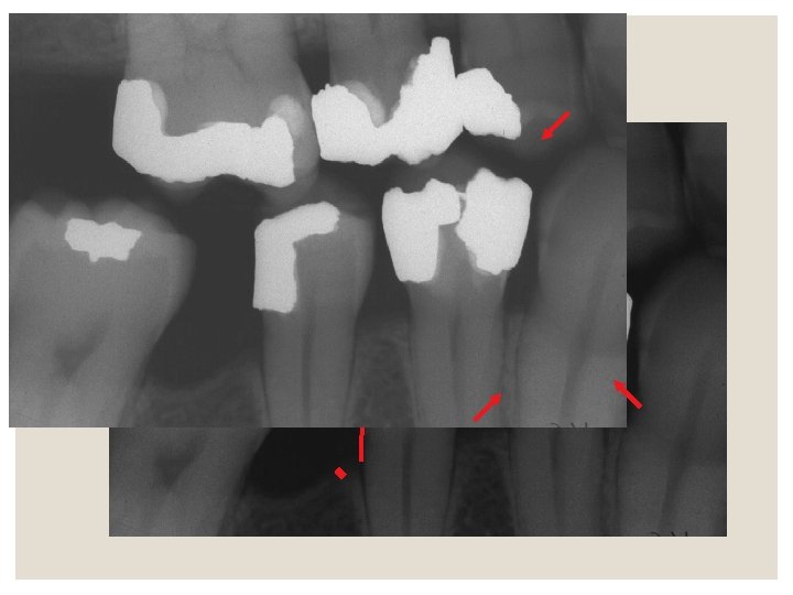
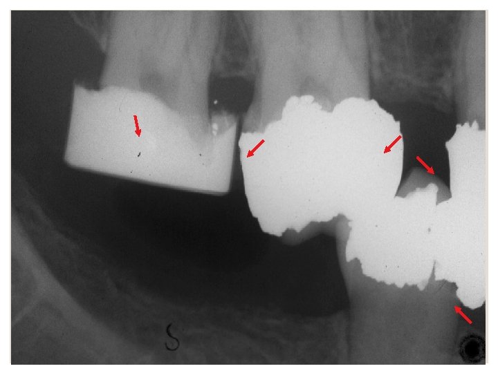
Predisposing conditions ◦ Tipping of teeth ◦ Open contacts ◦ Poor contours of restorations and overhang margins


- Slides: 52