Diagnosis and Treatment of Idiopathic Normal Pressure Hydrocephalus

")


 and brain imaging. Aging causes atrophy")


- Slides: 11
Diagnosis and Treatment of Idiopathic Normal Pressure Hydrocephalus Rohini Selvarajah 2007 Neuroscience presentation
Introduction First described by Hakim and Adams in 1965, idiopathic normal pressure hydrocephalus (INPH) is an enlargement of cerebral ventricles without evidence of chronic increased intracranial pressure (ICP). INPH is associated with a triad of symptoms including gait disturbance, dementia, and urinary incontinence In 2000, the costs of treating INPH exceeded $1 billion. There were 27, 870 patients with INPH treated that year, and more than 8, 000 new cases diagnosed. INPH is clinically diagnosed in most patients during the sixth or seventh decade of life. Many older adults are incorrectly diagnosed with disorders such as Parkinson's disease and dementia when their symptoms are actually caused by INPH. The diagnosis of INPH is challenging and requires a combination of clinical signs and symptoms, radiographic findings, and diagnostic testing. The purpose of evaluation and testing of individuals with suspected INPH is to determine if surgical implantation of a ventriculoperitoneal (VP) shunt will be beneficial. VP shunting is now a common neurosurgical procedure, but it is one associated with risks and complications, which makes evaluation of "shunt-responsiveness" essential.
Presentation A 65 -year-old man, presented with a profile of progressively worsening symptoms, including gait and balance impairment, deterioration of memory and cognition, and recent urinary urgency and incontinence. A family member brought him to his physician for evaluation and treatment. Although his signs and symptoms could be attributed to the effects of aging, a careful and thorough evaluation was necessary to accurately diagnose his disorder. His evaluation included careful review of his medical history, as well as a thorough systems examination. Special attention was paid to any specific brain disease or brain trauma that could precipitate his presenting symptomology. Any adult, especially one over 60 years of age that demonstrates an insidious onset of the triad of symptoms of gait disturbance, cognitive impairment, and urinary incontinence, should be evaluated by a geriatrician or internist, psychiatrist, neurologist, and neurosurgeon. The characteristic profile of signs and symptoms in an older individual unexplained by any specific cause, along with a high degree of clinical suspicion by the physician, will focus the diagnosis towards INPH. Many conditions affecting older individuals can mimic the symptom profile of INPH, including Parkinson's disease, Alzheimer's disease, metabolic and psychiatric disorders, endocrine dysfunction, infections, trauma, vascular and neurodegenerative disorders, and incontinence from urinary tract disorders. These conditions must be ruled out as causes of symptoms before a diagnosis of INPH can be accurately made.
The diagnostic evaluation also includes lumbar puncture (LP) and brain imaging. Aging causes atrophy of brain tissue which can lead to ventriculomegaly. The degree of ventriculomegaly common in INPH is disproportionate to the amount of brain atrophy, and is measured via brain imaging. Atrophic changes in the older adult's brain may be imaged with computed tomography (CT) or magnetic resonance imaging (MRI). Either technique can identify the ventriculomegaly necessary for the diagnosis of INPH, but MRI can also identify other pathology responsible for symptoms similar to INPH. The MRI scan in patients with INPH reveals dilated ventricles with preserved cerebral parenchyma, which is in contrast to the ventricular dilation and significant loss of brain tissue seen in patients with Alzheimer's disease. An MRI scan is also useful in identifying the amount of white matter changes around the ventricles. Although MRI is the most accurate diagnostic imaging modality, non-contrast CT remains the diagnostic imaging technique of choice for rapid and accurate diagnosis of hydrocephalus. The brain CT scan in INPH reveals enlargement of two lateral and third ventricles and relative sparing of the fourth ventricle. There is no single test or imaging study that can be used to conclusively diagnose INPH. It must be diagnosed clinically, because the symptoms caused by INPH overlap many other conditions that affect older adults.
Diagnosis. . Mr. M's CT scan revealed dilation of both lateral ventricles and the third ventricle, with sparing of the fourth ventricle, findings commonly associated with INPH unless the circulation of CSF is obstructed, which it is not in INPH, the fourth ventricle is relatively spared from no specific cause for his symptoms was identified. Blood tests were performed to rule out other diseases and conditions that may cause similar symptoms, such as neurodegenerative disorders, cerebrovascular disease, urological disorders or other metabolic dysfunctions. LP was performed. An LP is done only when the clinician performing the procedure is certain that intracranial pressures are not elevated. Performing an LP when ICP is elevated can result in catastrophic brain stem herniation. Elevated ICP can be determined noninvasively by viewing the CT scan and assessing for the presence of papilledema; if present, then this finding is consistent with elevated ICP. (Papilledema is a swollen and distorted optic disc with a characteristic reddish hue due to increased intracranial pressure transmitted through the subarachnoid space along the optic nerve) Mr. M's LP showed an opening pressure of 160 mm H 2 O, which is less than the upper limit of the normal range of 240 mm H 2 O, and within the range of 105– 190 mm H 2 O consistent with a diagnosis of probable INPH. Higher opening pressures correlate with the probability of INPH Mr. M's ICP measurement is associated with his signs and symptoms, including his ventriculomegaly evident on CT. Based on his symptoms, evaluation, imaging studies, and LP results, Mr. M was diagnosed with INPH.
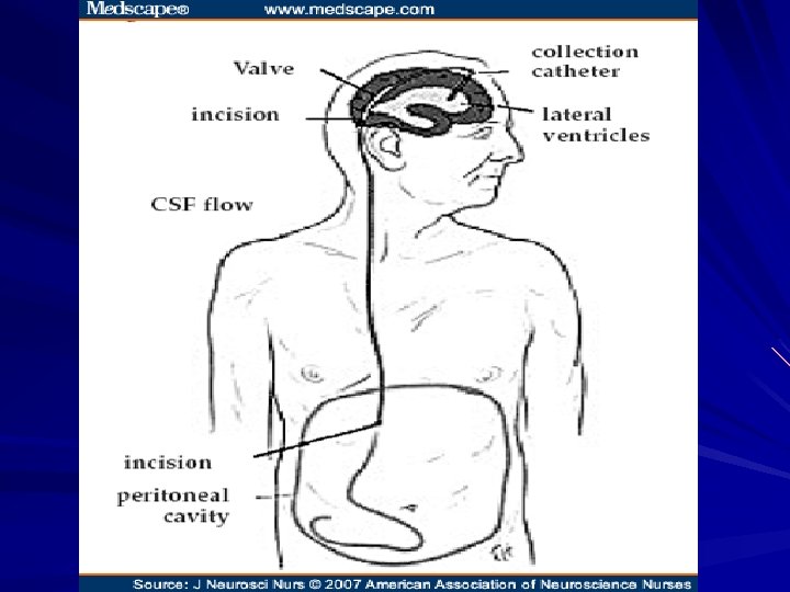
Treatment Considerations The next step was to attempt to determine his responsiveness to ventriculoperitoneal (VP) shunting Although VP shunting is prone to complications and not appropriate for every patient with INPH, it remains the most commonly recommended therapy for INPH for patients who demonstrate a favorable risk to benefit ratio recipients over time suggests that VP shunts do not always halt the progression of INPH. As other comorbidities independent of INPH worsen, shunted individuals can experience overall deterioration. Therefore, every effort should be invested in attempting to predict the probability of shunt responsiveness in each patient prior to surgical implantation.
A positive response to removal of 30– 50 ml of CSF, assessed by measuring improvements in gait stability and urinary control, is a positive prognostic indicator for VP shunting. The surgeon must estimate abdominal back pressure in the peritoneal cavity that will receive CSF drainage, because underdrainage may occur due to absorption incompetence Proper VP shunt function is dependent on the pressure differences between the ICP and abdominal cavity. Pressure in the abdominal cavity is normally lower than ICP so that CSF will flow into the abdomen. Any decrease in CSF flow into the peritoneal cavity must be investigated, focusing on conditions within the abdomen. Consideration must be given to the effects of obesity, constipation, small bowel obstruction, or ileus.
Complications There are many shunt-related complications and risks that make proper patient selection an even more important challenge. Intracerebral hemorrhage, or bleeding along the tract of the shunt, is the primary procedure-related risk. Other complications include infection, with staphylococcus responsible for 90% of all shunt infections. Seizures, shunt obstruction, subdural fluid collections causing increased ICP, and headaches due to overdrainage can occur. The most serious complication of VP shunting is overdrainage of CSF resulting in stress and tension on cerebral vasculature, which causes a potentially lethal subdural hematoma.
Thank you for your attention…