DIAGNOSIS AND MANAGEMENT OF RHEUMATIC FEVER BY Dr

DIAGNOSIS AND MANAGEMENT OF RHEUMATIC FEVER BY Dr. LALY D C SR CARDIOLOGY MCH CALICUT

GAS INFECTION Acute rheumatic fever is a non suppurative complication of Group A beta hemolytic streptococcal sore throat. It affects joints, skin, subcutaneous tissue, brain and heart. Except heart, all other effects are reversible Cardiac complications are significant resulting in life threatening valvular heart disease
 � Despite")
ACUTE RHEUMATIC FEVERCURRENT SCENARIO IN INDIA (A study by IB Vijayalakshmi, Bangaluru) � Despite the tremendous progress made in cardiology, the morbidity and mortality due to acute rheumatic fever and its consequences remain very high in india � In india, rheumatic fever accounts formerly 25 -45% of acquired heart disease � Annual incidence of rheumatic fever is 100 -200 %greater than that of developed countries � Recent data suggest that large no: of cases are still frequently in younger children under the age of 10 years

� Clinical diagnosis of carditis was noted in atleast 50% rheumatic fever in the past, seems to be declining now � In our country, many patients who presented with established RHD had a history of mono arthraligia/arthritis rather than classical polyarthritis � Fleating type of migratory polyarthritis was present in 52. 87 % where as migratory polyarthralgia in 47. 12 % � Out of 213 cases of arthralgia, 17. 84% had clinical evidence of carditis

NON-CARDIAC MANIFESTATIONS OF RHEUMATIC FEVER ARTHRITIS � Most frequent major manifestation. Occurs in 66 %-75% of children experiencing first attack of disease. � Frequently the presenting compliant. � In adolescents and adults it is often the sole major manifestation in first attack. Clinical features : Joint involvement varies from mild arthralgia to disabling arthritis. � Larger joints knees and ankles commonly affected. � Wrist, elbows, shoulders, hips may also be involved. � Less frequently smaller joints of hands, feet and neck. � Involved joints are warm, red and swollen. � Exquisite tenderness out of proportion to objective findings. �

� MIGRATORY- Doesn’t mean that inflammation resolves completely in one joint before striking the other rather joints may be involved sequentially so that some may be resolving while others are beginning. � MONOARTHRITIS – Also occurs. inflammatory fully expressed. - Frequency is increased in which antitherapy is initiated before disease is � Several joints may be affected simultaneously. � Arthritis may be addictive rather than migratory. � Inflammation in a joint usually last not longer than 2 -3 weeks, entire bout of poly-arthritis not more than a month. � Poly-arthritis and Sydenham’s chorea never occurs simultaneously. � Chorea may occur after arthritis has subsided. � Carditis and arthritis do frequently co-exist, with a striking inverse relationship. � Jaccouds arthopathy

PATHOLOGY • Acute exudative joint effusion. • Swelling of articular and periarticular structures, thickened synovium. • No pannus formation. • Focal lymphocytic and polymorphonuclar infiltrates. • Although fibrinoid lesions and histiocytic granulomas occur in later stages they heal without permanent damage. • Decrease in complement components in joint fluid.
. � May")
POSTSTREPTOCOCCAL REACTIVE ARTHRITIS Occurs after a short latent period (7 -10 days). � May be persistent or relapsing. � Does not respond as dramatically to anti-inflammatory agents as rheumatic polyarthritis. � Unassociated with other major manifestations of the disease. �

DIFFERENTIAL DIAGNOSIS CONFIRMATORY STUDY Septic arthritis Synovial fluid and blood culture Bacterial endocarditis Blood culture Lyme disease Serological studies Mycobacterial and fungal arthritis Culture or biopsy Viral arthritis Serological studies Enteric infection Culture or serological studies Urogenital infection culture

DIAGNOSIS CONFIRMATORY STUDY Inflammatory bowel disease clinical findings Rheumatoid arthritis and Stills disease “ Systemic vasculitis Biopsy or angiography SLE Serological studies Gout and pseudogout Polarising microscopy cancers biopsy sarcoidosis biopsy dermatomyocitis Behcets disease Henoch-Scholen purpura Kawasaki’s disease

SYDENHAM’S CHOREA First described by Sir Thomas Sydenham and Guillaime de Bailou in the late seventeenth century. � Occurs primarily in children between the age group 5 and 15, rarely after age 20. � There is a female preponderance. � CLINICAL MANIFESTATIONS- is characterized by; emotional lability in co-ordination muscular weakness Child may be fretful, irritable, inattentive to school work, fidgety or even severely disturbed with clumsiness and tendency to drop objects. � On physical examination, movements are abrupt and erratic. � Facial movements include grimaces, grins and frowns. � Tongue when protruded resembles a “bag of worms”. � Speech is jerky and staccato. � Handwriting deteriorates and may become illegible. �

Spoon or dish hands � Pronator sign � Milkmaid’s grip � Hemichorea � The choreiform movements disappear during sleep � Decrease with rest and sedation � Suppressed by volition only for a few seconds. � No sensory or pyramidal tract involvement. �


EEG AND BRAIN IMAGING Most common EEG abnormality is paroxysmal 3 Hz slowwave complex in parieto-occipital region. � MRI abnormalities- hyper intencities in left caudate nucleus, putamen, lateral pallidum, substantia nigra. RELATION TO OTHER MANIFESTATIONS OF RF � May be pure chorea. � Because of the long latent period (1 -7 months) of chorea, simultaneous occurrence of chorea and polyarthritis do not occur. � When carditis and chorea appear together it is often due to clinically inapparent carditis called to attention by the onset of chorea. � Antibody titers may have declined towards normal when choreiform movements appear. � Recurrences of chorea are not uncommon. �

DIFFERENTIAL DIAGNOSIS � Atypical seizures � CVA � Collagen vascular disease � Drug intoxication-phenytoin, amitriptyline, metoclopramide, fluphenazine. � Familiar chorea � Hormonally induced chorea � Hyperthyroidism � Hypoparathyroidism � Lyme disease � Wilson’s disease PROGNOSIS- Duration of chorea is variable from one week to more than two years. Three-fourth of patients recover within 6 months. Manifestations may wax and wane during the course. Sequelae- convulsions, decreased learning ability, behavior problems and psychosis.

Subcutaneous nodules Incidence -<1 -21% � Cinical manifestation-round, firm, freely movable, painless, size varies-. 5 -2 cm � Occur in crops over bony prominences or extensor tendons � Common locations are elbows, wrists, ankles&achilles tendons, scalp, spinous processes �

Subcutaneos nodules No varies from one to few dozen, usually 3 to 4 Persist from days to 1 -2 wks, rarely more than 1 month Are not pathognomic of RF Lesions in SLE, RA tend to larger

Relation to other manifestations Rare as isolated manifestation of RF � They are associated with carditis in most cases � Usually appears several weeks after the onset of cardiac findings. � Found more frequently in patients with severe carditis and may appear in recurrent crops � Pathology Histologicaly consist of a central zone of fibrinoid necrosis surrounded by cellular reaction of histiocytes and fibroblasts � Perivascular accumulations of lymphocytes and polymorphonuclear leukocytes � Do not exibit palisading.

Erythema Marginetum EM occurs in 15 persentage or less of patients with acute RF. � Evanescent in nature, maybe missed in particularry in dark skinned patients. � Rash appears first as bright pink macule or papule , that spreads outward in circular or serpiginous pattern. � Multiple appearing on the trunk or proximal extremities rarely on the distal extremities never on the face. �

Nonpruritic, nonpainful, blanch on pressure, only rarely raised � Individual lesions may come and go in min. at times changing shapes before observer’s eyes, so described as “smoke rings” beneath the skin � Usually occurs early in the course of rheumatic attack. � May persist or recur for months or even years. � Not influenced by anti inflammatory therapy. � Associated with carditis but not necessarily � Nodules and EM do occur together. � Sepsis, drug reactions, glomeronephritis can have EM. � Circinate rash of lyme disease may resemble EM.

RHEUMATIC PNEUMONIA � Acute inflammatory pneumonitis has been described, usually with severe carditis. -Chest xray –hilar or patchy Distribution or consolidation Deposition of RBCs, fibrin, hemosiderin

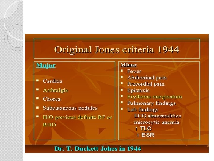
MINOR manifestations � Arthralgia –involves large joints , may be mild or incapacitating , present for days-weeks, verying in severity. � Epistaxis-mild or severe enough to require transfusions. � Abdominal pain-epigastric or periumbilical, may be indistinguishable from acute appendicitis � Fever-usually from 101 -104 F, no characteristic fever pattern � ESR > or = 60 � CRP > or = 3 � Prolonged PR intrevel

ACTIVE RHEUMATIC CARDITIS FREQUENCY OF CARDITIS IN ACUTE RHEUMATIC FEVER INDIA- 90% in all attacks. 30% in first attacks

MODE OF ONSET 0 f carditis Subclinical cardiac involvement � Indolent � Subacute � Acute � Fulminant congestive heart failure � MR, AR of varying severity � Pericarditis � Conduction disturbanes Younger patients- present with history of fever loss of apetite lassitude lethargy easy fatiguability vague pains over a period of few weeks Patients who have significant arthritis less commonly have severe carditis. �

CLINICAL RECOGNISION OF CARDITIS � Rheumatic carditis is an early manifestation of RF. � 80% of patients develop in the first two weeks. Pancarditis Myocarditis- Soft S 1 Gallop sounds Unexplained cardiomegaly Unexplained congestive heart failure

PERICARDITIS Pericardial rubs- transient � Serous or hemorrhagic effusion � Rare reports of pericardial tamponade and even pericardial constriction � Differential diagnosis- Tubercular pericarditis Normal valves No regurgitation of valves
 Mitral regurgitation-Soft blowing, pansystolic murmur of MR at the apex. Mirtal valve")
REGURGITATION (ENDOCARDITIS) Mitral regurgitation-Soft blowing, pansystolic murmur of MR at the apex. Mirtal valve involvement in 92 -95%of patients with carditis 70 -75% of patients, only valve affected Mitral valvulitis-Low-pitched mid-diastolic Carey Coombs murmur best appreciated at the apex with no presystolic accentuation and dissappears on follow-up.

AORTIC VALVE INVOLVEMENT � Presence as aortic regurgitation � Aortic stenosis is not a feature of acute rheumatic carditis. � AS with multiple attacks of carditis and nearly always associated with mitral valve disease. � High-pitched soft decrescento murmur of AR is easy to miss. � Aortic valve involvement in 20 -25%of patients with carditis, isolated finding in 5 -8%.

DISTRIBUTION OF VALVULITIS IN RHEUMATIC FEVER 70 -75% Almost never, rare pathologically 20 -25% 5 -8% 30 -50% at autopsy Tricuspid valve involvement is uncommon in first attack but in recurrent disease present as organic TR, less commonly organic TS.

MECHANISM OF VALVE INVOLVEMENT � Primary effects of rheumatic fever are inflammatory valvulitis, annular dysfunction/dilatation. � Valvulitis results in rheumatic verrucous vegetations, seen as nodules on ecocardiography.

� Mild to moderate MR- Have left ventricular dilatation with mild or no annular dilatation and no mitral valve prolapse in acute cases. Severe MR commonly have annular dilatation, chordal elongation and anterior mitaral leaflet prolapse. In majority of patients with mild to moderate regurgitation, carditis disappears on follow-up.

MECHANISM OF VALVE INVOLVEMENT � Ashoff nodules have a predilection for semilunar and aortic valve rings resulting in annular dysfuction and dilatation which stresses the chordae and papillary muscles resulting in chordal lengthening and mitral valve prolapse. � Mitral regurgitation begets mitral regurgitation and rarely stretched chordae may rupture resulting in flail leaflets and severe acute regurgitation. � Valvulitis and rheumatic involvement of aortic ring causes aortic regurgitation sometimes with aortic valve prolapse.

DIAGNOSIS OF ACUTE RHEUMATIC CARDITIS- IN RECURRENCES � Valvulitis Change in murmur New onset murmur � Myocarditis Evanescent murmurs Worsening cardiomegaly Worsening CHF � Pericarditis Pericardial rubs Pericardial effusion

CONDUCTION ABNORMALITIES � Increased vagal activity is involved in conduction abnormality, has low sensitivity and specificity for the diagnosis of carditis. � Elongation of PR interval Mobitz type 1 and 2 (rare) Complete heart block- has been mentioned Occasionally junctional rhythm and atrial or Ventricular premature beats.

Silent carditis Carditis may be clinically silent or not suspected. � Follow-up of isolated chorea for 20 years showed 23%to have mitral stenosis. � A number of patients normal clinically eventually showed evidence of carditis at histology. � Recently Doppler echo has been proposed for identifying subclinical carditis. Insidious or late onset carditis � Some patients in developing countries especially young children present with vague symptoms like malaise, lathargy, vague ill health for a few weeks to months. � On examination, there will be features of rheumatic heart disease. � Elevated ESR and CRP. � ASO may not be elevated. � Natural history of this group is still not fully defined.

CARDITIS AND OTHER MANIFESTATIONS OF RHEUMATIC FEVER � Carditis is an early manifestation of rheumatic fever. � Carditis is the most prominent manifestation in younger patients while arthritis is the more common presentation in older children. � Subcutaneous nodules are a late manifestation and their presence usually suggest an underlined carditis. � Presence of erythema marginatum is almost always indicative of underlined carditis. � Erythema marginatum, unlike subcutaneous nodules can be early or late manifestation. � Chorea is a late manifestation of rheumatic fever, it may be the presenting feature in some patients. � Rheumatic chorea may or may not be associated with carditis at the time of presentation. � Pure rheumatic chorea on follow-up has shown rheumatic MS in 23% in a 20 year and 27% in a 30 -year period.

PROGNOSIS OF CARDITIS � Depends on the severity of first attack and number of recurrences. � In the absence of pericardial effusion, anemia the presence of cardiomegaly is indicative of the severity of carditis. � The larger the heart, the more severe the carditis. � Presence of CCF is associated with severe acute MR or AR and severe carditis. � Pericarditis with or without effusion indicates severe carditis (controversial) � Echocardiography is useful in assessing the severity of rheumatic carditis. � An episode of rheumatic activity last for 8 -12 weeks in 95% of patients.
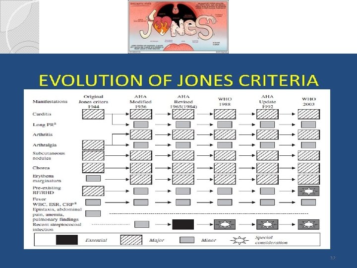

Revision of jones criteria -10 points may 08 2015 ---1. RF remains as one of most imp cause of morbidity & mortality in socially and economically disadvantaged populations all over the world ECHO-have evolved & has become the cornerstone in the management of RF

2 nd point �The global distribution of ARF&RHD is hetrogeneous �Certain geographical regions, ethnic groups&socioeconomic gps experience very high rates of RF

3 -Epidemiological implications �It is reasonable to consider individuals at low risk if they come from a population with known low rates of ARF &RHD(II-a, level of evidence-c) �Low risk is defined as<2 per 100, ooo in age gps 5 -14 �All age < or = 1 per 1000 population per yr (Class IIa, Level of Evidence –c) �Children not clearly from a low risk population are at moderate to high risk depending on their reference population
")
4 -Clinical manifestations of. ARF �Carditis �Arthritis �Chorea �s/c nodules �Erhythema marginatum(uncommon but specific)

5 th point Carditis diagnosis in an era of widely available echocardiography: Numerous studies over the past 20 years have addressed the role of echocardiography (compared with purely clinical assessment) in the diagnosis of ARF. More than 25 studies have reported echocardiography/Doppler evidence of mitral or aortic valve regurgitation in patients with ARF despite the absence of classic auscultatory findings. Echocardiography with Doppler should be performed in all cases of confirmed and suspected ARF (Class I, Level of Evidence B). It is reasonable to consider performing serial echocardiography/Doppler studies in any patient with diagnosed or suspected ARF, even if documented carditis is not present on diagnosis (Class IIa, Level of Evidence C).

Echocardiography/Doppler should be performed to assess whether carditis is present in the absence of auscultatory findings, particularly in moderate- to high-risk populations and when ARF is considered likely (Class I, Level of Evidence B). � Echocardiography/Doppler findings not consistent with carditis should exclude that diagnosis in patients with a heart murmur otherwise thought to indicate rheumatic carditis (Class I, Level of Evidence B). � Specific criteria exist for Doppler findings in rheumatic valvulitis: 1) Mitral regurgitation (all four): seen in ≥ 2 views, jet length ≥ 2 cm, peak velocity >3 m/s, pansystolic; and 2) Aortic regurgitation (all four): seen in ≥ 2 views, jet length ≥ 1 cm, peak velocity >3 m/s, pandiastolic. �

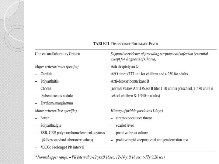
6 -th point Evidence of preceding Streptococcal infection: Because other illnesses may closely resemble ARF, laboratory evidence of antecedent group A streptococcal infection is needed whenever possible, and the diagnosis is in doubt when such evidence is not available. Any one of the following can serve as evidence of preceding infection: Increased or rising antistreptolysin O titer or other streptococcal antibodies (anti. DNASE B) (Class I, Level of Evidence B). A rise in titer is better evidence than a single titer result. � A positive throat culture for group A β-hemolytic streptococci (Class I, Level of Evidence B). � A positive rapid group A streptococcal carbohydrate antigen test in a child whose clinical presentation suggests a high pretest probability of streptococcal pharyngitis (Class I, Level of Evidence B). �

7 th point Revised Jones criteria, low-risk populations: Major and minor criteria are as follows: Major criteria: carditis (clinical and/or subclinical), arthritis (polyarthritis), chorea, Erythema marginatum, and subcutaneous nodules � Minor criteria: polyarthralgia, fever (≥ 38. 5° F), sedimentation rate ≥ 60 mm and/or C-reactive protein (CRP) ≥ 3. 0 mg/dl, and prolonged PR interval (unless carditis is a major criterion). �

8 th point �Revised Jones criteria, moderate- and high-risk populations: Major and minor criteria are as follows: Major criteria: carditis (clinical and/or subclinical), arthritis (monopolyarthritis or polyarthritis, or polyarthralgia), chorea, Erythema marginatum, and subcutaneous nodules �Minor criteria: fever (≥ 38. 5° c), sedimentation rate ≥ 30 mm and/or CRP ≥ 3. 0 mg/dl, and prolonged PR interval (unless carditis is a major criterion)
: The diagnosis of an initial episode of")
9 th point �ARF diagnosis (initial episode): The diagnosis of an initial episode of ARF requires two major criteria, � or one major plus two minor criteria.
: Patients with a history of ARF or")
10 th point ARF diagnosis (subsequent episode): Patients with a history of ARF or RHD are at high risk for recurrent attacks if reinfected with group A streptococci. � With a reliable past history of ARF or established RHD, and in the face of documented group A streptococcal infection, � two major, one major and two minor, or three minor manifestations may be sufficient for a presumptive diagnosis (Class IIb, Level of Evidence C). � When minor manifestations alone are present, the exclusion of other more likely causes of the clinical presentation is recommended before a diagnosis of an ARF recurrence is made (Class I, Level of Evidence C). �

LAB TEST IN RHEUMATIC FEVER � Identifying streptococci in the throat Throat culture- The gold standard for diagnosing streptococcal pharyngitis. Throat swabs should be inoculated on culture media within 5 hours. Limitations; It does not differentiate between a carrier state and active infection. 1/3 of patients with rheumatic fever give no history of a preceeding pharyngeal infection. delay in obtaining results of throat cultures is a problem of primary care physician

Rapid antigen detection It is the detection of gas antigen from throat swab using latex agglutination or immunoassay � Reliable, cost effective � Specificity 95% � Sensitivity 33% � Equivalent to positive throat culture Streptococcal antibody test �Antistreptolysin O test Described by Todd in 1932. Most widely used. Best standardised test

Antistreptolysin O test � Sreptolysin o is a oxygen labile toxin capable of lysing mammalian cells not bacteria demonstrated the presence of antisreptolysin –o antibodies in the sera of patients with sreptocccal infections by neutrolising streptolysin o with serial amount of their sera, the excess unneutralized sreptolysin is revealed by adding erythrocytes as an indicator End point is the highest dilution of serum having no hemolysis with ASO expressed as Todd units, equivalent to the reciprocal of dilution � Factors influencing antibody levels Age Geographical location Season of the year Genetic factors

� Antideoxyribonuclease B The second streptococcal antibody test after ASO detection. Not associated with false positive results. Have both seasonal and geographic variations. Also vary with patient’s age. Also varies with stage of rheumatic fever. Useful for the diagnosis of streptococcal infection and itscomplications. Has a positive detection rate than ASO. � Antihyaluronidase test � Antistreptococcal Esterase � Streptosyme test

ROLE OF ECHO IN DIAGNOSIS AND FOLLOW-UP OF RHEUMATIC CARDITIS Goals of echo; Help in precise and early diagnosis of carditis in ARF. Timely management can make the heart normal or can prevent the recurrences. Can prevent overdiagnosis of carditis. Regular checkup can evaluate the status of RSD and timely decision for valvuloplasty, valve repair/replacement.

ECHOCARDIOGRAPHIC INVESTIGATIONS M-mode interrogation � Dimension of LA, aorta and their ratio. LV dimensions in diastole and systole.

Cross-sectional interoggation � Thickness of valve- less than 3 mm-normal More than 4 mm-thickened. Beaded appearance, especially of mitral, tricuspid and aortic valves.

� Prolapse of mitral valve particularly the aortic leaflet. Decreased or increased mobility of the valves

� Hyperechogenicity of the thickened submitral apparatus Chordal tears to mitral leaflets

� Pericardial effusion � End diastolic volume, end systolic volume and ejection Fraction

Colour Doppler interrogation � Establishment of mitral, aortic and tricuspid regurgitation.

Mitral regurgitation

VIJAYA’S ECHO CRITERIA Echofeatures Score Mitral valve and aortic valve thickness ≥ 4 mm. 2 Increased echogenicity of submitral structures 2 Rheumatic nodules ( beaded appearance) 2 Mitral valve prolapse/aortic valve prolapse/tricuspid valve prolapse 2 Mitral regurgitation and/ aortic regurgitation/tricuspid regurgitation 2 Reduced mobility of valves Chordal tear Pericardial effusion Total score 2 2 2 16

ENDOMYOCARDIAL BIOPSY FOR THE DIAGNOSIS OF RHUEMATIC CARDITIS Aschoff nodules and histiocytic aggregates are considered diagnostic of rheumatic cardits These lesions occurred in 2/5 th of cases with clinically confirmed rheumatic carditis Sensitivity of endomyocardial biopsy is 43% and specificity 100%

Explanation of low prevalence of aschoff nodule 1. Aschoff nodule is a relatively mature lesion occuring later in course of rheumatic carditis 2. Sampling error of biopsy procedure 3. Non uniform distribution of aschoff nodules in the myocardium 4. Interventricular septum and pulmonary conus are the frequently involved locations Endomyocardial biopsy is not recommended as a routine diagnostic or prognostic tool
 are enlarged macrophages found within granuloma(aschoff bodies), is associated with rheumatic")
Anitschkow cells(Caterpillar cells) are enlarged macrophages found within granuloma(aschoff bodies), is associated with rheumatic fever Larger anitschkow cells may coalesce to form aschoff giant cells

RADIONUCLIDE IN RHEUMATIC FEVER Inflammation is associated with locally increased capillary permeability resulting in exudation of transferrin Gallium binds to transferrin which results in localization of the site of inflammation Pericardial uptake is described viral, bacterial, tubercular and rheumatoid diseases Myocardial uptake has been seen with viral, amyloid, sarcoid, spirochetal involvement, MI, dialated cardiomyopathy, metastatic disease, bacterial endocarditis As rheumatic carditis is predominantly infiltrative rather than degenerative in nature, radionuclide imaging is not a tool which can be used in usual clinical settings and may be reserved for research purposes.

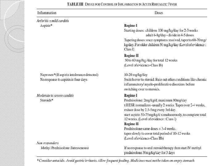
IAP GUIDELINES ON ARF & RHD General measures and symptomatic relief Pain relief-codiene and paracetamol till diagnosis confirmed and aspirin after diagnosis � Hosptalization-moderate to severe carditis, severe arthritis, chorea � Adequate rest-arthritis-2 weeks, carditis without CCF-4 -6 weeks, carditis with CCF-till CCF controlled � Appropriate diet for a growing child �
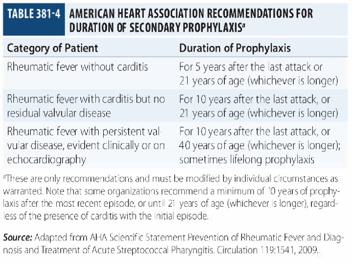
 No carditis: 5 years/18 yrs of age,")
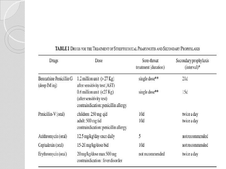
IAP GUIDELINES Duration of secondary prophylaxis (i) No carditis: 5 years/18 yrs of age, whichever is longer. (ii) Mild to moderate carditis and healed carditis: 10 yrs/25 yrs of age, whichever is longer. (iii) Severe disease or post intervention patients: Life long. One may opt for secondary prophylaxis up to the age of 40 years (Table I).

TERMINOLOGIES Recurrence � A new episode of rheumatic fever following another GABHS occurring 8 weeks following stopping treatment Rebound � Manifestations of rheumatic fever occurring within 4 -6 weeks of stopping treatment or while tapering drug Relapse � Worsening of rheumatic fever while under treatment and often with carditis

Subclinical carditis � When clinical examination is normal and echo is abnormal � Around 30% of rheumatic chorea presents with subclinical carditis Indolent carditis � Persistent features of CHF, murmer and cardiomegaly but with no or very few features of carditis

Management of chorea � Treated in a quiet environment � Sedatives like oral pheno/diazepam � If there is no response, haloperdol, sodium valproate or carbemazepine used � Resistant cases treated with plasmapheresis or pimozide � Treatment continued for 2 -4 weeks after clinical improvement

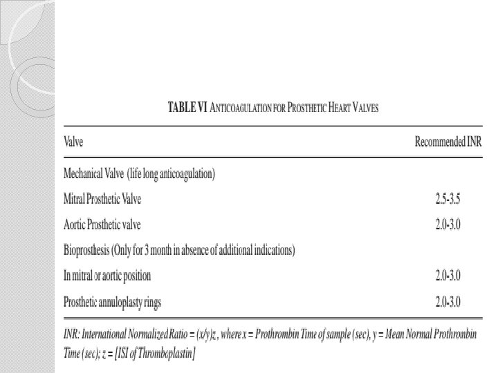
Interventions in valvular heart disease Mitral stenosis � Suitable cases with pure mitral stenosis treated with balloon mitral valvuloplasty(BMV) � Patients unsuitable for BMV-require valve repair or replacement Mitral regurgitation � Acute rheumatic fever with acute severe MR and uncontrolled CCF secondary to cordal rupture is a class 1 indication for urgent surgical intervention � Symptomatic chronic MR is treated with either valve repairment or replacement

�Aortic stenosis � Isolated aortic stenosis is rare � Treatment with balloon procedures not usually effective � Surgical intervention done in symptomatic patients �Aortic regurgitation � Presenting as isolated or combined lesion is treated with prosthetic valve replacement

Endocarditis and Thromboembolism � For unexplained fever, blood culture using atleast 3 samples from 3 different sites at an interval of half to one hour � Echocardiography in addition to routine blood examination � Endocarditis prophylaxis and management includes adequate antibiotics, family counselling regarding dental hygiene

PREVENTION-VACCINE � Effort to design a vaccine is there for more than 40 years, but unlikely to succeed � M protein is strain specific and produces type specific antibody response � According to M protein based classification, 250 strains of streptococcus is known � Infection with one strain does not confer immunity against other strains � Many streptococcal diseases in developing countries are caused by non typable strains

THANK YOU
- Slides: 84