
DIAGNOSIS AND MANAGEMENT OF PERINEAL AND ANAL SPHINCTER
- Slides: 56
DIAGNOSIS AND MANAGEMENT OF PERINEAL AND ANAL SPHINCTER INJURIES
AIMS OF THIS PRESENTATION To learn how to identify, repair and manage perineal and primary obstetric anal sphincter injuries. - Understand the anatomy and physiology of the anal sphincter. -Recognise and classify perineal and anal sphincter injury - Understand the dilemmas regarding prevention and management of subsequent pregnancies - Have insight into the complications of anal sphincter trauma
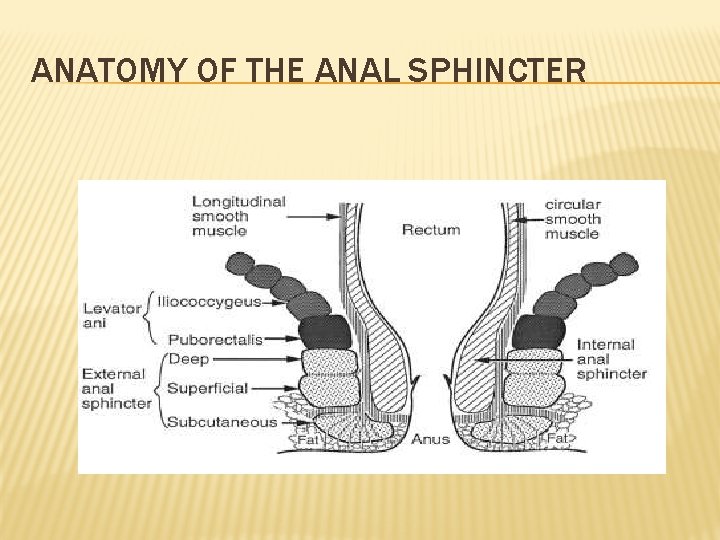
APPLIED ANATOMY AND PHYSIOLOGY OF THE PERINEUM AND ANORECTUM ANATOMY OF THE ANORECTUM The anorectum is the most distal part of the gastrointestinal tract and consists of two parts: the anal canal and rectum. The anal canal measures about 3. 5 cms and lies below the anorectal junction formed by the puborectalis muscle.
The striated external anal sphincter (EAS) is made up of three parts (subcutaneous, superficial and deep) and is inseparable from the puborectalis dorsally. The internal anal sphincter (IAS) is a thickened continuation of the circular smooth muscle of the rectum. It is separated from the EAS by the conjoint longitudinal coat which is a continuation of the longitudinal smooth muscle of the rectum
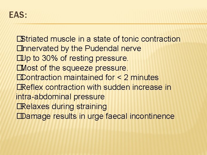
EAS: �Striated muscle in a state of tonic contraction �Innervated by the Pudendal nerve �Up to 30% of resting pressure. �Most of the squeeze pressure. �Contraction maintained for < 2 minutes �Reflex contraction with sudden increase in intra-abdominal pressure �Relaxes during straining �Damage results in urge faecal incontinence
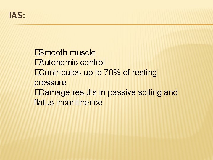
IAS: �Smooth muscle �Autonomic control �Contributes up to 70% of resting pressure �Damage results in passive soiling and flatus incontinence
ANATOMY OF THE ANAL SPHINCTER
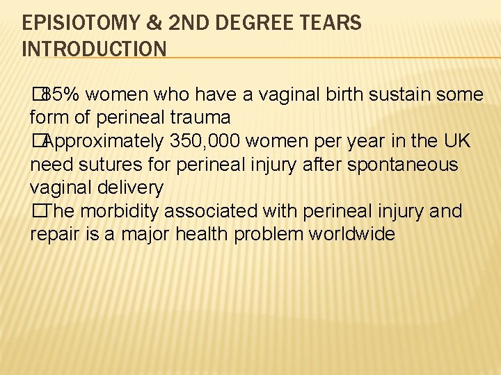
EPISIOTOMY & 2 ND DEGREE TEARS INTRODUCTION � 85% women who have a vaginal birth sustain some form of perineal trauma �Approximately 350, 000 women per year in the UK need sutures for perineal injury after spontaneous vaginal delivery �The morbidity associated with perineal injury and repair is a major health problem worldwide
INDICATIONS FOR EPISIOTOMY �Minimise multiple & extensive tears �Thick & inelastic perineum �Forceps delivery �Expedite delivery �Suspected fetal distress �Shoulder dystocia �Breech
BENEFITS OF MIDLINE EPISIOTOMY �Decreased blood loss �Easier to recognise OASIS �Easier to repair �Better anatomical result �Reduced pain �Decreased risk of infection �Decreased dyspareunia Disadvantage of midline episiotomy ��OASIS
EPISIOTOMY ANDREWS V ET AL BJOG 2004, ANDREWS V ET AL BIRTH 2006 � 254 primips �No midwife and only 13 (22%) doctors performed a truly mediolateral episiotomy (between 40 to 60 degrees from the midline) �Episiotomies angled closer to the midline significantly associated with OASIS (26 vs 37 degrees
EPISIOTOMY EOGAN ET AL BJOG 2006 �Case-control study (54 versus 46 controls) �Mean angle of episiotomy smaller �(30% versus 38% p<0. 001) � 50% risk reduction for every 6°from midline �The relationship of episiotomy angle with risk of OASIS was sig (p<0. 001)
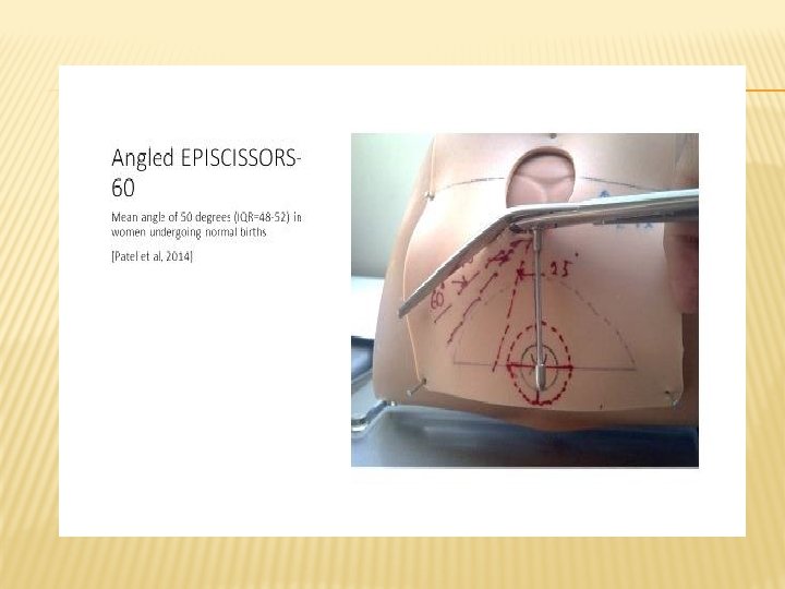
ANGLE OF EPISIOTOMY BEFORE AND AFTER REPAIR KALIS V ET AL 2008 (IJGO) � 50 women undergoing first delivery �Mediolateral episiotomy during crowning at 40 degrees away from midline �Angle of scar measured after delivery = 22. 5 degrees �Should aim for 60 degrees at crowning
EPISIOTOMY & 2 ND DEGREE TEARS SUTURE MATERIALS DEXON VSVICRYL KETTLE C, DOWSWELL T, ISMAIL K 2010 �Cochrane systematic review of 9 RCT’s (n = 4017) �Absorbable synthetic materials (Dexon and Vicryl) versus catgut �Perineal pain ��Analgesic use �Dehiscence wounds �Resuturing
Vicryl Rapide vs Vicryl Suture Material � 5 RCT (n = 2349 women) �Similar rates of short and long-term pain �Fewer women in the rapidly absorbing suture group reported the need for pain relief at 10 days �More women in the standard suture material group required suture removal
Suture material �Standard polyglactin 910 (Vicryl)- not totally absorbed from the wound until 60– 90 days. �Rapid absorption polyglactin 910 (Vicryl Rapide)- completely absorbed from the tissue by 42 days
Evidence based practice Repair Techniques Kettle C, Dowswell T, Ismail K 2012 �Cochrane systematic review - 16 RCT’s (n = 8184) found that continuous stitches compared to interrupted is associated with : �Less short term pain at 10 days �Reduction in analgesia use �Reduction in suture removal �No significant difference in dyspareunia �Reduction in pain is even greater if continuous technique used for all layers compared to only skin
Technique of repair Prior to commencing the repair �Check extent of perineal trauma – perform per vaginal and per rectal examination �Check equipment - suture pack, materials �If needed ensure that appropriate supervision/support is available prior to commencing the repair �Ensure that the wound is adequately anaesthetised (10 -20 mls Lignocaine 1%) don’t inject local through the skin
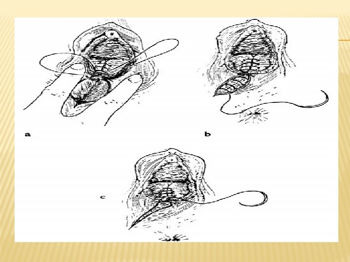
Step 1 - suturing the vagina �Identify the apex of the vaginal wound �Close the vaginal trauma with a loose continuous stitch �Continue to suture the vagina until the hymenal remnants are reached and reapproximated �At the fourchette insert the needle through the skin to emerge in the centre of the perineal trauma
Step 2 - suturing the muscle layer �Check the depth of the trauma - it may be necessary to insert two layers of sutures �Continue to close the perineal muscle with a continuous non-locking stitch - taking care not to leave any dead space
Step 3 - suturing the perineal skin �At the inferior end of the wound bring needle out under the skin surface �The stitches are placed below the skin surface in the subcutaneous layer - thus avoiding the profusion of nerve endings �Continue taking bites of tissue from each side of the wound until the hymenal remnants are reached �Secure the finished repair with a loop knot tied in the vagina
Finally �Check the finished repair is anatomically correct �No bleeding �PV - insert two fingers �PR �Check swabs & instruments �Complete documentation
Conclusion �It is imperative that women receive high quality evidenced based care wherever childbirth takes place �Practices that reduce the adverse effects of perineal trauma and make vaginal birth more desirable are to be encouraged �Improved perineal care may decrease the escalating interest in caesarean section as an alternative mode of delivery
DIAGNOSIS OF OBSTETRIC ANAL SPHINCTER INJURIES (OASIS �Until the advent of anal ultrasound, the development of anal incontinence was attributed largely to pelvic neuropathy. �However prospective studies before and after childbirth have shown that up to one third of women sustain anal sphincter damage that is not recognised at delivery (Sultan AH et al 1993).
�Andrews et al (2006) performed a study in which 241 women having their first vaginal delivery had their perineum re-examined by an experienced research fellow and endoanal ultrasound was performed immediately after delivery and repeated 7 weeks postpartum. When OASIS were identified by the research fellow, the injuries were confirmed and repaired by the duty registrar or consultant. The prevalence of clinically diagnosed OASIS increased from 11% to 25% (n=59). Every clinically diagnosed injury was identified by postpartum endoanal ultrasound.
At 7 weeks no de novo defects were identified by ultrasound. This study concluded that most if not all sphincter defects that have previously been designated as “occult” injuries were in fact injuries that should have been recognisable at delivery. It was alarming to find that 87% and 27% of OASIS were not identified by midwives and doctors respectively. Although it is likely that some of these would have been detected at the time of suturing the tear, it is of concern that clinical recognition of OASIS is suboptimal.
�This finding is not unique as Groom and Patterson found that the rate of third degree tears rose to 15% when all “ 2 nd degree tears” were re -examined by a second experienced person. �It has been shown that only 16% of doctors and 39% of midwives feel that they were trained adequately to identify OASIS (Sultan et al 1995).
On the other hand it is possible that the sphincter tear had been recognised but classified as a second-degree tear. A questionnaire sent to all UK consultants (Fernando et al 2002) and trainees (Sultan et al 1995) confirmed that up to 40% are still classifying partial and even complete disruption of the sphincter as a second degree.
The reason for this confusion is partly due to previous teachings (Sultan & Thakar 2002) and therefore for the sake of clarification and consistency Sultan (1999) proposed a comprehensive classification that is now accepted by RCOG (Greentop guideline 2007), NICE (Nice. org. uk) and the International Consultation on Incontinence (Norton et al 2002)
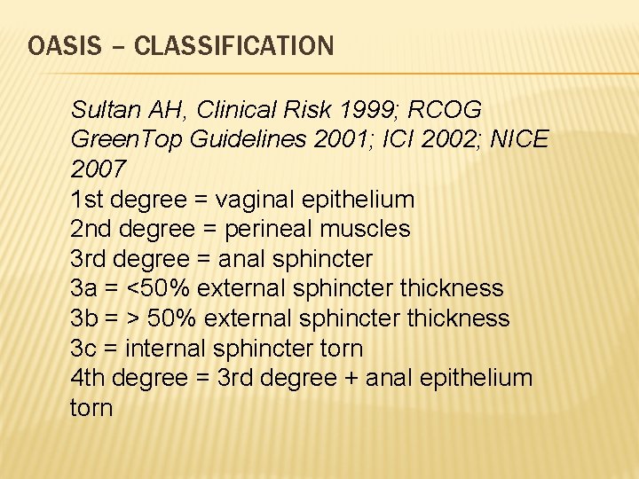
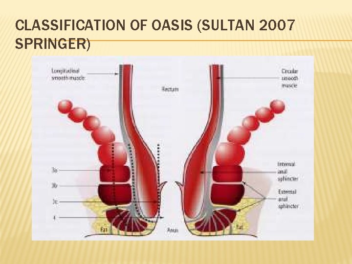
OASIS – CLASSIFICATION Sultan AH, Clinical Risk 1999; RCOG Green. Top Guidelines 2001; ICI 2002; NICE 2007 1 st degree = vaginal epithelium 2 nd degree = perineal muscles 3 rd degree = anal sphincter 3 a = <50% external sphincter thickness 3 b = > 50% external sphincter thickness 3 c = internal sphincter torn 4 th degree = 3 rd degree + anal epithelium torn
CLASSIFICATION OF OASIS (SULTAN 2007 SPRINGER)
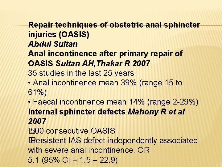
Repair techniques of obstetric anal sphincter injuries (OASIS) Abdul Sultan Anal incontinence after primary repair of OASIS Sultan AH, Thakar R 2007 35 studies in the last 25 years • Anal incontinence mean 39% (range 15 to 61%) • Faecal incontinence mean 14% (range 2 -29%) Internal sphincter defects Mahony R et al 2007 � 500 consecutive OASIS � Persistent IAS defect independently associated with severe anal incontinence. OR 5. 1 (95% CI = 1. 5 – 22. 9)
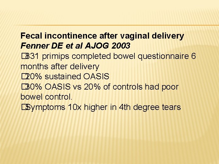
Fecal incontinence after vaginal delivery Fenner DE et al AJOG 2003 � 831 primips completed bowel questionnaire 6 months after delivery � 20% sustained OASIS � 30% OASIS vs 20% of controls had poor bowel control. �Symptoms 10 x higher in 4 th degree tears
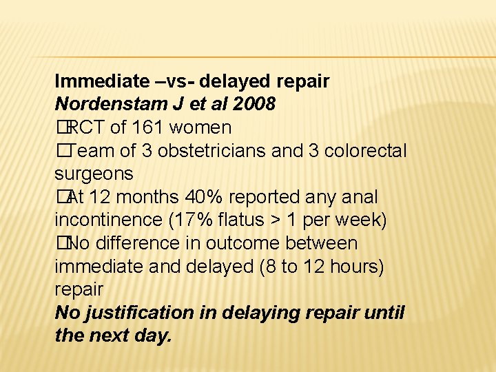
Immediate –vs- delayed repair Nordenstam J et al 2008 �RCT of 161 women �Team of 3 obstetricians and 3 colorectal surgeons �At 12 months 40% reported any anal incontinence (17% flatus > 1 per week) �No difference in outcome between immediate and delayed (8 to 12 hours) repair No justification in delaying repair until the next day.
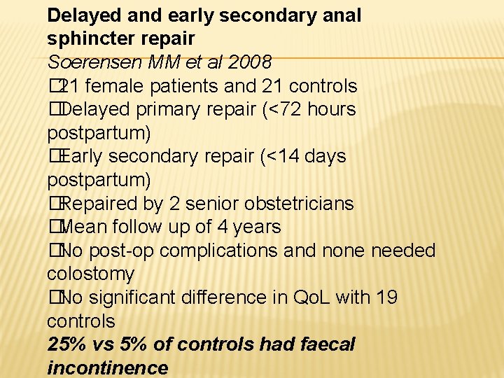
Delayed and early secondary anal sphincter repair Soerensen MM et al 2008 � 21 female patients and 21 controls �Delayed primary repair (<72 hours postpartum) �Early secondary repair (<14 days postpartum) �Repaired by 2 senior obstetricians �Mean follow up of 4 years �No post-op complications and none needed colostomy �No significant difference in Qo. L with 19 controls 25% vs 5% of controls had faecal incontinence
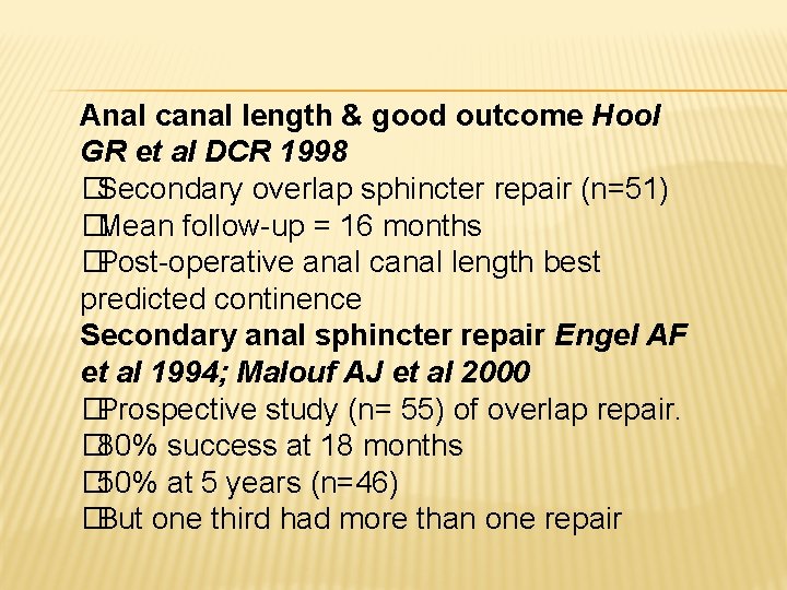
Anal canal length & good outcome Hool GR et al DCR 1998 �Secondary overlap sphincter repair (n=51) �Mean follow-up = 16 months �Post-operative anal canal length best predicted continence Secondary anal sphincter repair Engel AF et al 1994; Malouf AJ et al 2000 �Prospective study (n= 55) of overlap repair. � 80% success at 18 months � 50% at 5 years (n=46) �But one third had more than one repair
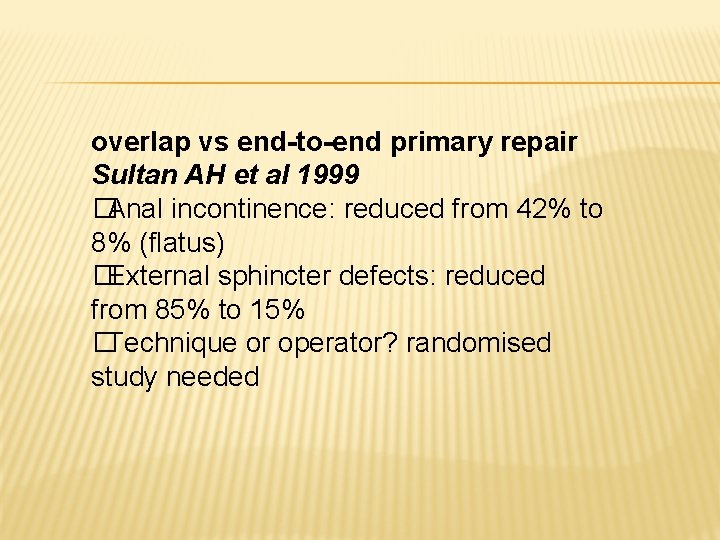
overlap vs end-to-end primary repair Sultan AH et al 1999 �Anal incontinence: reduced from 42% to 8% (flatus) �External sphincter defects: reduced from 85% to 15% �Technique or operator? randomised study needed
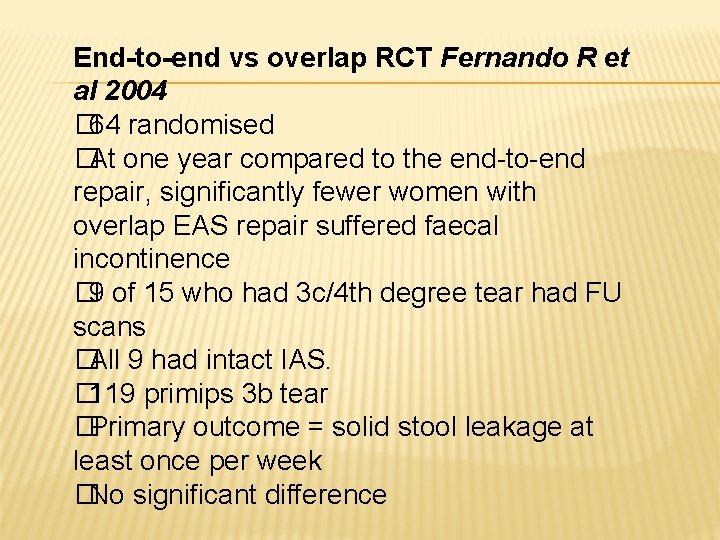
End-to-end vs overlap RCT Fernando R et al 2004 � 64 randomised �At one year compared to the end-to-end repair, significantly fewer women with overlap EAS repair suffered faecal incontinence � 9 of 15 who had 3 c/4 th degree tear had FU scans �All 9 had intact IAS. � 119 primips 3 b tear �Primary outcome = solid stool leakage at least once per week �No significant difference
End-to-end vs overlap RCT �Farrell SA et al 2012 � 3 year follow-up �No significant difference
Suture materials �Anal Mucosa - Vicryl 3 -0 �Internal Anal Sphincter - Mattress end-toend PDS 3 -0 �External Anal Sphincter Mattress/Overlap PDS 3 -0 Suture material Williams et al 2006 � 112 women – randomised study �No difference in suture related morbidity between Vicryl and PDS – But 70% were 3 a tears and only 54% 12 month follow-up
Operating Theatre �Sterile environment �Good lighting �Good exposure �Appropriate instrument tray, sutures �Anaesthesia – spinal, epidural, General �Assistance Antibiotic prophylaxis for OASIS Duggal N et al 2008 � Prospective placebo controlled RCT (n=147) � Single IV dose of cephalosporin � Perineal wound infection 8% vs 24% in placebo
OASIS repair - recommended practice Sultan AH, Thakar R 2007 �Experienced obstetrician �Repair in operating theatre �Regional or general anaesthesia �IV antibiotics �EAS �End-to-end for 3 a �End-to-end or overlap for full thickness and full length 3 b
�IAS �End–to-end mattress �Monofilament sutures (PDS) for the sphincter (Vicryl 2 -0 can also be used) �Rectal examination before and after repair �Foleys catheter for 12 hours �Lactulose 15 mls bd for 7 to 10 days �Clinic Follow up in 2 to 3 months
Management of OASIS after subsequent pregnancy Recurrence risks with previous OASIS Peleg D et al 1999 �Primips, ceph, term, 3 o/4 o (n=704); Incidence = 19% (midline episiotomy) �Recurrence rate = 12% vs 7 % if no previous OASIS (P=0. 001)
Previous OASIS - is recurrence predictable? Harkin R et al 2003 �Mediolateral episiotomy � 2 of 45 (4. 4%) in subsequent vaginal deliveries developed a repeat OASIS Previous OASIS Poen AC et al 1998 � 43 of 110 women studied �Anal incontinence 56% -v- 34% in women with no subsequent delivery. (RR = 1. 6, CI = 1. 1 -2. 5)
Previous OASIS Sangalli MR et al 2000 � 177 women 13 years FU �Faecal Incontinence in 114 subsequent deliveries (3 o tears = 2. 5% ; 4 o tears = 26. 5%
Performing mediolateral episiotomy Andrews et al 2004; Andrews et al 2006 � 254 primips, 41% mediolateral episiotomy �No midwife and only 13 (22%) doctors performed a truly mediolateral episiotomy (between 40 to 60 degrees from the midline) �Episiotomies angled closer to the midline significantly associated with OASIS (26 vs 37 degrees) Episiotomy Eogan et al 2006 � 50% risk reduction of third degree tears for every 6°away from midline
Management of subsequent pregnancy after OASIS Scheer I et al 2009 � 56 deliveries 2002 – 2006 � 38 (70%) vaginal deliveries �No significant deterioration in: – bowel or bladder symptoms – quality of life – resting and squeeze pressures �No significant new scan defects
Recurrence rate after previous OASIS • Midline episiotomy 12% Peleg et al 1999 • Mediolateral episiotomy – 4. 4% Harkin R 2003 – 4. 4% Elfaghi I 2004 – 5. 6% Baghestan L et al 2011 – 6. 8% Scheer I et al 2009 – 7. 1% Jango H et al 2012 – 13. 4% Ali A et al 2014 – ( 2 flatus incontinence at follow up)
Take Home Messages �Every woman who has a vaginal delivery has a 3 rd or 4 th degree tear until proved otherwise �A 3 rd or 4 th degree tear (as well as an isolated buttonhole tear of the rectum) cannot be excluded without a rectal examination �Therefore a careful digital rectal examination with good exposure, adequate lighting and analgesia is mandatory.
�There is Level 1 a evidence that second degree tears and episiotomies should be repaired using the continuous technique of repair for all three layers with Vicryl Rapide �Restoration of normal sphincter length best predicts continence and therefore the full length of the torn sphincter must be repaired �The torn internal sphincter should be identified and repaired separately using an end- to-end repair technique �Partially torn external sphincter tears must be repaired using the end-to-end technique.
�Full thickness and full length external sphincter tears can be repaired by either the overlap or end-to-end technique. However the superiority of the overlap repair has only been demonstrated in one study where all repairs were conducted by only two operators. The Cochrane review does not recommend one technique over the other. �The anal mucosa and anal sphincter muscles must not be repaired using figureof- eight sutures as they can cause ischaemia and necrosis of tissue
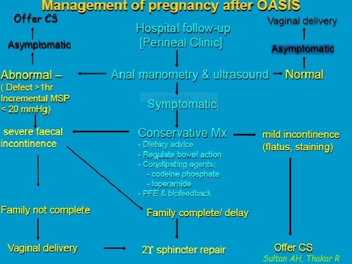
�The best chance of successful repair of OASIS is at the time of delivery; secondary sphincter repair (especially of the internal sphincter) is comparatively poor �In terms of subsequent pregnancy management: o Compare like with like ie. the worst scenarios of CS and VD o Risks following CS is not just for one CS but cumulative with each subsequent CS o The risk of recurrence of OASIS is about 7% o Outcome of vaginal delivery after OASIS is good in selected patients