
Diagnosis and Management of Osteomyelitis https commons wikimedia


- Slides: 40
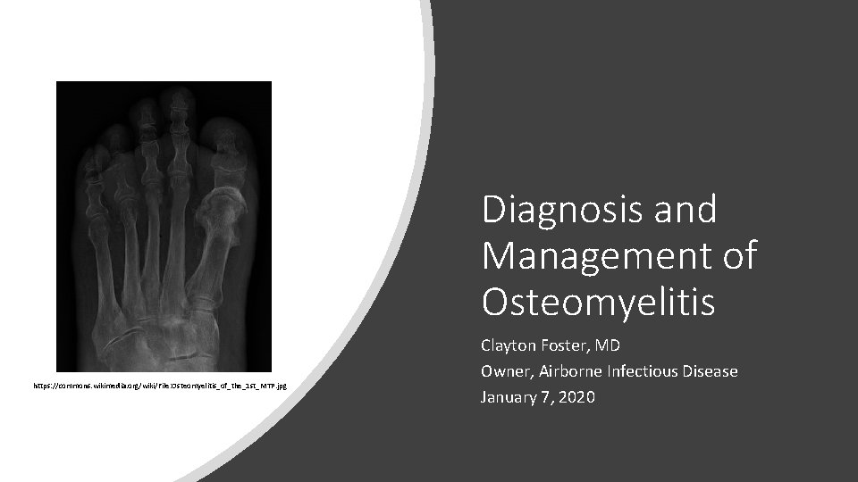
Diagnosis and Management of Osteomyelitis https: //commons. wikimedia. org/wiki/File: Osteomyelitis_of_the_1 st_MTP. jpg Clayton Foster, MD Owner, Airborne Infectious Disease January 7, 2020
Disclosures • No relevant financial disclosures • I will be discussing off‐label and investigational therapies
Objectives • Outline the epidemiology and classification of adult osteomyelitis • Recognize clinical manifestations of adult osteomyelitis • Apply evidence‐based treatment strategies in the management of adult osteomyelitis • Review orthopedic infections involving hardware and describe how this affects management
History • Chronic osteomyelitis was first described by Hippocrates (460‐ 370 BC) among patients with open fractures 1 • Acute hematogenous osteomyelitis was not characterized until 1773 (“abscessus in medulla”)2 • The term “osteomyelitis” was first introduced in 18443 • Early treatment was all surgical – debridement of diseased bone and packing of the remaining cavity 4
History • In the pre‐antibiotic era, mortality from osteomyelitis was estimated to be 33% 5 • Cause of death was typically either hematogenous dissemination of infection (acute OM) or secondary amyloidosis (chronic OM)6 • Almost immediately after the introduction of penicillin in 1940, mortality from osteomyelitis dropped to 10% or less 5 • After 1950, incidence and severity of osteomyelitis cases started increasing again with the rise of penicillin‐resistant Staphylococcus aureus 7 • The modern treatment approach of prolonged antibiotics with or without surgery, depending on the clinical context, was first outlined in the early 1970 s 8, 9
Current Understanding of Osteomyelitis • Classified by duration and route of infection: 10 • • Acute OM – symptoms for days to weeks Chronic OM – symptoms for months to years “Subacute” OM only exists in medical coding (M 86. 2 XX) Key difference between acute and chronic OM is development of sequestrum (dead bone) • Hematogenous • Nonhematogenous
Epidemiology • Etiology 11 • • Trauma (open fractures, pressure ulcers, penetrating injuries) Contiguous spread from infected joints or hardware Surgery Bacteremia • Risk Factors • Diabetes • Vascular disease • Peripheral neuropathy
Microbiology • We will focus on typical bacterial OM, though it can be caused by mycobacteria, fungi and even viruses (smallpox) • The most common pathogens are Staphylococcus aureus, coagulase‐negative staphylococci and aerobic Gram‐negative rods 12 • Staph species account for >50% of nonhematogenous OM • GNRs make up ~20% • Other pathogens include streptococci (12%), enterococci (8%) and anaerobes (5%) • Hematogenous OM is most often caused by Staph aureus, with aerobic GNRs causing an estimated 30% of cases 13 • Hematogenous OM is usually monomicrobial
Diagnosis • Positive bone culture is considered the gold standard (for organisms that fit the clinical picture) • Bone biopsy for pathology can make the diagnosis even with a negative culture • However, a negative biopsy does not rule out osteomyelitis 14 • MRI is the most sensitive imaging modality for detecting OM • CT may miss acute OM because it primarily picks up bony erosion • For patients who cannot get CT or MRI (or whose hardware precludes quality images), nuclear studies (3‐phase bone scan, PET/CT) have good sensitivity • Plain radiographs are least sensitive and only pick up advanced changes of OM (erosion, cortical disruption), which can be seen with other diseases
Diagnosis • You don’t need any of the previous tests to make a diagnosis of OM • Exposed bone = infected bone • The “probe‐to‐bone” test has high sensitivity and specificity for OM in meta‐analysis 15 • The presence of a sinus tract is pathognomonic of chronic OM 16 • Always suspect OM in non‐healing wounds, even in the absence of exposed bone • In studies of diabetic foot infections, OM often developed before exposed bone was present on exam 17 • Per the above study, “the majority of diabetic foot ulcers have an underlying osteomyelitis that is clinically unsuspected. ” • Ulcers larger than 2 cm 2 in diabetic foot wounds predicted OM as well at the probe‐to‐bone test in one study 18 • These rules apply to hardware infections as well
Diagnosis • You cannot rely on typical findings of infection to make (or exclude) a diagnosis of OM • A majority of patients with OM do not have fever (70% in one study)19 • A majority of patients with OM do not have leukocytosis (65% in one study) 20 • Local symptoms of redness, warmth, swelling and pain are not always present 21 • No pus ≠ no infection • Drainage from a focus of OM may be serous, serosanguinous, bloody or purulent 22 • ESR and CRP are usually elevated, but not always – especially with chronic OM 23 • Procalcitonin is not a reliable marker for OM 24
Diagnosis • Examples of when to be suspicious of OM: • A patient appears to be healing well after orthopedic surgery, but 3 weeks later, the incision dehisces and starts to drain • A venous stasis ulcer over the lateral malleolus has not healed after several months of going to wound clinic • An ischial pressure ulcer heals after intensive outpatient treatment, then breaks down dramatically as soon as the patient is released • Non‐union in the setting of orthopedic hardware • “The hardware is always infected!” (until proven otherwise)
Treatment • If at all possible, remove hardware and necrotic bone • Even without these complicating factors, I often advocate for surgical debridement to get deep cultures – surface cultures do not correlate well with bone cultures 25 • If there is any concern for residual infection following surgery (or if there is no surgery), the patient needs a minimum of 4 weeks of appropriate antibiotics 26 • My opinion: most osteomyelitis is undertreated in terms of antibiotic duration • My approach: treat to normalization of CRP and wound closure (if feasible) • Inadequately debrided chronic OM can often be cured by antibiotics alone, but it may require courses ranging from 3 months to over a year 27, 28, 29
Treatment • For patients who initially present without sepsis or overt wound infection, hold off on antibiotics until cultures can be obtained • The “antibiotic holiday” is common practice, although I personally disagree in many cases • A Cochrane review found no difference in outcomes between IV and oral antibiotic therapy in treating chronic OM 30 • However, it is still common practice to treat OM with Gram positive organisms (Staph, Strep, Enterococcus) with IV antibiotics, at least for the first few weeks • Antibiotic selection should try to account for differences in oral absorption and bone penetration
Antibiotic selection • According to a recent review, essentially all of the common IV antibiotics used to treat osteomyelitis should provide adequate bone penetration (tissue concentration >> MIC) 31 • Best bets for oral antibiotics (high oral bioavailability plus good bone penetration): • • • Fluoroquinolones Tetracyclines Clindamycin Bactrim Linezolid Metronidazole (although the review above found that bone concentration did not reliably exceed typical MICs) • Agents with notably poor bone penetration include penicillin (G/V), macrolides, imipenem and flucloxacillin (not available in the US)
Antibiotic selection • What if I don’t have a positive culture, but I’m pretty sure they have OM? • Strategy: cover all Staph and Strep species plus common Gram negatives • Common/reasonable regimens: • IV vancomycin (OFF‐LABEL) plus IV ceftriaxone 2 gm daily • IV vancomycin plus high‐dose PO Cipro (750 mg PO BID or renally‐dosed equivalent) • IV daptomycin in place of vancomycin in one of the regimens above (OFF‐LABEL) • PO linezolid or doxycycline for Gram positive coverage (both OFF‐LABEL) PLUS • High‐dose PO Cipro or Augmentin 875 mg PO BID (OFF‐LABEL) for Gram negative coverage • Probably don’t need to cover empirically for anaerobes • Look back at old cultures
Adjunctive treatments • Rifampin is the best‐studied adjunctive treatment for staphylococcal hardware infections (primarily Staph aureus, technically OFF‐LABEL) • For select cases of Staph aureus prosthetic joint infection (PJI) where the prosthesis is debrided and retained, the addition of rifampin to primary therapy can increase eradication rates from 50‐ 60% to almost 90%32‐ 36 • Data supporting use in PJI with Co. NS and Strep species are limited 37 • Rifampin is a messy drug – interacts with everything (induces multiple CYP 450 enzymes); can cause hypersensitivity reaction, hepatitis and severe dermatologic changes (SJS/TEN) • Make sure your isolate actually tests susceptible to rifampin • Do not use rifampin as monotherapy!
Adjunctive treatments • Hyperbaric oxygen (HBO) therapy is often employed to treat non‐healing wounds, but data are mostly limited to small series • A systematic review of HBO in 2009 concluded that it 38 • Reduces the chance of amputation from infected diabetic foot ulcers requiring surgery (OR 0. 24, high level of evidence) • Improves chances of healing an infected diabetic foot ulcer (OR 9. 99, high level of evidence) • Contributes to remission in about 85% of cases of refractory lower extremity OM (moderate level of evidence) • Efficacy tends to correlate with documenting hyperoxygenation of at‐risk tissue by transcutaneous oximetry
Adjunctive treatments • Local antibiotic delivery is frequently employed for OM and PJI – usually polymethylmethacrylate (PMMA) cement impregnated with antibiotics +/‐ antifungals • PMMA can be formed into beads, molded into a bony defect or used to coat hardware • Vancomycin and aminoglycosides are the most commonly used antibiotics for local delivery 39. Antifungal coverage, if added, is typically achieved with amphotericin (all OFF‐ LABEL) • Mixed results have been observed in small series using local antibiotic delivery 40‐ 43 • A number of issues have arisen in implementing this strategy
Adjunctive treatments • Problems with local antibiotic delivery: • No standardization for the type and amount of antimicrobials added • Little standardization in how cement is prepared • Elution of antibiotics is not consistent – most commonly, there is an early burst of drug into surrounding tissues, followed by a prolonged period of subtherapeutic concentrations 44 • Effects of varying the type and quantity of antimicrobials on cement strength and elution properties are not well‐described • Systemic toxicity has been reported – usually nephrotoxicity 45‐ 49 • Prolonged exposure to low‐level antibiotic concentrations can lead to bacterial resistance • Non‐absorbable media that are no longer eluting antibiotic provide surfaces for biofilm formation 50
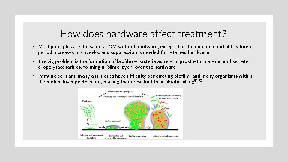
How does hardware affect treatment? • Most principles are the same as OM without hardware, except that the minimum initial treatment period increases to 6 weeks, and suppression is needed for retained hardware • The big problem is the formation of biofilm – bacteria adhere to prosthetic material and secrete exopolysaccharides, forming a “slime layer” over the hardware 51 • Immune cells and many antibiotics have difficulty penetrating biofilm, and many organisms within the biofilm layer go dormant, making them resistant to antibiotic killing 51‐ 52
How does hardware affect treatment? • At this time, our only reliable method of removing biofilm is to remove the hardware that allowed it to form • The idea behind prolonged antibiotics is to “wait out” the dormant bacteria in the biofilm • The current standard is at least 3 months of oral suppression following the initial (usually parenteral) antibiotic course 53. With non‐union, continue until the fracture heals. • Beyond that, duration is individualized based on • • Likelihood of recurrence Potential consequences of recurrence Tolerance of suppressive antibiotics Patient preference • We have no way of knowing if/when hardware is completely sterilized. Some experts think maybe it never is 54
Surgical options for hardware infection • For fixation hardware, allow the underlying bone to heal, then remove hardware in its entirety • Prosthetic joint infections have more options: • Debridement and retention of prosthesis • One‐stage exchange arthroplasty (not really done in the US) • Two‐stage exchange arthroplasty – replace the prosthesis with a temporary spacer, treat with antibiotics until you’re convinced infection is gone, then put in a new prosthesis • Permanent resection arthroplasty – Girdlestone for hips, arthrodesis for knees • Amputation
When to worry • Wound(s) over the infection site won’t heal • Associated fractures won’t heal • A draining sinus forms and won’t close • Persistent, or especially worsening local erythema, swelling and pain • CRP won’t normalize or, worse, starts increasing • Baseline ESR increases with age more so than CRP, so it often doesn’t normalize 55
I’m worried…what now? • When OM and hardware infections do not resolve with initial therapy, the problem is almost always one of three things: • Source control (hardware or necrotic bone needs to come out) • Inadequate antibiotic coverage (the organism has become resistant or, more often, a new organism has been introduced through the open wound) • Vascular insufficiency (the affected tissues aren’t getting enough antibiotics and immune cells delivered) • My bias: I don’t care how much prednisone they take, how poorly‐controlled their diabetes is or how non‐compliant they are with their dressings – if you address all the factors above, you will eventually heal the wound
What if I can’t control the source? • Examples: chronic osteomyelitis of the skull, extensive prosthesis, unacceptable surgical risk • Pick an oral antibiotic that • Is confirmed to have activity against the organism(s) involved • Has good bone penetration • The patient can tolerate long‐term • …And don’t stop • If you don’t think you have any oral options, call your ID consultant
Prognosis • For chronic osteomyelitis without hardware, reported success rates range from 60% to 90% 56 • Hardware outcomes are even harder to predict because they depend on the organism, type and extent of hardware, duration of infection and surgical approach (debridement and retention or explant) • Reported success rates with hardware infection range from 32% with debridement, antibiotics and implant retention (DAIR) in RA patients up to 90% in one study of DAIR in Staph aureus PJI with adjunctive rifampin 32, 57 • The largest and longest study of Staph aureus PJI treated with DAIR (345 patients over 8 years) reported a long‐term success rate of only 55%32 • Two‐stage arthroplasty is estimated to eradicate infection in >85% of patients 36, 58, 59
What’s on the horizon? • Oritavancin and dalbavancin – super long‐acting injectable lipoglycopeptides (OFF‐LABEL) • Half‐lives of 245 and 346 hours, respectively • Three simultaneous mechanisms of action – development of clinically significant resistance has not been observed • Activity against stationary (dormant) phase bacteria (oritavancin > dalbavancin) 60, 61 • Ability to penetrate biofilms 60, 62 • Growing body of evidence to support efficacy in osteomyelitis 63‐ 66 • Fusidic acid (investigational) • • • Oral protein synthesis inhibitor with a unique target (elongation factor G) Limited data show efficacy against difficult‐to‐treat MRSA infections when combined with rifampin 67 Most data on treating OM are in Staph aureus, in children and with combination therapy Low barrier to resistance Not available in the US
What’s on the horizon? • Organic acids for biofilm • Acetic acid and citric acid have been used to help clear biofilm in other contexts, including • Bladder irrigation to prevent CAUTI 68 • Lock flushes to prevent infection of long‐term vascular catheters 69 • Topical application in the setting of burns 70 • We are starting to see data in PJI suggesting a significant reduction in biofilm in vitro after a 20 ‐minute soak in 3‐ 5% acetic acid 71, 72 • Phototherapy • Blue light (405 nm) appears to kill a number of clinically significant pathogens within biofilm 73 • Effective application times range from 15 to 60 minutes • This wavelength does not appear to damage human tissue 74
What’s on the horizon? • Bacteriophages • Shown to penetrate biofilms in the setting of pulmonary and vascular catheter infections 75, 76 • Evidence of clinical efficacy thus far comes mostly from animal models and case reports of treating XDR organisms • Lots of work to be done on potential for toxicity, dose, route, formulation…
Recap • Osteomyelitis is primarily a clinical diagnosis – you cannot rely solely on culture, path, imaging or typical findings of infection • Hardware infection behaves very much like OM. The key difference is that you are much less likely to sterilize infected hardware • We probably underrecognize and undertreat most osteomyelitis and hardware infection • When it comes to treatment duration, don’t “set it and forget it. ” Use clinical endpoints. • Remove infected hardware and necrotic bone whenever possible • Use adjunctive rifampin with susceptible staphylococcal hardware infections • If it won’t heal, it needs source control, a change in antibiotics or revascularization • There is a time and place for indefinite antibiotics
Questions?
1. 2. 3. 4. 5. 6. 7. 8. 9. 10. 11. 12. 13. 14. References Eid AM. Osteomyelitis: historical review. Pan Arab J Orthop Trauma 2003; 7: 95‐ 109. Klenerman L. A history of osteomyelitis from the Journal of Bone and Joint Surgery: 1948 to 2006. The Journal of bone and joint surgery. British volume. 2007 May; 89(5): 667‐ 70. Nelaton A. Elements de pathologie chirurgical. Paris, Germer Bailliere 1844; 586: 595– 7. Orr HW. The treatment of acute osteomyelitis by drainage and rest. J Bone Joint Surg 1927; 9: 733– 9. Dennison WM. Haematogenous osteitis in children. J Bone Joint Surg [Br] 1948; 39‐B: 110– 23. Ikpeme IA, Ngim NE, Ikpeme AA. Diagnosis and treatment of pyogenic bone infections. Afr Health Sci. 2010; 10: 82– 8. Gilmour WN. Acute haematogenous osteomyelitis. J Bone Joint Surg [Br] 1962; 44‐B: 841– 53. Blockey NJ, Watson JT. Acute osteomyelitis in children. J Bone Joint Surg [Br] 1970; 52‐B: 77– 87. Blockey NJ, Mc. Allister TA. Antibiotics in acute osteomyelitis in children. J Bone Joint Surg [Br] 1972; 54‐B: 299– 309. Lew DP, Waldvogel FA. Osteomyelitis. N Engl J Med. 1997; 336(14): 999. Waldvogel FA, Medoff G, Swartz MN. Osteomyelitis: a review of clinical features, therapeutic considerations and unusual aspects. N Engl J Med. 1970; 282(4): 198. Senneville E, Melliez H, Beltrand E et al. Culture of percutaneous bone biopsy specimens for diagnosis of diabetic foot osteomyelitis: concordance with ulcer swab cultures. Clin Infect Dis. 2006; 42(1): 57. Mader JT, Ortiz M, Calhoun JH. Update on the diagnosis and management of osteomyelitis. Clin Podiatr Med Surg. 1996; 13(4): 701. White LM, Schweitzer ME, Deely DM, Gannon F. Study of osteomyelitis: utility of combined histologic and microbiologic evaluation of percutaneous biopsy samples. Radiology. 1995; 197(3): 840.
15. 16. 17. 18. 19. 20. 21. 22. 23. References Lam K, van Asten SA, Nguyen T, La Fontaine J, Lavery LA. Diagnostic Accuracy of Probe to Bone to Detect Osteomyelitis in the Diabetic Foot: A Systematic Review. Clin Infect Dis. 2016; 63(7): 944. Pereira PJDG, Duarte L, Silva MDS, kase M, Olivetti BC, Murakoshi R, sarpi MDO, Gomes RLE. Radiological manifestations of osteomyelitis in the head and neck. Poster presented at: European Society of Radiology 2016. http: //dx. doi. org/10. 1594/ecr 2016/C‐ 1220 Newman LG, Waller J, Palestro CJ et al. Unsuspected osteomyelitis in diabetic foot ulcers. Diagnosis and monitoring by leukocyte scanning with indium in 111 oxyquinoline. JAMA. 1991; 266(9): 1246. Butalia S, Palda VA, Sargeant RJ, Detsky AS, Mourad O. Does this patient with diabetes have osteomyelitis of the lower extremity? JAMA. 2008; 299(7): 806. Torda AJ, Gottlieb T, Bradbury R. Pyogenic vertebral osteomyelitis: analysis of 20 cases and review. Clin Infect Dis. 1995; 20(2): 320. Unkila‐Kallio L, Kallio MJ, Eskola J, Peltola H. Serum C‐reactive protein, erythrocyte sedimentation rate, and white blood cell count in acute hematogenous osteomyelitis of children. Pediatrics. 1994; 93(1): 59. Sia IG, Berbari EF. Osteomyelitis. Best practice & research Clinical rheumatology. 2006 Dec 1; 20(6): 1065‐ 81. Scholl L, Chang E, Reitz B, Chang J. Sternal osteomyelitis: use of vacuum‐assisted closure device as an adjunct to definitive closure with sternectomy and muscle flap reconstruction. Journal of cardiac surgery. 2004 Sep; 19(5): 453‐ 61. Perry M. Erythrocyte sedimentation rate and C reactive protein in the assessment of suspected bone infection‐‐are they reliable indices? J R Coll Surg Edinb. 1996; 41(2): 116.
24. 25. 26. 27. 28. 29. 30. 31. 32. References Faesch S, Cojocaru B, Hennequin C, Pannier S, Glorion C, Lacour B, Chéron G. Can procalcitonin measurement help the diagnosis of osteomyelitis and septic arthritis? A prospective trial. Italian Journal of Pediatrics. 2009 Dec 1; 35(1): 33. Senneville E, Melliez H, Beltrand E et al. Culture of percutaneous bone biopsy specimens for diagnosis of diabetic foot osteomyelitis: concordance with ulcer swab cultures. Clin Infect Dis. 2006; 42(1): 57. Lipsky BA, Berendt AR, Cornia PB et al. 2012 Infectious Diseases Society of America clinical practice guideline for the diagnosis and treatment of diabetic foot infections. Clinical infectious diseases. 2012 Jun 15; 54(12): e 132‐ 73. Lázaro‐Martínez JL, Aragón‐Sánchez J, García‐Morales E. Antibiotics versus conservative surgery for treating diabetic foot osteomyelitis: a randomized comparative trial. Diabetes Care. 2014; 37(3): 789. Epub 2013 Oct 15. Senneville E, Lombart A, Beltrand E, Valette M, Legout L, Cazaubiel M, Yazdanpanah Y, Fontaine P. Outcome of diabetic foot osteomyelitis treated nonsurgically: a retrospective cohort study. Diabetes Care. 2008 Apr 1; 31(4): 637‐ 42. Venkatesan P, Lawn S, Macfarlane RM, Fletcher EM, Finch RG, Jeffcoate WJ. Conservative management of osteomyelitis in the feet of diabetic patients. Diabet Med. 1997; 14(6): 487– 490. Conterno LO, Turchi MD. Antibiotics for treating chronic osteomyelitis in adults. Cochrane Database of Systematic Reviews 2013, Issue 9. Art. No. : CD 004439. DOI: 10. 1002/14651858. CD 004439. pub 3. Thabit AK, Fatani DF, Bamakhrama MS, Barnawi OA, Basudan LO, Alhejaili SF. Antibiotic penetration into bone and joints: an updated review. International Journal of Infectious Diseases. 2019 Feb 14. Lora‐Tamayo J, Murillo O, Iribarren JA et al. A large multicenter study of methicillin‐susceptible and methicillin‐resistant Staphylococcus aureus prosthetic joint infections managed with implant retention. Clin Infect Dis. 2013; 56(2): 182. Epub 2012 Aug 31
33. 34. 35. 36. 37. 38. 39. 40. 41. References Deirmengian C, Greenbaum J, Lotke PA, Booth RE Jr, Lonner JH. Limited success with open debridement and retention of components in the treatment of acute Staphylococcus aureus infections after total knee arthroplasty. J Arthroplasty. 2003; 18(7 Suppl 1): 22. Berbari EF, Osmon DR, Duffy MC et al. Outcome of prosthetic joint infection in patients with rheumatoid arthritis: the impact of medical and surgical therapy in 200 episodes. Clin Infect Dis. 2006; 42(2): 216. Marculescu CE, Berbari EF, Hanssen AD et al. Outcome of prosthetic joint infections treated with debridement and retention of components. Clin Infect Dis. 2006; 42(4): 471. Sia IG, Berbari EF, Karchmer AW. Prosthetic joint infections. Infect Dis Clin North Am. 2005 Dec; 19(4): 885‐ 914. Lora‐Tamayo J, Senneville É, Ribera A, Bernard L, Dupon M, Zeller V, Li HK, Arvieux C, Clauss M, Uçkay I, Vigante D. The not‐ so‐good prognosis of streptococcal periprosthetic joint infection managed by implant retention: the results of a large multicenter study. Clinical infectious diseases. 2017 Jun 15; 64(12): 1742‐ 52. Goldman RJ. Hyperbaric oxygen therapy for wound healing and limb salvage: a systematic review. PM R. 2009; 1(5): 471. Hake ME, Young H, Hak DJ, Stahel PF, Hammerberg EM, Mauffrey C. Local antibiotic therapy strategies in orthopaedic trauma: Practical tips and tricks and review of the literature. Injury. 2015; 46(8): 1447. Patzakis M. J. , Mazur K. , Wilkins J. , Sherman R. , and Holtom P. : Septopal beads and autogenous bone grafting for bone defects in patients with chronic osteomyelitis. Clin Orthop Relat Res 1993; undefined: pp. 112‐ 118 Calhoun J. H. , Henry S. L. , Anger D. M. , Cobos J. A. , and Mader J. T. : The treatment of infected nonunions with gentamicin– polymethylmethacrylate antibiotic beads. Clin Orthop Relat Res 1993; undefined: pp. 23‐ 27
42. 43. 44. 45. 46. 47. 48. 49. 50. 51. References Blaha J. D. , Calhoun J. H. , Nelson C. L. , et al: Comparison of the clinical efficacy and tolerance of gentamicin PMMA beads on surgical wire versus combined and systemic therapy for osteomyelitis. Clin Orthop Relat Res 1993; undefined: pp. 8‐ 12 Walenkamp G. H. , Kleijn L. L. , and De leeuw M. : Osteomyelitis treated with gentamicin‐PMMA beads: 100 patients followed for 1– 12 years. Acta Orthop Scand 1998; 69: pp. 518‐ 522 Guelcher SA, Brown KV, Li B, Guda T, Lee BH, Wenke JC. Dual‐purpose bone grafts improve healing and reduce infection. Journal of orthopaedic trauma. 2011 Aug 1; 25(8): 477‐ 82. Van raaij T. M. , Visser L. E. , Vulto A. G. , and Verhaar J. A. : Acute renal failure after local gentamicin treatment in an infected total knee arthroplasty. J Arthroplasty 2002; 17: pp. 948‐ 950 Curtis J. M. , Sternhagen V. , and Batts D. : Acute renal failure after placement of tobramycin‐impregnated bone cement in an infected total knee arthroplasty. Pharmacotherapy 2005; 25: pp. 876‐ 880 Patrick B. N. , Rivey M. P. , and Allington D. R. : Acute renal failure associated with vancomycin‐ and tobramycin‐laden cement in total hip arthroplasty. Ann Pharmacother 2006; 40: pp. 2037‐ 2042 Dovas S. , Liakopoulos V. , Papatheodorou L. , et al: Acute renal failure after antibiotic‐impregnated bone cement treatment of an infected total knee arthroplasty. Clin Nephrol 2008; 69: pp. 207‐ 212 Menge T. J. , Koethe J. R. , Jenkins C. A. , et al: Acute kidney injury after placement of an antibiotic‐impregnated cement spacer during revision total knee arthroplasty. J Arthroplasty 2012; 27 Barth RE, Vogely HC, Hoepelman AI, Peters EJ. ‘To bead or not to bead? ’Treatment of osteomyelitis and prosthetic joint‐ associated infections with gentamicin bead chains. International journal of antimicrobial agents. 2011 Nov 1; 38(5): 371‐ 5. Donlan RM. Biofilm formation: a clinically relevant microbiological process. Clin Infect Dis. 2001; 33(8): 1387.
52. 53. 54. 55. 56. 57. 58. 59. References Anwar H, Strap JL, Chen K, Costerton JW. Dynamic interactions of biofilms of mucoid Pseudomonas aeruginosa with tobramycin and piperacillin. Antimicrob Agents Chemother. 1992; 36(6): 1208. Keller SC, Cosgrove SE, Higgins Y, Piggott DA, Osgood G, Auwaerter PG. Role of Suppressive Oral Antibiotics in Orthopedic Hardware Infections for Those Not Undergoing Two‐Stage Replacement Surgery. Open Forum Infect Dis. 2016; 3(4): ofw 176. Epub 2016 Aug 30. Tande AJ, Gomez‐Urena EO, Berbari EF, Osmon DR. Management of Prosthetic Joint Infection. Infect Dis Clin North Am. 2017; 31(2): 237. Epub 2017 Mar 30. Siemons L, ten Klooster PM, Vonkeman HE, van Riel PL, Glas CA, van de Laar MA. How age and sex affect the erythrocyte sedimentation rate and C‐reactive protein in early rheumatoid arthritis. BMC musculoskeletal disorders. 2014 Dec; 15(1): 368. Spellberg B, Lipsky BA. Systemic antibiotic therapy for chronic osteomyelitis in adults. Clinical infectious diseases. 2011 Dec 12; 54(3): 393‐ 407. Berbari EF, Osmon DR, Duffy MC, Harmssen RW, Mandrekar JN, Hanssen AD, Steckelberg JM. Outcome of prosthetic joint infection in patients with rheumatoid arthritis: the impact of medical and surgical therapy in 200 episodes. Clinical Infectious Diseases. 2006 Jan 15; 42(2): 216‐ 23. Hsieh PH, Shih CH, Chang YH, Lee MS, Shih HN, Yang WE. Two‐stage revision hip arthroplasty for infection: comparison between the interim use of antibiotic‐loaded cement beads and a spacer prosthesis. JBJS. 2004 Sep 1; 86(9): 1989‐ 97. Mont MA, Waldman BJ, Hungerford DS. Evaluation of preoperative cultures before second‐stage reimplantation of a total knee prosthesis complicated by infection: a comparison‐group study. JBJS. 2000 Nov 1; 82(11): 1552.
60. 61. 62. 63. 64. 65. 66. 67. References Belley A, Neesham‐Grenon E, Mc. Kay G, Arhin FF, Harris R, Beveridge T, Parr TR, Moeck G. Oritavancin kills stationary‐phase and biofilm Staphylococcus aureus cells in vitro. Antimicrobial agents and chemotherapy. 2009 Mar 1; 53(3): 918‐ 25. Belley A, Seguin DL, Arhin F, Moeck G. Comparative in vitro activities of oritavancin, dalbavancin, and vancomycin against methicillin‐resistant Staphylococcus aureus isolates in a nondividing state. Antimicrobial agents and chemotherapy. 2016 Jul 1; 60(7): 4342‐ 5. Fernández J, Greenwood‐Quaintance KE, Patel R. In vitro activity of dalbavancin against biofilms of staphylococci isolated from prosthetic joint infections. Diagnostic microbiology and infectious disease. 2016 Aug 1; 85(4): 449‐ 51. Rappo U, Puttagunta S, Shevchenko V, Shevchenko A, Jandourek A, Gonzalez PL, Suen A, Mas Casullo V, Melnick D, Miceli R, Kovacevic M. Dalbavancin for the treatment of osteomyelitis in adult patients: a randomized clinical trial of efficacy and safety. In Open forum infectious diseases 2018 Dec 10 (Vol. 6, No. 1, p. ofy 331). US: Oxford University Press. Tobudic S, Forstner C, Burgmann H, Lagler H, Steininger C, Traby L, Vossen MG, Winkler S, Thalhammer F. Real‐world experience with dalbavancin therapy in gram‐positive skin and soft tissue infection, bone and joint infection. Infection. 2019 Sep 13: 1‐ 8. Delaportas DJ, Estrada SJ, Darmelio M. Successful treatment of methicillin susceptible Staphylococcus aureus osteomyelitis with oritavancin. Pharmacotherapy: The Journal of Human Pharmacology and Drug Therapy. 2017 Aug; 37(8): e 90‐ 2. Ruggero MA, Ziegler MJ, Tebas P, Binkley A, Kelly BJ. Successful treatment of methicillin‐resistant Staphylococcus aureus vertebral osteomyelitis with outpatient oritavancin therapy. Infectious Diseases in Clinical Practice. 2018 May 1; 26(3): 141‐ 4. Howden BP, Grayson ML. Dumb and dumber‐‐the potential waste of a useful antistaphylococcal agent: emerging fusidic acid resistance in Staphylococcus aureus. Clin Infect Dis. 2006; 42(3): 394. Epub 2005 Dec 15.
68. 69. 70. 71. 72. 73. 74. 75. 76. References Doles W, Wilkerson G, Morrison S, Richmond RG (2015). Glacial acetic acid adverse events: case reports and review of the literature. Hosp Pharm 50: 304– 309. Grudzinski A, Agarwal A, Bhatnagar N, Nesrallah G (2015). Benefits and harms of citrate locking solutions for hemodialysis catheters: a systematic review and meta‐analysis. Can J Kidney Health Dis 2: 13. Halstead FD, Rauf M, Moiemen NS, Bamford A, Wearn CM, Fraise AP et al. (2015). The antibacterial activity of acetic acid against biofilm‐producing pathogens of relevance to burns patients. PLo. S One 10: e 0136190. Tsang ST, Gwynne PJ, Gallagher MP, Simpson AH. The biofilm eradication activity of acetic acid in the management of periprosthetic joint infection. Bone & Joint Research. 2018 Aug; 7(8): 517‐ 23. Williams RL, Ayre WN, Khan WS, Mehta A, Morgan‐Jones R. Acetic acid as part of a debridement protocol during revision total knee arthroplasty. The Journal of arthroplasty. 2017 Mar 1; 32(3): 953‐ 7. Halstead FD, Thwaite JE, Burt R, Laws TR, Raguse M, Moeller R et al. (2016). Antibacterial activity of blue light against nosocomial wound pathogens growing planktonically and as mature biofilms. Appl Environ Microbiol 82: 4006– 4016. Keemss K, Pfaff S, Bornm M, Liebmann J, Merk H, Felbert VV (2016). Prospective, randomized study on the efficacy and safety of local UV‐free blue light treatment of eczema. Dermatology 232: 496– 502. Carmody LA, Gill JJ, Summer EJ, Sajjan US, Gonzalez CF, Young RF et al. (2010). Efficacy of bacteriophage therapy in a model of Burkholderia cenocepacia pulmonary infection. J Infect Dis 201: 264– 271. Fu W, Forster T, Mayer O, Curtin JJ, Lehman SM, Donlan RM (2010). Bacteriophage cocktail for the prevention of biofilm formation by Pseudomonas aeruginosa on catheters in an in vitro model system. Antimicrob Agents Chemother 54: 397– 404.